Skip to main content Accessibility help
×
  • Cited by 5
Publisher:
Cambridge University Press
Online publication date:
December 2009
Print publication year:
2006
Online ISBN:
9780511544941

Book description

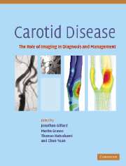
Stroke is a major cause of morbidity and mortality, with carotid disease representing an important contributory risk factor. This book is about the pathogenesis and management of carotid disease with specific focus on the role imaging has to play in the early recognition of symptomatic and asymptomatic disease as well as the treatment of the developed condition. Technological advances in imaging modalities now allow detailed analysis of the disease progression, the prediction of critical events leading to a stroke, as well as the identification of the most effective surgical or other interventional treatments. This book should be read by neurologists, cardiologists, vascular surgeons, neurosurgeons and radiologists involved in the care of patients with carotid disease, and also by researchers involved in the development of new therapeutic techniques and drugs.

Reviews

'This is a very useful book. … an excellent monograph on a rapidly developing topic which it covers well.'

Source: Neuroradiology

'Another quality of the book is the adequate balance among the 35 chapters. The editors should be praised for producing a pleasing consistency of style. Production is excellent with helpful tables and good illustrations. A welcome list of abbreviations used through the work can be found at the beginning of the book.'

Source: Pediatric Radiology

'This book provides a comprehensive review of the current state of imaging both for the assessment of carotid stenosis, and particularly of the methods and rationale for plaque imaging. The reviews are well-written and up to date with comprehensive references. … I think this book is of particular value to departments and practitioners that have a large carotid practice, particularly those who want to develop or research imaging of plaque morphology.'

Source: Clinical Radiology

Refine List

Actions for selected content:

Select all | Deselect all
  • View selected items
  • Export citations
  • Download PDF (zip)
  • Save to Kindle
  • Save to Dropbox
  • Save to Google Drive

Save Search

You can save your searches here and later view and run them again in "My saved searches".

Please provide a title, maximum of 40 characters.
×

Contents


Page 2 of 2


  • 22 - Carotid magnetic resonance direct thrombus imaging
    pp 302-312
    • By Alan Moody, Sunnybrook Health Sciences Centre, Toronto ON, Canada
  • View abstract

    Summary

    Atherosclerosis is the basis of the majority of carotid artery disease which, via occlusive/stenotic disease and subsequent thromboembolic events, results in end organ (brain) damage. An element of the atheromatous plaque now becoming appreciated as potentially important in progression and rupture is intraplaque hemorrhage (IPH). Applying T1-weighted magnetic resonance imaging (MRI) to patients suspected of suffering deep vein thrombosis or pulmonary embolism, the identification of high signal within the deep veins of the legs or the arteries of the lungs was shown to accurately identify intravascular thrombus. At the same time as intravascular methemoglobin was under investigation there was increasing interest in using MRI to directly image atherosclerotic disease within the vessel wall. Exploiting the already proven ability of MRI to generate high signal from methemoglobin, the possibility exists to specifically target hemorrhage within plaque using this technique.
  • Plaque modelling
  • View abstract

    Summary

    This chapter concentrates on computational simulation based on magnetic resonance imaging (MRI) and ultrasound imaging. It explores the flow structure and wall shear stress distributions, and describes the relationship with arterial disease patterns. An accurate description of 3D vessel geometry is essential for accurate modelling of blood flow using computational fluid dynamics (CFD), and magnetic resonance angiography (MRA) has been the most popular technique for obtaining the information in vivo. However, for superficial vessels such as the carotid and femoral arteries, extravascular 3D ultrasound can be a cost-effective alternative to MRA. Extravascular 3D ultrasound has potential to become a relatively inexpensive, fast and accurate alternative to MRI for CFD-based hemodynamics studies of superficial arteries. Standardized imaging protocols with high quality images will certainly help to reduce the manpower needed for model reconstruction and preparation, and to minimize operator dependence of the reconstruction process.
  • 24 - Mechanical image analysis using finite element method
    pp 324-340
    • By Dalin Tang, Worcester Polytechnic Institute, Worcester, MA, USA, Chun Yang, Worcester Polytechnic Institute, Worcester, MA, USA; Beijing Normal University, Beijing, China, Chun Yuan, University of Washington, Seattle, WA 98195, USA
  • View abstract

    Summary

    This chapter describes the mechanical image analysis using finite element (FE) method for atherosclerotic plaques. Q. Long combined computational fluid dynamics (CFD) modeling and MRI techniques together to perform patient-specific flow analysis based on in vivo magnetic resonance imaging (MRI) images from real patients. Phase contrast (PC) MRI was used to measure time varying inlet/outlet flow rates which are used as boundary conditions (BC) for the subject-specific CFD model. Image-based computational modeling is adding a new dimension (mechanical analysis) to atherosclerostic plaque image analysis. With advanced imaging and computational modeling techniques, atherosclerotic plaques can be analyzed under fluid-structure interaction environment and critical flow, and stress/strain conditions can be identified and used for more accurate plaque rupture risk assessment and predictions. Combination of imaging analysis, computational modeling, and clinical studies may lead to establishment of new industrial standard, gold standard for arterial disease assessment, and new noninvasive diagnostic and screening procedures.
  • Monitoring the local and distal effects of carotid interventions
  • View abstract

    Summary

    Transcranial Doppler (TCD) utilizes the Doppler principle to determine the direction and velocity of blood flow. Most TCDs use long sample volumes in order to improve the signal-to-noise ratio and ease the detection of the basal cerebral arteries. Most TCDs use the fast Fourier transform (FFT) method of spectral analysis which produces the typical visual representation of blood flow velocity. The FFT method of spectral analysis is used in most TCD systems because it allows almost instantaneous detection and display of information in a form which is understandable to most observers. Pulsatility and resistance indices reflect characteristics of the Doppler shift velocity waveforms, and indicate the degree of pulsatility of the waveform. TCD is able to detect two of the major causes of neurological deficits that are abnormalities in blood flow and cerebral embolization. These have made it a valuable practical tool for treating patients in diverse clinical disciplines.
  • 26 - Imaging carotid disease: MR and CT perfusion
    pp 358-371
  • View abstract

    Summary

    Determination of the actual cerebral hemodynamical status at the tissue level in patients with carotid artery disease remains one of the potential methods to determine the risk for recurrent, hemodynamically induced, symptoms in patients with carotid artery disease. Magnetic resonance imaging (MRI) offers two different approaches to measure the cerebral perfusion. The first technique, arterial spin labeling (ASL), exploits the water in blood as an endogenous tracer. Dynamic susceptibility contrast MRI (DSC-MRI or MR bolus tracking) is the alternative method. This method uses an intravenous injection of contrast agent, applied as an exogenous tracer. Both techniques have been used to study the CBF in patients with stenotic artery disease, showing their ability to provide important information on the hemodynamic status. Perfusion computed tomography (CT) is useful to evaluate the hemodynamic state of patients with chronic major cerebral artery occlusive disorders.
  • 27 - Near infrared spectroscopy in carotid endarterectomy
    pp 372-386
  • View abstract

    Summary

    The use of near infrared spectroscopy (NIRS) to evaluate cerebral oxygenation changes has been reported in a variety of medical and neurosurgical conditions. This chapter looks at the application of NIRS during carotid endarterectomy. It demonstrates our own use of this technique in combination with other monitoring modalities. The NIRO 300 is a noninvasive bedside monitor which gives simultaneous measurement of tissue oxygen index (TOI) and hemoglobin concentration changes. The instrument is based on technology employed in the earlier NIRO 500 and a prototype spatially resolved spectrometer. The reliable detection of compromised regional cerebral perfusion therefore plays an important role in the management of patients undergoing carotid surgery. During carotid endarterectomy, segregation of the NIRS signal between intra- and extracranial vascular territories is possible, by staged application of vascular clamps to the external and internal carotid arteries.
  • 28 - Single photon emission computed tomography (SPECT)
    pp 387-395
  • View abstract

    Summary

    This chapter discusses the utility of single-photon emission computed tomography (SPECT) in the evaluation of carotid disease and interventions. A prospective study demonstrated that reduced regional cerebrovascular reactivity (rCVR) to acetazolamide determined quantitatively using 133Xe SPECT is significantly associated with an increased risk of stroke recurrence in patients with symptomatic occlusion of the middle cerebral artery (MCA) or internal carotid artery. Post-operative hyperperfusion is observed only in patients with reduced preoperative cerebrovascular reactivity to acetazolamide. Preoperative assessment of cerebral blood flow (CBF) with acetazolamide challenge using SPECT can identify patients at risk for postcarotid endarterectomy hyperperfusion. A SPECT study performed between the first and third post-operative day could be advocated for the timely and reliable identification of patients at risk for hyperperfusion syndrome. CBF measurements can determine appropriate withdrawal of blood pressure control in patients with ischemic events related to other atherosclerotic steno-occlusive lesions.
  • 29 - Monitoring carotid interventions with xenon CT
    pp 396-417
  • View abstract

    Summary

    Direct measurements of cerebral blood flow (CBF) by 133xenon or stable xenon computerized tomography (CT) or other methods appear to be the most reliable measure of the adequacy of flow. Other techniques including transcranial Doppler (TCD), cerebral angiography, and various types of magnetic resonance imaging (MRI) are proxy measures for this value. Positron emission tomography (PET) measures CBF and several variables that are interrelated in a consistent and predictable manner to decreasing perfusion pressure. J. P. Witt analyzed quantitative CBF data obtained with Xenon CT CBF technology prior to and during BTO, and classified 11 separate response patterns based on symmetry and degree of CBF change after balloon test occlusion (BTO). Metabolic response of the brain in symptomatic carotid occlusion has been characterized by MR spectroscopy. The hemodynamic physiology of carotid occlusion has been extensively studied in many disparate clinical situations.
  • Monitoring pharmaceutical interventions
  • View abstract

    Summary

    This chapter discusses the use of vascular imaging data to advance the development of new therapies from the perspective of the pharmaceutical industry. It discusses the technologies currently used in large phase-3 studies, and addresses the technical, clinical, and regulatory considerations for the purpose of drug development and regulatory approval. M. A. Espeland reviewed the utility of carotid ultrasound as a clinical research tool to monitor atherosclerosis progression, and discussed the concept of surrogacy with respect to imaging data, and using the clinical and statistical criteria that are required for research data to be considered clinically meaningful. The chapter presents the angiography and intra- and extravascular ultrasound, to support their use in clinical trials for the development of antiatherosclerotic therapies. The technical validation elements to be addressed in a regulatory submission should have background information on the hardware, analytical software, training, data processing, data transfer, and quality control.
  • 31 - Monitoring pharmaceutical interventions with conventional ultrasound (IMT)
    pp 430-450
    • By John R. Crouse, Wake Forest University School of Medicine, Winston-Salem NC, USA
  • View abstract

    Summary

    This chapter deals with established correlates of intima-media thickness (IMT) as well as new findings and formulations of data derived from this technology. The reliability and validity of the method are well described as are the relationships with risk factors, although new risk factors continue to be tested for associations with IMT. Differences in progression are now routinely used to define surrogate outcomes for clinical trials of behavioral and pharmacologic intervention; the Food and Drug Administration (FDA) accepts this as a valid marker of atherosclerosis change. Of considerable interest are insights that B-mode ultrasound provides into the process of vascular remodeling. The chapter discusses the associations of IMT with prevalent as well as incident coronary artery and cerebrovascular disease. Thus B-mode ultrasound has clearly become a mature technology for quantification and monitoring of the extent and progression of IMT of the extracranial carotid arteries.
  • 32 - Monitoring pharmaceutical interventions with IVUS
    pp 451-463
  • View abstract

    Summary

    The application of intravascular ultrasound (IVUS) within the coronary arteries has allowed for an enhanced characterization of the size and distribution of atheroma and the natural history of the arterial wall's response to its accumulation. IVUS is performed safely at the time of diagnostic coronary angiography and involves the placement of a high frequency ultrasound transducer on the tip of a catheter within the major epicardial coronary arteries. IVUS has been used to estimate the extent of atheroma throughout segments of coronary arteries. The ability to visualize the entire arterial wall in vivo has allowed IVUS to be employed to characterize the accumulation of atherosclerotic plaque within the arterial wall. A continuous relationship was demonstrated between changes in low density lipoprotein (LDL) cholesterol and atheroma volume. IVUS has been employed to investigate the formation of neointimal hyperplasia in a range of clinical settings.
  • 33 - Monitoring of pharmaceutical interventions: MR plaque imaging
    pp 464-470
  • View abstract

    Summary

    Magnetic resonance imaging (MRI) detects significant changes in the morphology of atherosclerotic plaque during the course of lipid-lowering therapy, and demonstrates the promise of this noninvasive imaging modality for use in clinical trials. Images obtained from the placebo-control arm of a five-site carotid MRI study were analyzed to assess the variability of MRI for measuring lesion size and composition, and to provide sample size calculations for a variety of imaging endpoints. MRI is capable of quantifying atherosclerotic lesion size and plaque composition in the setting of a multicenter trial with low interscan variability. Sample-size calculations based on reproducibility data for carotid MRI indicate that clinical trials involving approximately 150 subjects per treatment arm would be sufficiently powered to detect a 5% difference in treatment effect. MRI may provide valuable insight into pathophysiology and mechanisms of pharmacologic activity.
  • Future directions in carotid plaque imaging
  • View abstract

    Summary

    Developments in both magnetic resonance imaging (MRI) technology, computer software and a huge array of potential cellular and molecular targets are a step toward the identification of high-risk carotid disease. This chapter covers the recent advances with particular respect to MRI. The carotid artery plaque is ideally suited for imaging by multicontrast MRI. The chapter discusses a variety of molecular targets that may provide improved imaging of carotid atherosclerotic plaque using MRI. The presence of neovessels is strongly associated with plaque inflammation and likelihood of rupture, presumably by allowing an alternative route for entry of monocytes and lymphocytes into the plaque. Histological studies have demonstrated that superficial thrombus superimposed on a ruptured atherosclerotic plaque characterizes those plaques at high risk of ischemic events. The chapter describes the role of matrix metalloproteinases (MMPs) in plaque instability and matrix remodeling in atherosclerotic plaques.
  • 34 - Molecular imaging of carotid artery disease
    pp 471-483
    • By James H. F. Rudd, The Zena and Michael A. Wiener Cardiovascular Institute, The Marie-Josée and Henry R. Kravis Cardiovascular Health Center, Mount Sinai School of Medicine, New York NY, USA, Michael J. Lipinski, The Zena and Michael A. Wiener Cardiovascular Institute, The Marie-Josée and Henry R. Kravis Cardiovascular Health Center, Mount Sinai School of Medicine, New York NY, USA, Fabien Hyafil, The Zena and Michael A. Wiener Cardiovascular Institute, The Marie-Josée and Henry R. Kravis Cardiovascular Health Center, Mount Sinai School of Medicine, New York NY, USA, Zahi A. Fayad, The Zena and Michael A. Wiener Cardiovascular Institute, The Marie-Josée and Henry R. Kravis Cardiovascular Health Center, Mount Sinai School of Medicine, New York NY, USA
  • 35 - Future technical developments
    pp 484-498
  • View abstract

    Summary

    Therapeutic strategies for treatment of atherosclerosis have changed focus from plaque reduction to plaque stabilization. Magnetic resonance imaging (MRI) is highly advantageous because it is capable of providing high-resolution images without requirement for invasive transducers or ionizing radiation, providing a desirable method for longitudinal studies of atherosclerosis. MRI has shown great potential for detailed analysis of plaque structure, cellular content, and even molecular content and activity, and together, these characteristics give MRI the most promising potential of all modalities for differentiating between stable and unstable plaque. The choice of pulse sequence(s) used for a given study should be considered carefully, as specific applications will benefit from certain pulse sequences. The three hardware subsystems that are important in the discussion of technical advancements are: the MR machine; the gradient coils; and the radiofrequency (RF) coils. A brief introduction to each component and the novelinnovations being developed is provided.

Page 2 of 2


Metrics

Full text views

Total number of HTML views: 0
Total number of PDF views: 0 *
Loading metrics...

Book summary page views

Total views: 0 *
Loading metrics...

* Views captured on Cambridge Core between #date#. This data will be updated every 24 hours.

Usage data cannot currently be displayed.