Skip to main content Accessibility help
×
Hostname: page-component-76fb5796d-5g6vh Total loading time: 0 Render date: 2024-04-28T12:28:48.629Z Has data issue: false hasContentIssue false

Chapter 6 - Optical coherence tomography and low-contrast acuity

Published online by Cambridge University Press:  05 May 2015

Peter A. Calabresi
Affiliation:
Department of Neurology, Johns Hopkins University Hospital, Baltimore
Laura J. Balcer
Affiliation:
Department of Neurology, NYU Langone Medical Center, New York
Elliot M. Frohman
Affiliation:
Department of Neurology, UT Southwestern Medical Center, Dallas
Get access

Summary

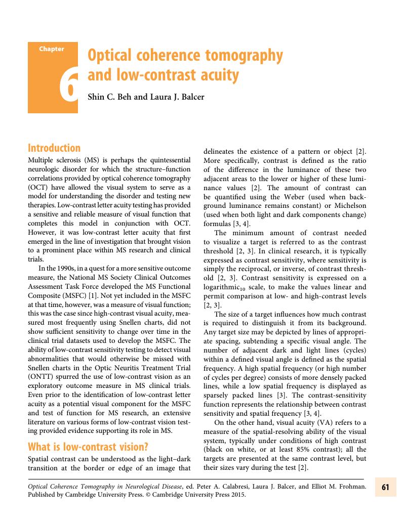
Image of the first page of this content. For PDF version, please use the ‘Save PDF’ preceeding this image.'
Type
Chapter
Information
Publisher: Cambridge University Press
Print publication year: 2015

Access options

Get access to the full version of this content by using one of the access options below. (Log in options will check for institutional or personal access. Content may require purchase if you do not have access.)

References

Sakai, RE, Feller, DJ, Galetta, KM, et al. Vision in multiple sclerosis: the story, structure-function correlations,and models for neuroprotection. J Neuroophthalmol 2011; 31: 362–73CrossRefGoogle ScholarPubMed
Owsley, C. Contrast sensitivity. Ophthalmol Clin N Am 2003; 16: 171–7CrossRefGoogle ScholarPubMed
Richman, J, Spaeth, GL, Wirotsko, B. Contrast sensitivity basics and a critique of currently available tests. J Cataract Refract Surg 2013; 39: 1100–6CrossRefGoogle Scholar
Pelli, DG, Bex, P. Measuring contrast sensitivity. Vision Res 2013; 90: 10–4CrossRefGoogle ScholarPubMed
Gouras, P. Identification of cone mechanisms in monkey ganglion cells. J Physiol 1968; 199: 533–47CrossRefGoogle ScholarPubMed
Levanthal, AG, Roedick, RW, Dreher, B. Retinal ganglion cell classes in the Old World monkey: morphology and central projections. Science 1981; 213: 1139–42Google Scholar
Shapley, R, Perry, VH. Cat and monkey retinal ganglion cells and their visual function roles. Trends Neurosci 1986; 9: 235CrossRefGoogle Scholar
Dreher, B, Fukada, Y, Roedick, RW. Identification, classification and anatomical segregation of cells with X-like and Y-like properties in the lateral geniculate nucleus of old-world primates. J Neurophysiol 1976; 258: 433–52Google ScholarPubMed
Hubel, DH, Wiesel, TN. Laminar and columnar distribution of geniculo-cortical fibers in the macaque monkey. J Comp Neurol 1972; 146: 421–50CrossRefGoogle ScholarPubMed
Hendrickson, AE, Wilson, JR, Ogren, MP. The neuroanatomical organization of pathways between the dorsal lateral geniculate nucleus and visual cortex in Old World and New World primates. J Comp Neurol 1978; 182: 123–36Google ScholarPubMed
Blasdel, GG, Lund, JS. Termination of afferent axons in macaque striate cortex. J Neurosci 1983; 3: 1389–413CrossRefGoogle ScholarPubMed
Hubel, DH, Wiesel, TN. Effects of varying stimulus size and color on single lateral geniculate cells in Rhesus monkeys. Proc Natl Acad Sci U S A 1966; 55: 1345–6CrossRefGoogle ScholarPubMed
Kaplan, E, Shapley, RM. X and Y cells in the lateral geniculate nucleus of macaque monkeys. J Physiol 1982; 330: 125–43CrossRefGoogle ScholarPubMed
Derrington, AM, Lennie, P. Spatial and temporal contrast sensitivities of neurons in lateral geniculate nucleus of macaque. J Physiol 1984; 357: 219–40CrossRefGoogle ScholarPubMed
Livingstone, MS, Hubel, DH. Psychophysical evidence for separate channels for the perception of form, color, movement, and depth. J Neurosci 1987; 7: 3416–68CrossRefGoogle ScholarPubMed
Lee, BB, Martin, PR, Valberg, A. Sensitivity of macaque retinal ganglion cells to chromatic and luminance flicker. J Physiol 1989; 414: 223–43Google ScholarPubMed
Lee, BB, Pokorny, J, Smith, VC, et al. Luminance and chromatic modulation sensitivity of macaque ganglion cells and human observers. J Opt Soc Am A 1990; 7: 2223–36CrossRefGoogle ScholarPubMed
Laycock, R, Crewther, SG, Crewther, DP. A role for the ‘magnocellular advantage’ in visual impairments in neurodevelopmental and psychiatric disorders. Neurosci Biobehav Rev 2007; 31: 363–76CrossRefGoogle ScholarPubMed
McKendrick, AM, Sampson, GP, Walland, MJ, Badcock, DR. Contrast sensitivity changes due to glaucoma and normal aging: low-spatial frequency losses in both magnocellular and parvocellular pathways. Invest Ophthalmol Vis Sci 2007; 48: 2115–22CrossRefGoogle ScholarPubMed
Risacher, SL, Wudunn, D, Pepin, SM, et al. Visual contrast sensitivity in Alzheimer’s disease, mild cognitive impairment, and older adults with cognitive complaints. Neurobiol Aging 2013; 34: 1133–44CrossRefGoogle ScholarPubMed
Anderson, AJ, Johnson, CA. Frequency-doubling technology perimetry. Opthalmol Clin North Am 2003; 16: 213–25Google ScholarPubMed
De Monasterio, FM, Gouras, P. Functional properties of ganglion cells of the rhesus monkey retina. J Physiol 1975; 251: 167–95Google ScholarPubMed
Wiesel, TN, Hubel, DH. Spatial and chromatic interactions in the lateral geniculate body of the rhesus monkey. J Neurophysiol 1966; 29: 1115–56CrossRefGoogle ScholarPubMed
Hicks, TP, Lee, BB, Vidyasagar, TR. The responses of cells in macaque lateral geniculate nucleus to sinusoidal gratings. J Physiol 1983; 337: 183200CrossRefGoogle ScholarPubMed
Hendry, SH, Yoshioka, T. A neurochemically distinct third channel in the macaque dorsal lateral geniculate nucleus. Science 1994; 264: 575–7CrossRefGoogle ScholarPubMed
Johnson, JK, Casagrande, VA. Distribution of calcium-binding proteins within the parallel visual pathways of a primate (Galago crassicaudatus). J Comp Neurol 1995; 356: 238–60CrossRefGoogle ScholarPubMed
Hendry, SH, Casagrande, VA. A common pattern for a third visual channel in the primate LGN. Soc Neurosci Abstr 1996; 22: 1605Google Scholar
Goodchild, AK, Martin, PR. The distribution of calcium-binding proteins in the lateral geniculate nucleus and visual cortex of a New World monkey, the marmoset, Callithrix jacchus. Vis Neurosci 1998; 15: 625–42CrossRefGoogle ScholarPubMed
Xu, X, Ichida, JM, Allison, JD, et al. A comparison of koniocellular, magnocellular, and parvocellular receptive field properties in the lateral geniculate nucleus of the owl monkey (Aotus trivirgatus). J Physiol 2001; 531: 203–18CrossRefGoogle ScholarPubMed
Pelli, DG, Robson, JG, Wilkins, AJ. Designing a new letter chart for measuring contrast sensitivity. Clin Vision Sci 1988; 2: 187–99Google Scholar
Leat, SJ, Woo, GC. The validity of current clinical tests of contrast sensitivity and their ability to predict reading speed in low vision. Eye 1997; 11: 893–9CrossRefGoogle ScholarPubMed
Balcer, LJ, Baier, ML, Pelak, VA, et al. New low-contrast vision charts: reliability and test characteristics in patients with multiple sclerosis. Mult Scler 2000; 6: 163–71CrossRefGoogle ScholarPubMed
Bodis-Wollner, I, Diamond, SP. The measurement of spatial contrast sensitivity in cases of blurred vision associated with cerebral lesions. Brain 1976; 99: 695710CrossRefGoogle ScholarPubMed
Regan, D, Silver, R, Murray, TJ. Visual acuity and contrast sensitivity in multiple sclerosis – hidden visual loss: an auxiliary diagnostic test. Brain 1977; 100: 563–79CrossRefGoogle ScholarPubMed
Kupersmith, MJ, Seiple, WH, Nelson, JI, Carr, RE. Contrast sensitivity loss in multiple sclerosis. Selectivity by eye, orientation, and spatial frequency measured with the evoked potential. Invest Ophthalmol Vis Sci 1984; 25: 632–9Google ScholarPubMed
Nordmann, JP, Saraux, H, Roullet, E. Contrast sensitivity in multiple sclerosis: a study in 35 patients with and without optic neuritis. Ophthalmologica 1987; 195: 199204CrossRefGoogle ScholarPubMed
Regan, D, Neima, D. Low-contrast letter charts as a test of visual function. Ophthalmology 1983; 90: 1192–200CrossRefGoogle ScholarPubMed
Regan, D, Neima, D. Low-contrast letter charts in early diabetic retinopathy, ocular hypertension, glaucoma, and Parkinson’s disease. Br J Ophthalmol 1984; 68: 885–9CrossRefGoogle ScholarPubMed
Baier, ML, Cutter, GR, Rudick, RA, et al. Low-contrast letter acuity testing captures visual dysfunction in patients with multiple sclerosis. Neurology 2005; 64: 992–5CrossRefGoogle ScholarPubMed
Bailey, IL, Lovie, JE. New design principles for visual acuity letter charts. Am J Optom Physiol Opt 1976; 53: 740–5CrossRefGoogle ScholarPubMed
Ferris, FL, Kassoff, A, Bresnick, GH, et al. New visual acuity charts for clinical research. Am J Ophthalmol 1982; 94: 91–6Google ScholarPubMed
Ferris, FL, Bailey, I. Standardizing the measurement of visual acuity for clinical research studies: guideline from the Eye Care Technology Forum. Ophthalmology 1996; 103: 181–2CrossRefGoogle ScholarPubMed
Balcer, LJ, Galetta, SL, Calabresi, PA, et al. Natalizumab reduces visual loss in patients with relapsing multiple sclerosis. Neurology 2007; 68: 1299–304CrossRefGoogle ScholarPubMed
Talman, LS, Bisker, ER, Sackel, DJ, et al. Longitudinal study of vision and retinal nerve fiber layer thickness in multiple sclerosis. Ann Neurol 2010; 67: 749–60CrossRefGoogle ScholarPubMed
Rubin, GS, Roche, KB, Prasada-Rao, P, Fried, LP. Visual impairment and disability in older adults. Optom Vis Sci 1994; 71: 750–60CrossRefGoogle ScholarPubMed
Szylick, JS, Seiple, W, Fishman, GA, et al. Perceived and actual performance of daily tasks: relationship to visual function tests in individuals with retinitis pigmentosa. Ophthalmology 2001; 108: 6575Google Scholar
Rubin, GS, Legge, GE. Psychophysics of reading. VI – The role of contrast in low vision. Vision Res 1989; 29: 7991CrossRefGoogle ScholarPubMed
Whittaker, SG, Lovie-Kitchin, J. Visual requirements for reading. Optom Vis Sci 1993; 70: 5465CrossRefGoogle ScholarPubMed
Crossland, MD, Rubin, GS. Text accessibility by people with reduced contrast sensitivity. Optom Vis Sci 2012; 89: 1276–81CrossRefGoogle ScholarPubMed
Marron, JA, Bailey, IL. Visual factors and orientation-mobility performance. Am J Optom Physiol Opt 1982; 59: 413–26CrossRefGoogle ScholarPubMed
Startzell, JK, Owens, DA, Mulfinger, LM, Cavanagh, PR. Stair negotiation in older people: a review. J Am Geriatr Soc; 48: 567–80CrossRefGoogle Scholar
Freeman, EE, Munoz, B, Turano, KA, West, SK. Measures of visual function and their association with driving modification in older adults. Invest Opthalmol Vis Sci 2006; 47: 514–20Google ScholarPubMed
Owsley, C, Sekuler, R, Boldt, C. Aging and low-contrast vision: face perception. Invest Ophthalmol Vis Sci 1981; 21: 362–5Google ScholarPubMed
Owsley, C, Sloane, ME. Contrast sensitivity, acuity, and the perception of “real world” targets. Br J Ophthalmol 1987; 71: 791–6CrossRefGoogle ScholarPubMed
McCulloch, DL, Loffler, G, Colguhoun, K, et al. The effects of visual degradation on face discrimination. Ophthalmic Physiol Opt 2011; 31: 240–8CrossRefGoogle ScholarPubMed
Elliott, DB, Hurst, MA, Weatherhill, J. Comparing clinical tests of visual function with the patient’s perceived visual disability. Eye 1990; 4: 712–7CrossRefGoogle ScholarPubMed
Lord, S, Dayhew, J. Visual factors for falls in older people. J Am Geriatr Soc 2001; 49: 508–15CrossRefGoogle ScholarPubMed
French, D, Campbell, R, Spehar, A, et al. Drugs and falls in community dwelling older people: a national veterans study. Clin Therapeutics 2006; 28: 619–30CrossRefGoogle ScholarPubMed
Leat, SJ, Legge, GE, Bullimore, MA. What is low vision? A re-evaluation of definitions. Optom Vis Sci 1999; 76: 198211CrossRefGoogle ScholarPubMed
Mowry, EM, Loquidice, MJ, Daniels, AB, et al. Vision related quality of life in multiple scleorsis: correlation with new measures of low and high contrast letter acuity. J Neurol Neurosurg Psychiatry 2009; 80: 767–72CrossRefGoogle Scholar
Elliot, DB, Whitaker, D. How useful are contrast sensitivity charts in optometric practice? Case reports. Optom Vis Sci 1992; 69: 378–85Google Scholar
Pineles, SL, Birch, EE, Talman, LS, et al. One eye or two: a comparison of binocular and monocular low-contrast acuity testing in multiple sclerosis. Am J Ophthalmol 2011; 152: 133–40CrossRefGoogle ScholarPubMed
Newman, NJ, Wolfe, JM, Steward, MI, Lessell, S. Binocular visual function in patients with a history of monocular optic neuritis. Clin Vision Sci 1991; 6: 95107Google Scholar
Gillespie-Gallery, H, Konstantakopoulou, E, Harlow, JA, Barbur, JL. Capturing age-related changes in functional contrast sensitivity with decreasing light levels in monocular and binocular vision. Invest Ophthalmol Vis Sci 2013; 54: 6093–103CrossRefGoogle ScholarPubMed
Pineles, SL, Velez, FG, Isenberg, SJ, et al. Functional burden of strabismus: decreased binocular summation and binocular inhibition. JAMA Ophthalmol 2013; 131: 1413–19CrossRefGoogle ScholarPubMed
Gallo, A, Esposito, F, Sacco, R, et al. Visual resting-state network in relapsing-remitting MS with and without previous optic neuritis. Neurology 2012; 79: 1458–65CrossRefGoogle ScholarPubMed
Collins, JW, Carney, LG. Visual performance in high myopia. Curr Eye Res 1990; 9: 217–23CrossRefGoogle ScholarPubMed
Hess, R, Woo, G. Vision through cataracts. Invest Ophthalmol Vis Sci 1978; 17: 428–35Google ScholarPubMed
Abrahamsson, M, Sjostrand, J. Impairment of contrast sensitivity function (CSF) as a measure of disability glare. Invest Ophthalmol Vis Sci 1986; 27: 1131–6Google ScholarPubMed
Gil, MA, Varon, C, Rosello, N, et al. Visual acuity, contrast sensitivity, subjective quality of vision, and quality of life with 4 different multifocal IOLs. Eur J Ophthalmol 2012; 22: 175–87CrossRefGoogle ScholarPubMed
Freedman, RD, Thibos, LN. Contrast sensitivity in humans with abnormal visual experience. J Physiol 1975; 247: 687710CrossRefGoogle Scholar
Rogers, SA, Khan-Lim, D, Manners, RM. Does upper lid blepharoplasty improve contrast sensitivity? Ophthal Plast Reconstr Surg 2012; 28: 163–5CrossRefGoogle ScholarPubMed
Coe, CD, Bower, KS, Brooks, DB, et al. Effect of blast trauma and corneal foreign bodies on visual performance. Optom Vis Sci 2010; 87: 604–11CrossRefGoogle ScholarPubMed
Sjostrand, J, Frisen, L. Contrast sensitivity in macular disease: a preliminary report. Acta Ophthalmol (Copenh) 197;55: 507–14CrossRefGoogle Scholar
Kleiner, RC, Enger, C, Alexander, MF, Fine, SL. Contrast sensitivity in age-related macular degeneration. Arch Ophthalmol 1988; 106: 55–7CrossRefGoogle ScholarPubMed
Owsley, C, Sloane, ME, Skalka, HW, et al. A comparison of the Regan Low-Contrast Letter Charts and contrast sensitivity testing in older patients. Clin Vis Sci 1990; 5: 325–34Google Scholar
Rolando, M, Lester, M, Macri, A, Calabria, G. Low spatial-contrast sensitivity in dry eyes. Cornea 1993; 17: 376–9Google Scholar
Kappel, PJ, Monnet, D, Yu, F, et al. Contrast sensitivity among patients with birdshot chorioretinopathy. Am J Ophthalmol 2009; 147: 351–6CrossRefGoogle ScholarPubMed
Lindberg, CR, Fishman, GA, Anderson, RJ, Vasquez, V. Contrast sensitivity in retinitis pigmentosa. Br J Ophthalmol 1981; 65: 855–8CrossRefGoogle ScholarPubMed
Yioti, GC, Kalogeropoulos, CD, Aspiotis, MB, Stefaniotou, MI. Contrast sensitivity and color vision in eyes with retinitis pigmentosa and good visual acuity: correlations with SD-OCT findings. Ophthalmic Surg Lasers Imaging 2012; 43: S44S53CrossRefGoogle ScholarPubMed
Midena, E, Segato, T, Bottin, G, et al. The effect on the macular function of laser photocoagulation for diabetic macular edema. Graefes Arch Clin Exp Ophthalmol 1992; 230: 162–5CrossRefGoogle ScholarPubMed
Greenstein, VC, Chen, H, Hood, DC, et al. Retinal function in diabetic macular edema after focal laser photocoagulation. Invest Ophthalmol Vis Sci 2000; 41: 3655–64Google ScholarPubMed
Stamper, RL. The effect of glaucoma on central visual function. Trans Am Ophthalmol Soc 1984; 82: 792826Google ScholarPubMed
Johnson, CA. Psychophysical measurement of glaucomatous damage. Surv Ophthalmol 2001; 45: S313S318CrossRefGoogle ScholarPubMed
Gandolfi, SA. Improvement in spatial contrast sensitivity threshold after surgical reduction of intraocular pressure in unilateral high-tension glaucoma. Invest Ophthalmol Vis Sci 2005; 46: 197201CrossRefGoogle ScholarPubMed
Skrandies, W, Gottlob, I. Alterations of visual contrast sensitivity in Parkinson’s disease. Hum Neurobiol 1986; 5: 255–9Google ScholarPubMed
Kupersmith, MJ, Shakin, E, Siegel, IM, et al. Visual system abnormalities in patients with Parkinson’s disease. Arch Neurol 1982; 39: 284–6CrossRefGoogle ScholarPubMed
Peppe, A, Stanzione, P, Pierelli, F, et al. Low contrast stimuli enhance PERG sensitivity to the visual dysfunction in Parkinson’s disease. Electroencephalogr Clin Neurophysiol 1992; 82: 453–7CrossRefGoogle Scholar
Langheinric, T, Tebartz van Elst, L, Lagreze, WA, et al. Visual contrast response functions in Parkinson’s disease: evidence from electroretinograms, visually evoked potentials and psychophysics. Clin Neurophysiol 2000; 111: 6674CrossRefGoogle Scholar
Cronin-Golomb, A, Corkin, S, Rizzo, JF, et al. Visual dysfunction in Alzheimer’s disease: relation to normal aging. Ann Neurol 1991; 29: 4152CrossRefGoogle ScholarPubMed
Gilmore, GC, Levy, JA. Spatial contrast sensitivity in Alzheimer’s disease: a comparison of two methods. Optom Vis Sci 1991; 68: 790–4Google ScholarPubMed
Sadun, AA, Borchert, M, DeVita, E, et al. Assessment of visual impairments in Alzheimer’s disease. Am J Ophthalmol 1987; 104: 113–20CrossRefGoogle Scholar
Barboni, MT, Nagy, BV, de Araujo Moura, AL, et al. ON and OFF electroretinography and contrast sensitivity in Duchenne muscular dystrophy. Invest Ophthalmol Vis Sci 2013; 54: 3195–204CrossRefGoogle ScholarPubMed
Ribeiro, MJ, Violante, IR, Bernardino, I, et al. Abnormal achromatic and chromatic contrast sensitivity in neurofibromatosis type 1. Invest Ophthalmol Vis Sci 2012; 53: 287–93CrossRefGoogle ScholarPubMed
Braunitzer, G, Rokszin, A, Kobor, J, Benedek, G. Is the development of visual contrast sensitivity impaired in children with migraine? An exploratory study. Cephalalgia 2010; 30: 991–5CrossRefGoogle ScholarPubMed
Shepherd, AJ, Beaumont, HM, Hine, TJ. Motion processing deficits in migraine are related to contrast sensitivity. Cephalalgia 2012; 32: 554–70CrossRefGoogle ScholarPubMed
Wall, M. Contrast sensitivity testing in pseudotumor cerebri. Ophthalmology 1986; 93: 47CrossRefGoogle ScholarPubMed
Verplanck, M, Kaufman, DI, Parson, T, et al. Electrophysiology versus psychophysics in the detection of visual loss in pseudotumor cerebri. Neurology 1988; 38: 1789–92CrossRefGoogle ScholarPubMed
Buncie, JR, Tytla, ME. Contrast sensitivity in shunted hydrocephalus. Invest Ophthalmol Vis Sci 1989; 30: 407Google Scholar
Wall, M, Conway, MD, House, PH, et al. Evaluation of sensitivity and specificity of spatial resolution and Humphrey automated perimetry in pseudotumor cerebri patients and normal subjects. Invest Ophthalmol Vis Sci 1991; 32: 3306–12Google ScholarPubMed
Lynch, DR, Farmer, JM, Rochestie, D, Balcer, LJ. Contrast letter acuity as a measure of visual dysfunction in patients with Friedreich ataxia. J Neuroophthalmol 2002; 22: 270–4CrossRefGoogle ScholarPubMed
Bodis-Wollner, I. Visual acuity and contrast sensitivity in patients with cerebral lesions. Science 1972; 178: 769–71CrossRefGoogle ScholarPubMed
Halasz, I, Levy-Gigi, E, Kelemen, O, et al. Neuropsychological functions and visual contrast sensitivity in schizophrenia: the potential impact of comorbid posttraumatic stress disorder (PTSD). Front Psychol 2013; 4: 136CrossRefGoogle ScholarPubMed
Bubl, E, Kern, E, Ebert, D, et al. Seeing gray when feeling blue? Depression can be measured in the eye of the diseased. Biol Psychiatry 2010; 68: 205–8CrossRefGoogle ScholarPubMed
Bubl, E, Ebert, D, Kern, E, et al. Effect of antidepressive therapy on retinal contrast processing in depressive disorder. Br J Psychiatry 2012; 201: 151–8CrossRefGoogle Scholar
Santaella, RM, Fraunfelder, FW. Ocular adverse effects associated with systemic medications: recognition and management. Drugs 2007; 67: 7593CrossRefGoogle ScholarPubMed
Sorri, I, Kalviainen, R, Mantyjarvi, M. Color vision and contrast sensitivity in epilepsy patients treated with initial tiagabine monotherapy. Epilepsy Res 2005; 67: 101–7CrossRefGoogle ScholarPubMed
Boeckelmann, I, Pfister, EA. Influence of occupational exposure to organic solvent mixtures on contrast sensitivity in printers. J Occup Environ Med 2003; 45: 2533CrossRefGoogle ScholarPubMed
Gong, Y, Kishi, R, Kasai, S, et al. Visual dysfunction in workers exposed to a mixture of organic solvents. Neurotoxicology 2003; 24: 703–10CrossRefGoogle ScholarPubMed
Costa, TL, Barboni, MT, Moura, AL, et al. Long-term occupational exposure to organic solvents affects color vision, contrast sensitivity, and visual fields. PLoS One 2012; 7: e42961CrossRefGoogle ScholarPubMed
Owsley, C. Aging and vision. Vision Res 2011; 51: 1610–22CrossRefGoogle ScholarPubMed
Mateus, C, Lemos, R, Silva, MF, et al. Aging of low and high level vision: from chromatic and achromatic contrast sensitivity to local and 3D object motion perception. PLoS One 2013; 8: e55348CrossRefGoogle ScholarPubMed
Jackson, GR, Owsley, C. Visual dysfunction, neurodegenerative diseases, and aging. Neurol Clin 2003; 21: 709–28CrossRefGoogle ScholarPubMed
Curcio, CA, Milican, CL, Allen, KA, et al. Aging of the human photoreceptor mosaic: evidence for selective vulnerability of rods in central retina. Invest Ophthalmol Vis Sci 1993; 34: 3278–96Google ScholarPubMed
Zhang, J, Wang, X, Wang, Y, et al. Spatial and temporal sensitivity degradation of primary visual cortical cells in senescent rhesus monkeys. Eur J Neurosci 2008; 28: 201–7CrossRefGoogle ScholarPubMed
Yang, Y, Liang, Z, Liang, Z, et al. Aging affects contrast response functions and adaptation of middle temporal visual area neurons in rhesus monkeys. Neuroscience 2008; 156: 748–57CrossRefGoogle ScholarPubMed
Adams, CW, Bullimore, MA, Wall, M, et al. Normal aging effects for frequency doubling technology perimetry. Optom Vis Sci 1999; 76: 582–7CrossRefGoogle ScholarPubMed
Anderson, AJ, Johnson, CA, Fingeret, M, et al. Characteristics of the normal database for the Humphrey Matrix Perimeter. Invest Ophthalmol Vis Sci 2005; 46: 1540–8CrossRefGoogle ScholarPubMed
Harnois, C, Di Paolo, T. Decreased dopamine in the retinas of patients with Parkinson’s disease. Invest Ophthalmol Vis Sci 1990; 31: 2473–5Google ScholarPubMed
Nightingale, S, Mitchell, KW, Howe, JW. Visual evoked cortical potentials and pattern electroretinograms in Parkinson’s disease and control subjects. J Neurol Neurosurg Psychiatry 1986; 49: 1280–7CrossRefGoogle ScholarPubMed
Gottlob, I, Scneider, E, Heider, W, et al. Alteration of visual evoked potentials and electroretinograms in Parkinson’s disease. Electroencephalogr Clin Neurophysiol 1987; 66: 349–57CrossRefGoogle ScholarPubMed
Reader, TA, Quesney, LF. Dopamine in the visual cortex of the cat. Experimentia 1986; 42: 1242–4CrossRefGoogle ScholarPubMed
Parkinson, D. Evidence for a dopaminergic innervations of cat primary visual cortex. Neuroscience 1989; 30: 171–9CrossRefGoogle ScholarPubMed
Papadopoulos, GC, Parnavelas, JG. Distribution and synaptic organization of dopaminergic axons in the lateral geniculate nucleus of the rat. J Comp Neurol 1990; 294: 356–61Google ScholarPubMed
Regan, D, Maxner, C. Orientation-selective visual loss in patients with Parkinson’s disease. Brain 1987; 110: 415–32CrossRefGoogle ScholarPubMed
Bulens, C, Meerwaldt, JD, Van der Wildt, GJ. Effect of stimulus orientation on contrast sensitivity in Parkinson’s disease. Neurology 1988; 38: 7681CrossRefGoogle ScholarPubMed
Katz, B, Rimmer, S, Iragui, V, et al. Abnormal pattern electroretinogram in Alzheimer’s disease: evidence for retinal ganglion cell degeneration. Ann Neurol 1989; 26: 221–5CrossRefGoogle ScholarPubMed
Trick, GL, Barris, MC, Bickler-Bluth, M. Abnormal pattern electroretinogram in patients with senile dementia of the Alzheimer’s type. Ann Neurol 1989; 26: 226–31CrossRefGoogle Scholar
Koronyo, Y, Salumbides, BC, Black, KL, Koronyo-Hamaoui, M. Alzeheimer’s disease in the retina: imaging retinal Aβ plaques for early diagnosis and therapy assessment. Neurodegener Dis 2012; 10: 19CrossRefGoogle Scholar
Armstrong, RA. Visual field defects in Alzheimer’s disease patients may reflect differential pathology in the primary visual cortex. Optom Vis Sci 1996; 73: 677–82CrossRefGoogle ScholarPubMed
Hof, PR, Morrison, JH. Quantitative analysis of a vulnerable subset of pyramidal neurons in Alzheimer’s disease: II. Primary and secondary visual cortex. J Comp Neurol 1990; 301: 5564CrossRefGoogle ScholarPubMed
Leudba, G, Saini, K. Pathology of subcortical visual centres in relation to cortical degeneration in Alzheimer’s disease. Neuropathol Appl Neurobiol 1995; 21: 410–22Google Scholar
McKee, AC, Au, R, Cabral, HJ, et al. Visual association pathology in preclinical Alzheimer disease. J Neuropathol Exp Neurol 2006; 65: 621–30CrossRefGoogle ScholarPubMed
Mielke, R, Kessler, J, Fink, G, et al. Dysfunction of visual cortex contributes to disturbed processing of visual information in Alzheimer’s disease. Int J Neurosci 1995; 82: 19CrossRefGoogle ScholarPubMed
Blanks, JC, Hinton, DR, Sadun, AA, Miller, CA. Retinal ganglion cell degeneration in Alzheimer’s disease. Brain Res 1989; 501: 364–72CrossRefGoogle ScholarPubMed
Curcio, CA, Drucker, DN. Retinal ganglion cells in Alzheimer’s disease and aging. Ann Neurol 1993; 33: 248–57CrossRefGoogle ScholarPubMed
Hinton, DR, Sadun, AA, Blanks, HC, Miller, CA. Optic-nerve degeneration in Alzheimer’s disease. N Engl J Med 1986; 315: 485–7CrossRefGoogle ScholarPubMed
Paquet, C, Boissonnot, M, Roger, F, et al. Abnormal retinal thickness in patients with mild cognitive impairment and Alzheimer’s disease. Neurosci Lett 2007; 420: 97–9CrossRefGoogle ScholarPubMed
Parisi, V, Restuccia, R, Fattapposta, F, et al. Morphological and functional retinal impairment in Alzheimer’s disease patients. Clin Neurophysiol 2001; 112: 1860–7CrossRefGoogle ScholarPubMed
Sadun, AA, Bassi, CJ. Optic nerve damage in Alzheimer’s disease. Ophthalmology 1990; 97: 917CrossRefGoogle ScholarPubMed
Tsai, CS, Ritch, R, Schwartz, B, et al. Optic nerve head and nerve fiber layer in Alzheimer’s disease. Arch Ophthalmol 1991; 109: 199204CrossRefGoogle ScholarPubMed
Valenti, DA. Neuroimaging of retinal nerve fiber layer in AD using optical coherence tomography. Neurology 2007; 69: 1060CrossRefGoogle ScholarPubMed
Valenti, DA. Alzheimer’s disease: visual system review. Optometry 2010; 81: 1221CrossRefGoogle ScholarPubMed
McDonald, WI, Barnes. The ocular manifestations of multiple sclerosis. 1. Abnormalities of the afferent visual system. J Neurol Neurosurg Psychiatry 1992; 55: 747–52CrossRefGoogle ScholarPubMed
Leibowitz, U, Alter, M. Optic nerve involvement and diplopia as initial manifestations of multiple sclerosis. Clin Neurosci 1994; 2: 180–8Google Scholar
Balcer, LJ. Optic Neuritis. N Engl J Med 2006; 354: 1273–80CrossRefGoogle ScholarPubMed
Ulrich, J, Groebke-Lorenz, W. The optic nerve in multiple sclerosis. A morphological study with retrospective clinic-pathological correlations. Neuro-ophthalmology 1983; 3: 149–59CrossRefGoogle Scholar
Lumsden, CE. The neuropathology of multiple sclerosis. In: Vinken, PJ, Bruyn, GW, editors. Handbook of Clinical Neurology New York: Elsevier 1970. p. 161216Google Scholar
Kurtzke, JF. Rating neurologic impairment in multiple sclerosis: an Expanded Disability Status Scale (EDSS). Neurology 1983; 33: 1444–52CrossRefGoogle ScholarPubMed
Rudick, R, Antel, J, Confavreux, C, et al. Recommendations from the National Multiple Sclerosis Society Clinical Outcomes Assessment Task Force. Ann Neurol 1997; 42: 379–82CrossRefGoogle ScholarPubMed
Miller, DM, Rudick, RA, Cutter, G, et al. Clinical significance of the multiple sclerosis functional composite: relationship to patient-reported quality of life. Arch Neurol 2000; 57: 1319–24CrossRefGoogle ScholarPubMed
Rudick, RA, Cutter, G, Reingold, S. The multiple sclerosis functional composite: a new clinical outcome measure for multiple sclerosis trials. Mult Scler 2002; 8: 359–65CrossRefGoogle Scholar
Balcer, LJ, Baier, ML, Cohen, JA, et al. Contrast letter acuity as a visual component for the Multiple Sclerosis Functional Composite. Neurology 2003; 61: 1367–73CrossRefGoogle ScholarPubMed
Fisher, JB, Jacobs, DA, Markowitz, CE, et al. Relation of visual function to retinal nerve fiber layer thickness in multiple sclerosis. Ophthalmology 2006; 113: 324–32CrossRefGoogle ScholarPubMed
Bock, M, Brandt, AU, Kuchenbecker, J, et al. Impairment of contrast visual acuity as a functional correlate of retinal nerve fibre layer thinning and total macular volume reduction in multiple sclerosis. Br J Ophthalmol 2012; 96: 62–7CrossRefGoogle ScholarPubMed
Zimmern, RL, Campbell, FW, Wilkinson, IMS. Subtle disturbances of vision after optic neuritis elicited by studying contrast sensitivity. J Neurol Neurosurg Psychiatry 1979; 42: 407–12CrossRefGoogle ScholarPubMed
Regan, D, Raymond, J, Ginsburg, AP, Murray, TJ. Contrast sensitivity, visual acuity and the discrimination of Snellen letters in multiple sclerosis. Brain 1981; 104: 333–50CrossRefGoogle ScholarPubMed
Patterson, VH, Foster, DH, Heron, J, Mason, RJ. Multiple sclerosis. Luminance threshold and measurements of temporal characteristics of vision. Arch Neurol 1981; 38: 687–9CrossRefGoogle ScholarPubMed
Kupersmith, MJ, Nelson, JI, Seiple, WH, Carr, RE, Weiss, PA. The 20/20 eye in multiple sclerosis. Neurology 1983; 33: 1015–20CrossRefGoogle ScholarPubMed
Pace, R, Woo, GC. Contrasta sensitivity function for vision testing in suspected demyelinating disease. Lancet 1984; 8373: 405–6Google Scholar
Ashworth, B, Aspinall, PA, Mitchell, JD. Visual function in multiple sclerosis. Doc Ophthalmology 1989; 73: 209–24Google ScholarPubMed
Honan, WP, Heron, JR, Foster, DH, et al. Visual loss in multiple sclerosis and its relation to previous optic neuritis, disease duration and clinical classification. Brain 1990; 113: 975–87CrossRefGoogle ScholarPubMed
Russell, MHA, Murray, IJ, Metcalfe, RA, Kulikowski, JJ. The visual defect in multiple sclerosis and optic neuritis. Brain 1991; 114: 2419–35CrossRefGoogle ScholarPubMed
Leys, MJ, Candaele, CM, De Rouck, AF, Odom, JV. Detection of hidden visual loss in multiple sclerosis. A comparison of pattern-reversal visual evoked potentials and contrast sensitivity. Doc Opthalmol 1991; 77: 255–64CrossRefGoogle ScholarPubMed
Trobe, JD, Beck, RW, Moke, PS, Cleary, PA. Contrast sensitivity and other vision tests in the Optic Neuritis Treatment Trial. Am J Ophthalmol 1996; 121: 547–53CrossRefGoogle ScholarPubMed
Beck, RW, Gal, RL, Bhatti, MT, et al. Optic Neuritis Study Group. Visual function more than 10 years after optic neuritis: experience of the Optic Neuritis Treatment Trial. Am J Ophthalmol 2004; 137: 7783Google Scholar
Wender, M. Value of Pelli-Robson contrast sensitivity chart for evaluation of visual system in multiple sclerosis patients. Neurol Neurochir Pol 2007; 41: 141–3Google ScholarPubMed
Wu, GF, Schwartz, ED, Lei, T, et al. Relation of vision to global and regional brain MRI in multiple sclerosis. Neurology 2007; 69: 2128–35CrossRefGoogle ScholarPubMed
Optic Neuritis Study Group. Visual function 15 years after optic neuritis: a final follow-up report from the Optic Neuritis Treatment Trial. Ophthalmology 2008; 115: 1079–82Google Scholar
Keltner, JL, Johnson, CA, Cello, KE, et al. Optic Neuritis Study Group. Visual field profile of optic neuritis: a final follow-up report from the Optic Neuritis Treatment Trial from baseline through 15 years. Arch Ophthalmol 2010; 128: 330–7CrossRefGoogle Scholar
Graves, J, Galetta, SL, Palmer, J, et al. Alemtuzumab improves contrast sensitivity in patients with relapsing-remitting multiple sclerosis. Mult Scler 2013; 19: 1302–9CrossRefGoogle ScholarPubMed
Reich, DS, Smith, SA, Gordon-Lipkin, EM, et al. Damage to the optic radiation in multiple sclerosis is associated with retinal injury and visual disability. Arch Neurol 2009; 66: 9981006CrossRefGoogle Scholar
Walter, SD, Ishikawa, H, Galetta, KM, Sakai, RE, Feller, DJ, Wilson, JA, Maguire, MG, Galetta, SL, Frohman, E, Calabresi, PA, Schuman, JS, Balcer, LJ. Ganglion cell loss in relation to visual disability in multiple sclerosis. Ophthalmology 119: 1250–57CrossRefGoogle Scholar
Burkholder, BM, Osborne, B, Loguidice, MJ, Bisker, E, Frohman, TC, Conger, A, Ratchford, JN, Warner, C, Markowitz, CE, Jacobs, DA, Galetta, SL, Cutter, GR, Maguire, MG, Calabresi, PA, Balcer, LJ, Frohman, EM. Macular volume determined by optical coherence tomography as a measure of neuronal loss in multiple sclerosis. Arch Neurol 2009; 66: 1366–72CrossRefGoogle ScholarPubMed

Save book to Kindle

To save this book to your Kindle, first ensure coreplatform@cambridge.org is added to your Approved Personal Document E-mail List under your Personal Document Settings on the Manage Your Content and Devices page of your Amazon account. Then enter the ‘name’ part of your Kindle email address below. Find out more about saving to your Kindle.

Note you can select to save to either the @free.kindle.com or @kindle.com variations. ‘@free.kindle.com’ emails are free but can only be saved to your device when it is connected to wi-fi. ‘@kindle.com’ emails can be delivered even when you are not connected to wi-fi, but note that service fees apply.

Find out more about the Kindle Personal Document Service.

Available formats
×

Save book to Dropbox

To save content items to your account, please confirm that you agree to abide by our usage policies. If this is the first time you use this feature, you will be asked to authorise Cambridge Core to connect with your account. Find out more about saving content to Dropbox.

Available formats
×

Save book to Google Drive

To save content items to your account, please confirm that you agree to abide by our usage policies. If this is the first time you use this feature, you will be asked to authorise Cambridge Core to connect with your account. Find out more about saving content to Google Drive.

Available formats
×