Skip to main content Accessibility help
×
Hostname: page-component-76fb5796d-5g6vh Total loading time: 0 Render date: 2024-04-28T02:11:07.007Z Has data issue: false hasContentIssue false

Chapter 4 - Optical coherence tomography in acute optic neuritis

Published online by Cambridge University Press:  05 May 2015

Peter A. Calabresi
Affiliation:
Department of Neurology, Johns Hopkins University Hospital, Baltimore
Laura J. Balcer
Affiliation:
Department of Neurology, NYU Langone Medical Center, New York
Elliot M. Frohman
Affiliation:
Department of Neurology, UT Southwestern Medical Center, Dallas
Get access

Summary

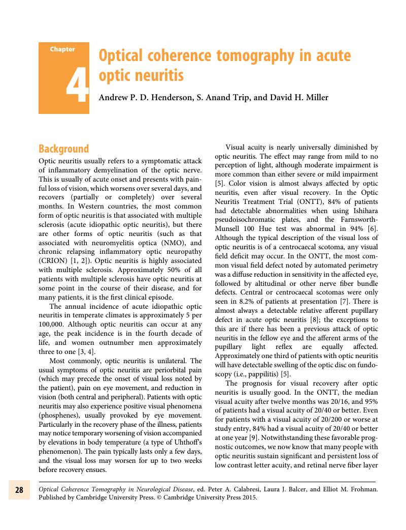
Image of the first page of this content. For PDF version, please use the ‘Save PDF’ preceeding this image.'
Type
Chapter
Information
Publisher: Cambridge University Press
Print publication year: 2015

Access options

Get access to the full version of this content by using one of the access options below. (Log in options will check for institutional or personal access. Content may require purchase if you do not have access.)

References

Kidd, D, Burton, B, Plant, GT, Graham, EM. Chronic relapsing inflammatory optic neuropathy (CRION). Brain. February 2003; 126(Pt 2): 276–84.CrossRefGoogle ScholarPubMed
Petzold, A, Pittock, S, Lennon, V, Maggiore, C, Weinshenker, BG, Plant, GT. Neuromyelitis optica-IgG (aquaporin-4) autoantibodies in immune mediated optic neuritis. J Neurol Neurosurg Psychiatr. January 2010; 81(1): 109–11.CrossRefGoogle ScholarPubMed
Rodriguez, M, Siva, A, Cross, SA, O’Brien, PC, Kurland, LT. Optic neuritis: a population-based study in Olmsted County, Minnesota. Neurology [Internet]. February 1995; 45(2): 244–50. Available from: http://www.neurology.org/content/45/2/244.short?sid=5d3bc217-394e-46d0-a720-df8704457287//www.neurology.orgCrossRefGoogle ScholarPubMed
Beck, RW, Cleary, PA, Anderson, MM, Keltner, JL, Shults, WT, Kaufman, DI, et al. A randomized, controlled trial of corticosteroids in the treatment of acute optic neuritis. The Optic Neuritis Study Group. N Engl J Med. February 27, 1992; 326(9): 581–8.Google ScholarPubMed
Optic Neuritis Study Group. The clinical profile of optic neuritis. Experience of the Optic Neuritis Treatment Trial. Arch Ophthalmol. December 1991; 109(12): 1673–8.Google Scholar
Trobe, JD, Beck, RW, Moke, PS, Cleary, PA. Contrast sensitivity and other vision tests in the optic neuritis treatment trial. Am J Ophthalmol. May 1996; 121(5): 547–53.CrossRefGoogle ScholarPubMed
Keltner, JL, Johnson, CA, Spurr, JO, Beck, RW. Baseline visual field profile of optic neuritis. The experience of the optic neuritis treatment trial. Optic Neuritis Study Group. Arch Ophthalmol. February 1993; 111(2): 231–4.CrossRefGoogle ScholarPubMed
Cox, TA, Thompson, HS, Corbett, JJ. Relative afferent pupillary defects in optic neuritis. Am J Ophthalmol. November 1, 1981; 92(5): 685–90.CrossRefGoogle ScholarPubMed
Beck, RW, Cleary, PA. Optic neuritis treatment trial. One-year follow-up results. Arch Ophthalmol. June 1993; 111(6): 773–5.CrossRefGoogle ScholarPubMed
la Cruz de, J, Kupersmith, MJ. Clinical profile of simultaneous bilateral optic neuritis in adults. British Journal of Ophthalmology. May 2006; 90(5): 551–4.Google Scholar
Beck, RW, Kupersmith, MJ, Cleary, PA, Katz, B. Fellow eye abnormalities in acute unilateral optic neuritis. Experience of the optic neuritis treatment trial. Ophthalmology. May 1993; 100(5): 691–8.CrossRefGoogle ScholarPubMed
Ormerod, IE, McDonald, WI. Multiple sclerosis presenting with progressive visual failure. J Neurol Neurosurg Psychiatr. September 1, 1984; 47(9): 943–6.CrossRefGoogle ScholarPubMed
Ashworth, B. Chronic retrobulbar and chiasmal neuritis. British Journal of Ophthalmology. October 1967; 51(10): 698702.CrossRefGoogle ScholarPubMed
Hoyt, WF, Schlicke, B, Eckelhoff, RJ. Fundoscopic appearance of a nerve-fibre-bundle defect. British Journal of Ophthalmology. August 1972; 56(8): 577–83.CrossRefGoogle ScholarPubMed
Frisén, L, Hoyt, WF. Insidious atrophy of retinal nerve fibers in multiple sclerosis. Funduscopic identification in patients with and without visual complaints. Arch Ophthalmol. August 1, 1974; 92(2): 91–7.CrossRefGoogle ScholarPubMed
Sharpe, JA, Sanders, MD. Atrophy of myelinated nerve fibres in the retina in optic neuritis. British Journal of Ophthalmology. Aprl 1, 1975; 59(4): 229–32.CrossRefGoogle ScholarPubMed
Elbøl, P, Work, K. Retinal nerve fiber layer in multiple sclerosis. Acta Ophthalmology. August 1, 1990; 68(4): 481–6.Google ScholarPubMed
MacFadyen, DJ, Drance, SM, Douglas, GR, Airaksinen, PJ, Mawson, DK, Paty, DW. The retinal nerve fiber layer, neuroretinal rim area, and visual evoked potentials in MS. Neurology [Internet]. September 1, 1988; 38(9): 1353–8. Available from: http://neurology.org/content/38/9/1353.shortCrossRefGoogle ScholarPubMed
Quigley, HA, Addicks, EM. Quantitative studies of retinal nerve fiber layer defects. Arch Ophthalmology. May 1982; 100(5): 807–14.CrossRefGoogle ScholarPubMed
Parisi, V, Manni, G, Spadaro, M, Colacino, G, Restuccia, R, Marchi, S, et al. Correlation between morphological and functional retinal impairment in multiple sclerosis patients. Invest Ophthalmol Vis Sci. October 1999; 40(11): 2520–7.Google ScholarPubMed
Trip, SA, Schlottmann, PG, Jones, SJ, Altmann, DR, Garway-Heath, DF, Thompson, AJ, et al. Retinal nerve fiber layer axonal loss and visual dysfunction in optic neuritis. Ann Neurol. September 2005; 58(3): 383–91.CrossRefGoogle ScholarPubMed
Costello, F, Hodge, W, Pan, Y, Eggenberger, E, Coupland, S, Kardon, R. Tracking retinal nerve fiber layer loss after optic neuritis: a prospective study using optical coherence tomography. Multiple Sclerosis. June 23, 2008; 14(7): 893905.CrossRefGoogle ScholarPubMed
Lange, AP, Sadjadi, R, Zhu, F, Alkabie, S, Costello, F, Traboulsee, AL. Spectral-domain optical coherence tomography of retinal nerve fiber layer thickness in NMO patients. J Neuroophthalmol [Internet]. September 2013; 33(3): 213–9. Available from: http://eutils.ncbi.nlm.nih.gov/entrez/eutils/elink.fcgi?dbfrom=pubmed&id=23863782&retmode=ref&cmd=prlinksCrossRefGoogle ScholarPubMed
Bichuetti, DB, Oliveira, EML, Souza, NA, Rivero, RLM, Gabbai, AA. Neuromyelitis optica in Brazil: a study on clinical and prognostic factors. Multiple Sclerosis. May 2009; 15(5): 613–9.CrossRefGoogle Scholar
Fisher, J, Jacobs, D, Markowitz, C, Galetta, S, Volpe, N, Nanoschiavi, M, et al. Relation of Visual Function to Retinal Nerve Fiber Layer Thickness in Multiple Sclerosis. Ophthalmology. February 2006; 113(2): 324–32.CrossRefGoogle ScholarPubMed
Costello, F, Coupland, S, Hodge, W, Lorello, GR, Koroluk, J, Pan, YI, et al. Quantifying axonal loss after optic neuritis with optical coherence tomography. Annals of Neurology. June 1, 2006; 59(6): 963–9.CrossRefGoogle ScholarPubMed
Albrecht, P, Fröhlich, R, Hartung, H-P, Kieseier, BC, Methner, A. Optical coherence tomography measures axonal loss in multiple sclerosis independently of optic neuritis. J Neurol. November 9, 2007; 254(11): 1595–6.CrossRefGoogle ScholarPubMed
Pulicken, M, Gordon-Lipkin, E, Balcer, LJ, Frohman, E, Cutter, G, Calabresi, PA. Optical coherence tomography and disease subtype in multiple sclerosis. Neurology. November 27, 2007; 69(22): 2085–92.CrossRefGoogle ScholarPubMed
Pueyo, V, Martin, J, Fernandez, J, Almarcegui, C, Ara, J, Egea, C, et al. Axonal loss in the retinal nerve fiber layer in patients with multiple sclerosis. Multiple Sclerosis. June 2008; 14(5): 609–14.CrossRefGoogle ScholarPubMed
Zaveri, MS, Conger, A, Salter, A, Frohman, TC, Galetta, SL, Markowitz, CE, et al. Retinal imaging by laser polarimetry and optical coherence tomography evidence of axonal degeneration in multiple sclerosis. Arch Neurol. July 2008; 65(7): 924–8.CrossRefGoogle ScholarPubMed
Klistorner, A, Arvind, H, Nguyen, T, Garrick, R, Paine, M, Graham, S, et al. Axonal loss and myelin in early ON loss in postacute optic neuritis. Ann Neurol. September 2008; 64(3): 325–31.CrossRefGoogle ScholarPubMed
Siger, M, Dziegielewski, K, Jasek, L, Bieniek, M, Nicpan, A, Nawrocki, J, et al. Optical coherence tomography in multiple sclerosis: thickness of the retinal nerve fiber layer as a potential measure of axonal loss and brain atrophy. J Neurol. October 2008; 255(10): 1555–60.CrossRefGoogle ScholarPubMed
Frohman, EM, Dwyer, MG, Frohman, T, Cox, JL, Salter, A, Greenberg, BM, et al. Relationship of optic nerve and brain conventional and non-conventional MRI measures and retinal nerve fiber layer thickness, as assessed by OCT and GDx: a pilot study. J Neurol Sci. July 15, 2009; 282(1–2): 96105.CrossRefGoogle ScholarPubMed
Ratchford, JN, Quigg, ME, Conger, A, Frohman, T, Frohman, E, Balcer, LJ, et al. Optical coherence tomography helps differentiate neuromyelitis optica and MS optic neuropathies. Neurology. July 28, 2009; 73(4): 302–8.CrossRefGoogle ScholarPubMed
Burkholder, BM, Osborne, B, Loguidice, MJ, Bisker, E, Frohman, TC, Conger, A, et al. Macular volume determined by optical coherence tomography as a measure of neuronal loss in multiple sclerosis. Arch Neurol. November 2009; 66(11): 1366–72.CrossRefGoogle ScholarPubMed
Garcia-Martin, E, Pueyo, V, Ara, J, Almarcegui, C, Martin, J, Pablo, L, et al. Effect of optic neuritis on progressive axonal damage in multiple sclerosis patients. Mult Scler [Internet]. June 20, 2011 Jun 20; 17(7): 830–7. Available from: http://eutils.ncbi.nlm.nih.gov/entrez/eutils/elink.fcgi?dbfrom=pubmed&id=21300734&retmode=ref&cmd=prlinksCrossRefGoogle ScholarPubMed
Syc, SB, Saidha, S, Newsome, SD, Ratchford, JN, Levy, M, Ford, E, et al. Optical coherence tomography segmentation reveals ganglion cell layer pathology after optic neuritis. Brain. February 2012; 135(Pt 2): 521–33.CrossRefGoogle ScholarPubMed
Costello, F, Hodge, W, Pan, YI, Burton, JM, Freedman, MS, Stys, PK, et al. Sex-specific differences in retinal nerve fiber layer thinning after acute optic neuritis. Neurology. October 2012; 79(18): 1866–72.CrossRefGoogle ScholarPubMed
Monteiro, MLR, Fernandes, DB, Apóstolos-Pereira, SL, Callegaro, D. Quantification of retinal neural loss in patients with neuromyelitis optica and multiple sclerosis with or without optic neuritis using Fourier-domain optical coherence tomography. Invest Ophthalmol Vis Sci [Internet]. June 2012; 53(7): 3959–66. Available from: http://eutils.ncbi.nlm.nih.gov/entrez/eutils/elink.fcgi?dbfrom=pubmed&id=22589446&retmode=ref&cmd=prlinksGoogle Scholar
Gelfand, JM, Nolan, R, Schwartz, DM, Graves, J, Green, AJ. Microcystic macular edema in multiple sclerosis is associated with disease severity. Brain. June 2012; 135(Pt 6): 1786–93.CrossRefGoogle ScholarPubMed
Saidha, S, Sotirchos, ES, Ibrahim, MA, Crainiceanu, CM, Gelfand, JM, Sepah, YJ, et al. Microcystic macular edema, thickness of the inner nuclear layer of the retina, and disease characteristics in multiple sclerosis: a retrospective study. Lancet Neurol. October 4, 2012; 11(11): 963–72.CrossRefGoogle ScholarPubMed
Balk, LJ, Killestein, J, Polman, CH, Uitdehaag, BMJ, Petzold, A. Microcystic macular edema confirmed, but not specific for multiple sclerosis. Brain. December 18, 2012; 135(12): e226–6.CrossRefGoogle Scholar
Kaufhold, F, Zimmermann, H, Schneider, E, Ruprecht, K, Paul, F, Oberwahrenbrock, T, et al. Optic neuritis is associated with inner nuclear layer thickening and microcystic macular edema independently of multiple sclerosis. PLoS ONE. 2013; 8(8): e71145.CrossRefGoogle ScholarPubMed
Abegg, M, Zinkernagel, M, Wolf, S. Microcystic macular degeneration from optic neuropathy. Brain. December 18, 2012; 135(12): 225–5.CrossRefGoogle ScholarPubMed
van Buren, JM. Trans-synaptic retrograde degeneration in the visual system of primates. J Neurol Neurosurg Psychiatr. October 1963; 26: 402–9.Google Scholar
Kaushik, M, Wang, CY, Barnett, MH, Garrick, R, Parratt, J, Graham, SL, et al. Inner nuclear layer thickening is inversley proportional to retinal ganglion cell loss in optic neuritis. PLoS ONE. 2013; 8(10): e78341.CrossRefGoogle ScholarPubMed
Kerrison, JB, Flynn, T, Green, WR. Retinal pathologic changes in multiple sclerosis. Retina (Philadelphia, Pa). 1994; 14(5): 445–51.CrossRefGoogle ScholarPubMed
Green, AJ, McQuaid, S, Hauser, SL, Allen, IV, Lyness, R. Ocular pathology in multiple sclerosis: retinal atrophy and inflammation irrespective of disease duration. Brain. June 2010; 133(Pt 6): 1591–601.CrossRefGoogle ScholarPubMed
Pro, MJ, Pons, ME, Liebmann, JM, Ritch, R, Zafar, S, Lefton, D, et al. Imaging of the optic disc and retinal nerve fiber layer in acute optic neuritis. J Neurol Sci. December 1, 2006; 250(1–2): 114–9.CrossRefGoogle ScholarPubMed
Henderson, APD, Altmann, DR, Trip, AS, Kallis, C, Jones, SJ, Schlottmann, PG, et al. A serial study of retinal changes following optic neuritis with sample size estimates for acute neuroprotection trials. Brain. September 2010; 133(9): 2592–602.CrossRefGoogle ScholarPubMed
Hickman, SJ, Toosy, AT, Jones, SJ, Altmann, DR, Miszkiel, KA, MacManus, DG, et al. A serial MRI study following optic nerve mean area in acute optic neuritis. Brain. November 2004; 127(Pt 11): 2498–505.CrossRefGoogle ScholarPubMed
Garas, A, Simó, M, Holló, G. Nerve fiber layer and macular thinning measured with different imaging methods during the course of acute optic neuritis. European Journal of Ophthalmology. October 22, 2010.CrossRefGoogle Scholar
Henderson, APD, Altmann, DR, Trip, SA, Miszkiel, KA, Schlottmann, PG, Jones, SJ, et al. Early factors associated with axonal loss after optic neuritis. Ann Neurol. December 21, 2011; 70(6): 955–63.CrossRefGoogle ScholarPubMed
Khaleeli, Z, Ciccarelli, O, Manfredonia, F, Barkhof, F, Brochet, B, Cercignani, M, et al. Predicting progression in primary progressive multiple sclerosis: a 10-year multicenter study. Ann Neurol. June 2008; 63(6): 790–3.CrossRefGoogle ScholarPubMed
Koch, M, Kingwell, E, Rieckmann, P, Tremlett, H, UBC MS Clinic Neurologists. The natural history of secondary progressive multiple sclerosis. J Neurol Neurosurg Psychiatr. September 2010; 81(9): 1039–43.CrossRefGoogle ScholarPubMed
Lucchinetti, CF, Mandler, RN, McGavern, D, Brück, W, Gleich, G, Ransohoff, RM, et al. A role for humoral mechanisms in the pathogenesis of Devic’s neuromyelitis optica. Brain. July 2002; 125(Pt 7): 1450–61.CrossRefGoogle ScholarPubMed
Parratt, JD, Prineas, JW. Neuromyelitis optica: a demyelinating disease characterized by acute destruction and regeneration of perivascular astrocytes. Mult Scler. September 30, 2010; 16(10): 1156–72.CrossRefGoogle ScholarPubMed
Lennon, VA, Wingerchuk, DM, Kryzer, TJ, Pittock, SJ, Lucchinetti, CF, Fujihara, K, et al. A serum autoantibody marker of neuromyelitis optica: distinction from multiple sclerosis. Lancet. November 2004; 364(9451): 2106–12.CrossRefGoogle ScholarPubMed
Lennon, VA, Kryzer, TJ, Pittock, SJ, Verkman, AS, Hinson, SR. IgG marker of optic-spinal multiple sclerosis binds to the aquaporin-4 water channel. J Exp Med. August 15, 2005; 202(4): 473–7.CrossRefGoogle Scholar
Wingerchuk, DM, Hogancamp, WF, O’Brien, PC, Weinshenker, BG. The clinical course of neuromyelitis optica (Devic’s syndrome). Neurology. 1999; 53(5): 1107–14.CrossRefGoogle ScholarPubMed
O’Riordan, JI, Gallagher, HL, Thompson, AJ, Howard, RS, Kingsley, DP, Thompson, EJ, et al. Clinical, CSF, and MRI findings in Devic’s neuromyelitis optica. J Neurol Neurosurg Psychiatr. April 1996; 60(4): 382–7.Google ScholarPubMed
Papais-Alvarenga, RM, Carellos, SC, Alvarenga, MP, Holander, C, Bichara, RP, Thuler, LCS. Clinical course of optic neuritis in patients with relapsing neuromyelitis optica. Arch Ophthalmol. January 2008; 126(1): 12–6.CrossRefGoogle ScholarPubMed
Rivera, JF, Kurtzke, JF, Booth, VJA, Corona, T V. Characteristics of Devic’s disease (neuromyelitis optica) in Mexico. J Neurol. May 2008; 255(5): 710–5.CrossRefGoogle ScholarPubMed
Nakamura, M, Nakazawa, T, Doi, H, Hariya, T, Omodaka, K, Misu, T, et al. Early high-dose intravenous methylprednisolone is effective in preserving retinal nerve fiber layer thickness in patients with neuromyelitis optica. Graefes Arch Clin Exp Ophthalmol. December 2010; 248(12): 1777–85.CrossRefGoogle ScholarPubMed
Bichuetti, DB, de Camargo, AS, Falcão, AB, Gonçalves, FF, Tavares, IM, de Oliveira, EML. The retinal nerve fiber layer of patients with neuromyelitis optica and chronic relapsing optic neuritis is more severely damaged than patients with multiple sclerosis. J Neuroophthalmol [Internet]. September 2013; 33(3): 220–4. Available from: http://eutils.ncbi.nlm.nih.gov/entrez/eutils/elink.fcgi?dbfrom=pubmed&id=23917443&retmode=ref&cmd=prlinksCrossRefGoogle ScholarPubMed
Fernandes, DB, Raza, AS, Nogueira, RGF, Wang, D, Callegaro, D, Hood, DC, et al. Evaluation of inner retinal layers in patients with multiple sclerosis or neuromyelitis optica using optical coherence tomography. Ophthalmology. February 2013; 120(2): 387–94.CrossRefGoogle ScholarPubMed
Sotirchos, ES, Saidha, S, Byraiah, G, Mealy, MA, Ibrahim, MA, Sepah, YJ, et al. In vivo identification of morphologic retinal abnormalities in neuromyelitis optica. Neurology [Internet]. April 8, 2013; 80(15): 1406–14. Available from: http://eutils.ncbi.nlm.nih.gov/entrez/eutils/elink.fcgi?dbfrom=pubmed&id=23516321&retmode=ref&cmd=prlinksCrossRefGoogle ScholarPubMed
de Seze, J, Blanc, F, Jeanjean, L, Zéphir, H, Labauge, P, Bouyon, M, et al. Optical coherence tomography in neuromyelitis optica. Arch Neurol. July 2008; 65(7): 920–3.CrossRefGoogle ScholarPubMed
Merle, H, Olindo, S, Donnio, A, Richer, R, Smadja, D, Cabre, P. Retinal peripapillary nerve fiber layer thickness in neuromyelitis optica. Invest Ophthalmol Vis Sci. October 2008; 49(10): 4412–7.CrossRefGoogle ScholarPubMed
Naismith, RT, Tutlam, NT, Xu, J, Klawiter, EC, Shepherd, J, Trinkaus, K, et al. Optical coherence tomography differs in neuromyelitis optica compared with multiple sclerosis. Neurology. March 24, 2009; 72(12): 1077–82.CrossRefGoogle ScholarPubMed
Green, AJ, Cree, BAC. Distinctive retinal nerve fibre layer and vascular changes in neuromyelitis optica following optic neuritis. J Neurol Neurosurg Psychiatr. September 2009; 80(9): 1002–5.CrossRefGoogle ScholarPubMed
Bouyon, M, Collongues, N, Zéphir, H, Ballonzoli, L, Jeanjean, L, Lebrun, C, et al. Longitudinal follow-up of vision in a neuromyelitis optica cohort. Mult Scler [Internet]. August 30, 2013; 19(10): 1320–2. Available from: http://eutils.ncbi.nlm.nih.gov/entrez/eutils/elink.fcgi?dbfrom=pubmed&id=23413296&retmode=ref&cmd=prlinksCrossRefGoogle Scholar
Compston, A. Mechanisms of axon-glial injury of the optic nerve. Eye. November 2004; 18(11): 1182–7.CrossRefGoogle ScholarPubMed
Kolappan, M, Henderson, APD, Jenkins, TM, Wheeler-Kingshott, CAM, Plant, GT, Thompson, AJ, et al. Assessing structure and function of the afferent visual pathway in multiple sclerosis and associated optic neuritis. J Neurol. March 2009; 256(3): 305–19.CrossRefGoogle ScholarPubMed
Esfahani, MR, Harandi, ZA, Movasat, M, Nikdel, M, Adelpour, M, Momeni, A, et al. Memantine for axonal loss of optic neuritis. Graefes Arch Clin Exp Ophthalmol. June 2012; 250(6): 863–9.CrossRefGoogle ScholarPubMed
Tsakiri, A, Kallenbach, K, Fuglø, D, Wanscher, B, Larsson, H, Frederiksen, J. Simvastatin improves final visual outcome in acute optic neuritis: a randomized study. Mult Scler. January 2012; 18(1): 7281.CrossRefGoogle ScholarPubMed
Jepson, S, Vought, B, Gross, CH, Gan, L, Austen, D, Frantz, JD, et al. LINGO-1, a transmembrane signaling protein, inhibits oligodendrocyte differentiation and myelination through intercellular self-interactions. J Biol Chem. June 22, 2012; 287(26): 22184–95.CrossRefGoogle ScholarPubMed
Mi, S, Pepinsky, RB, Cadavid, D. Blocking LINGO-1 as a therapy to promote CNS repair: from concept to the clinic. CNS Drugs. July 2013; 27(7): 493503.CrossRefGoogle ScholarPubMed
Canessa, CM, Horisberger, JD, Rossier, BC. Epithelial sodium channel related to proteins involved in neurodegeneration. Nature. February 4, 1993; 361(6411): 467–70.CrossRefGoogle ScholarPubMed
Black, JA, Waxman, SG. Phenytoin protects central axons in experimental autoimmune encephalomyelitis. J Neurol Sci. November 15, 2008; 274(1–2): 5763.CrossRefGoogle ScholarPubMed

Save book to Kindle

To save this book to your Kindle, first ensure coreplatform@cambridge.org is added to your Approved Personal Document E-mail List under your Personal Document Settings on the Manage Your Content and Devices page of your Amazon account. Then enter the ‘name’ part of your Kindle email address below. Find out more about saving to your Kindle.

Note you can select to save to either the @free.kindle.com or @kindle.com variations. ‘@free.kindle.com’ emails are free but can only be saved to your device when it is connected to wi-fi. ‘@kindle.com’ emails can be delivered even when you are not connected to wi-fi, but note that service fees apply.

Find out more about the Kindle Personal Document Service.

Available formats
×

Save book to Dropbox

To save content items to your account, please confirm that you agree to abide by our usage policies. If this is the first time you use this feature, you will be asked to authorise Cambridge Core to connect with your account. Find out more about saving content to Dropbox.

Available formats
×

Save book to Google Drive

To save content items to your account, please confirm that you agree to abide by our usage policies. If this is the first time you use this feature, you will be asked to authorise Cambridge Core to connect with your account. Find out more about saving content to Google Drive.

Available formats
×