Skip to main content Accessibility help
×
Hostname: page-component-76fb5796d-x4r87 Total loading time: 0 Render date: 2024-04-27T21:57:41.981Z Has data issue: false hasContentIssue false

Chapter 11 - Optical coherence tomography in neurodegenerative and other neurologic diseases

Published online by Cambridge University Press:  05 May 2015

Peter A. Calabresi
Affiliation:
Department of Neurology, Johns Hopkins University Hospital, Baltimore
Laura J. Balcer
Affiliation:
Department of Neurology, NYU Langone Medical Center, New York
Elliot M. Frohman
Affiliation:
Department of Neurology, UT Southwestern Medical Center, Dallas
Get access

Summary

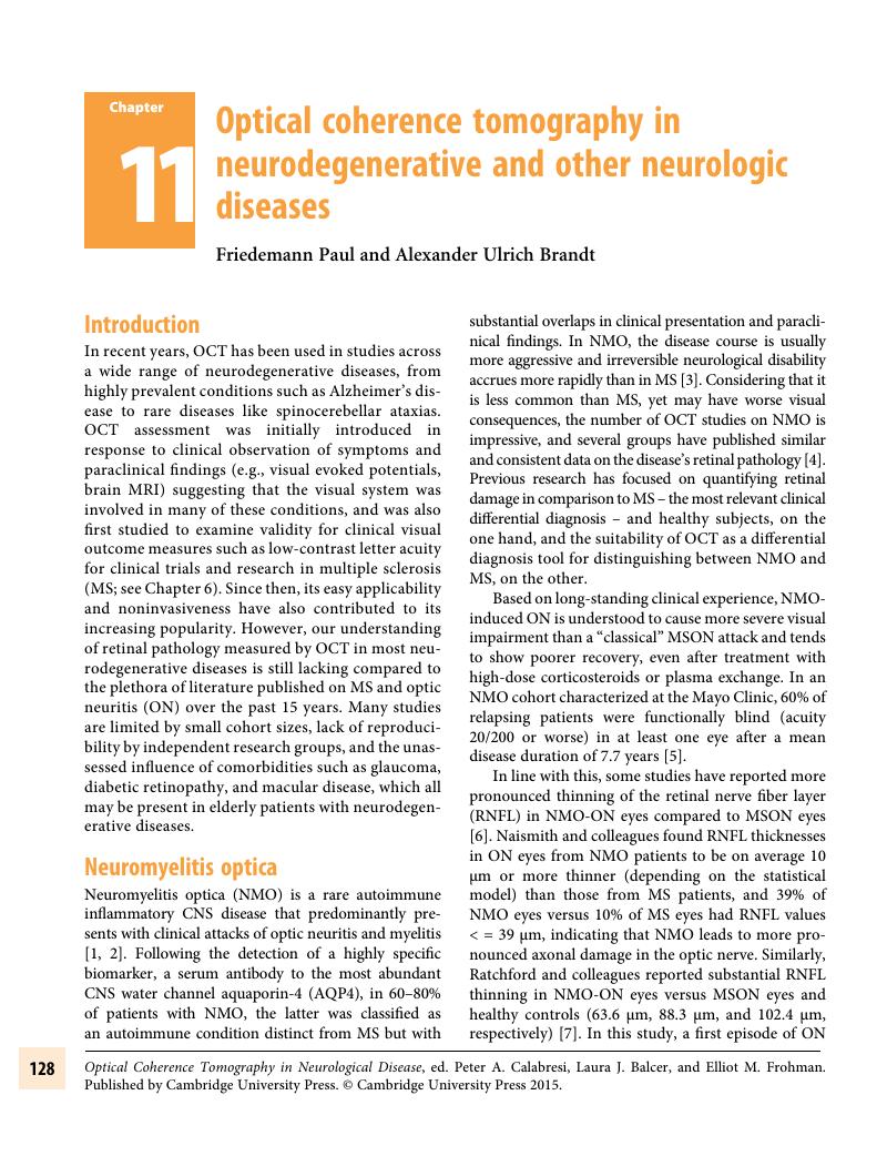
Image of the first page of this content. For PDF version, please use the ‘Save PDF’ preceeding this image.'
Type
Chapter
Information
Publisher: Cambridge University Press
Print publication year: 2015

Access options

Get access to the full version of this content by using one of the access options below. (Log in options will check for institutional or personal access. Content may require purchase if you do not have access.)

References

Jarius, S, Ruprecht, K, Wildemann, B, Kuempfel, T, Ringelstein, M, Geis, C, et al. Contrasting disease patterns in seropositive and seronegative neuromyelitis optica: A multicentre study of 175 patients. J Neuroinflammation 2012;9:14.CrossRefGoogle ScholarPubMed
Wingerchuk, DM, Lennon, VA, Pittock, SJ, Lucchinetti, CF, Weinshenker, BG. Revised diagnostic criteria for neuromyelitis optica. Neurology May 23, 2006;66(10):1485–9.CrossRefGoogle ScholarPubMed
Jarius, S, Paul, F, Franciotta, D, Waters, P, Zipp, F, Hohlfeld, R, et al. Mechanisms of disease: aquaporin-4 antibodies in neuromyelitis optica. Nat Clin Pract Neurol April 2008;4(4):202–14.CrossRefGoogle ScholarPubMed
Bennett, JL, Seze, J, Lana-Peixoto, M, Palace, J, Waldman, A, Schippling, S, et al. Neuromyelitis optica and multiple sclerosis: Seeing differences through optical coherence tomography. Mult Scler February 6, 2015. pii: 1352458514567216.CrossRefGoogle Scholar
Wingerchuk, DM, Hogancamp, WF, O’Brien, PC, Weinshenker, BG. The clinical course of neuromyelitis optica (Devic’s syndrome). Neurology September 22, 1999;53(5):1107–14.CrossRefGoogle ScholarPubMed
Naismith, RT, Tutlam, NT, Xu, J, Klawiter, EC, Shepherd, J, Trinkaus, K, et al. Optical coherence tomography differs in neuromyelitis optica compared with multiple sclerosis. Neurology. March 24, 2009;72(12):1077–82.CrossRefGoogle ScholarPubMed
Ratchford, JN, Quigg, ME, Conger, A, Frohman, T, Frohman, EM, Balcer, LJ, et al. Optical coherence tomography helps differentiate neuromyelitis optica and MS optic neuropathies. Neurology July 28, 2009;73(4):302–8.CrossRefGoogle ScholarPubMed
De Seze, J, Blanc, F, Jeanjean, L, Zéphir, H, Labauge, P, Bouyon, M, et al. Optical coherence tomography in neuromyelitis optica. Arch Neurol July 2008;65(7):920–3.CrossRefGoogle ScholarPubMed
Bock, M, Brandt, AU, Dörr, J, Pfueller, CF, Ohlraun, S, Zipp, F, et al. Time domain and spectral domain optical coherence tomography in multiple sclerosis: a comparative cross-sectional study. Mult Scler Houndmills Basingstoke Engl. July 2010;16(7):893–6.Google ScholarPubMed
Oberwahrenbrock, T, Ringelstein, M, Jentschke, S, Deuschle, K, Klumbies, K, Bellmann-Strobl, J, et al. Retinal ganglion cell and inner plexiform layer thinning in clinically isolated syndrome. Mult Scler December 2013;19(14): 1885–95. doi: 10.1177/1352458513489757. Epub May 23, 2013.CrossRefGoogle ScholarPubMed
Syc, SB, Saidha, S, Newsome, SD, Ratchford, JN, Levy, M, Ford, E, et al. Optical coherence tomography segmentation reveals ganglion cell layer pathology after optic neuritis. Brain J Neurol [Internet]. October 17, 2011 [cited December 6, 2011]; Available from: http://www.ncbi.nlm.nih.gov/pubmed/22006982Google Scholar
Sotirchos, ES, Saidha, S, Byraiah, G, Mealy, MA, Ibrahim, MA, Sepah, YJ, et al. In vivo identification of morphologic retinal abnormalities in neuromyelitis optica. Neurology April 9, 2013;80(15):1406–14.CrossRefGoogle ScholarPubMed
Monteiro, MLR, Fernandes, DB, Apóstolos-Pereira, SL, Callegaro, D. Quantification of retinal neural loss in patients with neuromyelitis optica and multiple sclerosis with or without optic neuritis using Fourier-domain optical coherence tomography. Invest Ophthalmol Vis Sci 2012;53(7):3959–66.CrossRefGoogle ScholarPubMed
Fernandes, DB, Raza, AS, Nogueira, RGF, Wang, D, Callegaro, D, Hood, DC, et al. Evaluation of inner retinal layers in patients with multiple sclerosis or neuromyelitis optica using optical coherence tomography. Ophthalmology. October 17, 2012;CrossRefGoogle Scholar
Schneider, E, Zimmermann, H, Oberwahrenbrock, T, Kaufhold, F, Kadas, EM, Petzold, A, et al. Optical coherence tomography reveals distinct patterns of retinal damage in neuromyelitis optica and multiple sclerosis. PLoS ONE. June 21, 2013;8(6):e66151.CrossRefGoogle ScholarPubMed
Gelfand, JM, Nolan, R, Schwartz, DM, Graves, J, Green, AJ. Microcystic macular edema in multiple sclerosis is associated with disease severity. Brain [Internet]. April 25, 2012 [cited May 30, 2012]; Available from: http://brain.oxfordjournals.org/content/early/2012/04/25/brain.aws098Google Scholar
Gelfand, JM, Cree, BA, Nolan, R, Arnow, S, Green, AJ. Microcystic inner nuclear layer abnormalities and neuromyelitis optica. JAMA Neurol. March 25, 2013;15.Google Scholar
Green, AJ, McQuaid, S, Hauser, SL, Allen, IV, Lyness, R. Ocular pathology in multiple sclerosis: retinal atrophy and inflammation irrespective of disease duration. Brain J Neurol June 2010;133(Pt 6):1591–601.CrossRefGoogle ScholarPubMed
Saidha, S, Sotirchos, ES, Ibrahim, MA, Crainiceanu, CM, Gelfand, JM, Sepah, YJ, et al. Microcystic macular edema, thickness of the inner nuclear layer of the retina, and disease characteristics in multiple sclerosis: a retrospective study. Lancet Neurol October 4, 2012;CrossRefGoogle Scholar
Kaufhold, F, Zimmermann, H, Schneider, E, Ruprecht, K, Paul, F, Oberwahrenbrock, T, et al. Optic Neuritis Is Associated with Inner Nuclear Layer Thickening and Microcystic Macular Edema Independently of Multiple Sclerosis. PLoS ONE Aug 6, 2013;8(8):e71145.CrossRefGoogle ScholarPubMed
Barboni, P, Carelli, V, Savini, G, Carbonelli, M, La Morgia, C, Sadun, AA. Microcystic macular degeneration from optic neuropathy: not inflammatory, not trans-synaptic degeneration. Brain J Neurol February 8, 2013;CrossRefGoogle Scholar
Abegg, M, Zinkernagel, M, Wolf, S. Microcystic macular degeneration from optic neuropathy. Brain J Neurol September 7, 2012;CrossRefGoogle Scholar
Brandt, AU, Oberwahrenbrock, T, Kadas, EM, Lagrèze, WA, Paul, F. Dynamic formation of macular microcysts independent of vitreous traction changes. Neurology July 1, 2014;83(1):73–7. doi: 10.1212/WNL.0000000000000545. Epub May 23, 2014.CrossRefGoogle ScholarPubMed
Wingerchuk, DM, Pittock, SJ, Lucchinetti, CF, Lennon, VA, Weinshenker, BG. A secondary progressive clinical course is uncommon in neuromyelitis optica. Neurology February 20, 2007;68(8):603–5.CrossRefGoogle ScholarPubMed
Jarius, S, Wildemann, B, Paul, F. Neuromyelitis optica: clinical features, immunopathogenesis and treatment. Clin Exp Immunol May 2014;176(2):149–64. doi: 10.1111/cei.12271. Review.CrossRefGoogle ScholarPubMed
Hinson, SR, Pittock, SJ, Lucchinetti, CF, Roemer, SF, Fryer, JP, Kryzer, TJ, et al. Pathogenic potential of IgG binding to water channel extracellular domain in neuromyelitis optica. Neurology December 11, 2007;69(24):2221–31.CrossRefGoogle ScholarPubMed
Levin, MH, Bennett, JL, Verkman, AS. Optic neuritis in neuromyelitis optica. Prog Retin Eye Res March 30, 2013;CrossRefGoogle Scholar
Biousse, V, Skibell, BC, Watts, RL, Loupe, DN, Drews-Botsch, C, Newman, NJ. Ophthalmologic features of Parkinson’s disease. Neurology January 27, 2004;62(2):177–80.CrossRefGoogle ScholarPubMed
Archibald, NK, Clarke, MP, Mosimann, UP, Burn, DJ. The retina in Parkinson’s disease. Brain J Neurol. May 2009;132(Pt 5):1128–45.CrossRefGoogle ScholarPubMed
Bodis-Wollner, I, Marx, MS, Mitra, S, Bobak, P, Mylin, L, Yahr, M. Visual dysfunction in Parkinson’s disease. Loss in spatiotemporal contrast sensitivity. Brain J Neurol December 1987;110 (Pt 6):1675–98.Google ScholarPubMed
Nightingale, S, Mitchell, KW, Howe, JW. Visual evoked cortical potentials and pattern electroretinograms in Parkinson’s disease and control subjects. J Neurol Neurosurg Psychiatry November 1986;49(11):1280–7.CrossRefGoogle ScholarPubMed
Tagliati, M, Bodis-Wollner, I, Yahr, MD. The pattern electroretinogram in Parkinson’s disease reveals lack of retinal spatial tuning. Electroencephalogr Clin Neurophysiol January 1996;100(1):111.CrossRefGoogle ScholarPubMed
Frederick, JM, Rayborn, ME, Laties, AM, Lam, DM, Hollyfield, JG. Dopaminergic neurons in the human retina. J Comp Neurol September 1, 1982;210(1):6579.CrossRefGoogle ScholarPubMed
Djamgoz, MB, Hankins, MW, Hirano, J, Archer, SN. Neurobiology of retinal dopamine in relation to degenerative states of the tissue. Vision Res December 1997;37(24):3509–29.CrossRefGoogle ScholarPubMed
Jackson, GR, Owsley, C. Visual dysfunction, neurodegenerative diseases, and aging. Neurol Clin August 2003;21(3):709–28.CrossRefGoogle ScholarPubMed
Harnois, C, Di Paolo, T. Decreased dopamine in the retinas of patients with Parkinson’s disease. Invest Ophthalmol Vis Sci November 1990;31(11):2473–5.Google ScholarPubMed
Hutton, JT, Morris, JL, Elias, JW. Levodopa improves spatial contrast sensitivity in Parkinson’s disease. Arch Neurol July 1993;50(7):721–4.CrossRefGoogle ScholarPubMed
Altintaş, O, Işeri, P, Ozkan, B, Cağlar, Y. Correlation between retinal morphological and functional findings and clinical severity in Parkinson’s disease. Doc Ophthalmol Adv Ophthalmol March 2008;116(2):137–46.CrossRefGoogle ScholarPubMed
Inzelberg, R, Ramirez, JA, Nisipeanu, P, Ophir, A. Retinal nerve fiber layer thinning in Parkinson disease. Vision Res November 2004;44(24):2793–7.CrossRefGoogle ScholarPubMed
Cubo, E, Tedejo, RP, Rodriguez Mendez, V, López Peña, MJ, Trejo Gabriel, Y Galán, JM. Retina thickness in Parkinson’s disease and essential tremor. Mov Disord Off J Mov Disord Soc. October 30, 2010;25(14):2461–2.CrossRefGoogle ScholarPubMed
Hajee, ME, March, WF, Lazzaro, DR, Wolintz, AH, Shrier, EM, Glazman, S, et al. Inner retinal layer thinning in Parkinson disease. Arch Ophthalmol. June 2009;127(6):737–41.CrossRefGoogle ScholarPubMed
Aaker, GD, Myung, JS, Ehrlich, JR, Mohammed, M, Henchcliffe, C, Kiss, S. Detection of retinal changes in Parkinson’s disease with spectral-domain optical coherence tomography. Clin Ophthalmol Auckl NZ 2010;4:1427–32.Google ScholarPubMed
Shrier, EM, Adam, CR, Spund, B, Glazman, S, Bodis-Wollner, I. Interocular asymmetry of foveal thickness in Parkinson disease. J Ophthalmol 2012;2012:728457.CrossRefGoogle ScholarPubMed
Archibald, NK, Clarke, MP, Mosimann, UP, Burn, DJ. Retinal thickness in Parkinson’s disease. Parkinsonism Relat Disord July 2011;17(6):431–6.CrossRefGoogle ScholarPubMed
Albrecht, P, Müller, A-K, Südmeyer, M, Ferrea, S, Ringelstein, M, Cohn, E, et al. Optical coherence tomography in parkinsonian syndromes. PloS One 2012;7(4):e34891.CrossRefGoogle ScholarPubMed
Garcia-Martin, E, Satue, M, Fuertes, I, Otin, S, Alarcia, R, Herrero, R, et al. Ability and reproducibility of Fourier-domain optical coherence tomography to detect retinal nerve fiber layer atrophy in Parkinson’s disease. Ophthalmology October 2012;119(10):2161–7.CrossRefGoogle ScholarPubMed
Roth, NM, Saidha, S, Zimmermann, H, Brandt, AU, Isensee, J, Benkhellouf-Rutkowska, A, et al. Photoreceptor layer thinning in idiopathic Parkinson’s disease. Mov Disord April 1, 2014;n/a–n/a.CrossRefGoogle Scholar
Müller, T, Woitalla, D, Peters, S, Kohla, K, Przuntek, H. Progress of visual dysfunction in Parkinson’s disease. Acta Neurol Scand April 2002;105(4):256–60.CrossRefGoogle ScholarPubMed
Saidha, S, Syc, SB, Durbin, MK, Eckstein, C, Oakley, JD, Meyer, SA, et al. Visual dysfunction in multiple sclerosis correlates better with optical coherence tomography derived estimates of macular ganglion cell layer thickness than peripapillary retinal nerve fiber layer thickness. Mult Scler J [Internet] August 24, 2011 [cited September 12, 2011]; Available from: http://www.ncbi.nlm.nih.gov/pubmed/21865411CrossRefGoogle Scholar
Nguyen-Legros, J. Functional neuroarchitecture of the retina: hypothesis on the dysfunction of retinal dopaminergic circuitry in Parkinson’s disease. Surg Radiol Anat SRA 1988;10(2):137–44.Google ScholarPubMed
Wulle, I, Wagner, HJ. GABA and tyrosine hydroxylase immunocytochemistry reveal different patterns of colocalization in retinal neurons of various vertebrates. J Comp Neurol June 1, 1990;296(1):173–8.CrossRefGoogle ScholarPubMed
Wu, Z, Huang, J, Dustin, L, Sadda, SR. Signal strength is an important determinant of accuracy of nerve fiber layer thickness measurement by optical coherence tomography. J Glaucoma March 2009;18(3):213–6.Google ScholarPubMed
Jack, CR Jr, Knopman, DS, Jagust, WJ, Petersen, RC, Weiner, MW, Aisen, PS, et al. Tracking pathophysiological processes in Alzheimer’s disease: an updated hypothetical model of dynamic biomarkers. Lancet Neurol February 2013;12(2):207–16.CrossRefGoogle ScholarPubMed
Koronyo-Hamaoui, M, Koronyo, Y, Ljubimov, AV, Miller, CA, Ko, MK, Black, KL, et al. Identification of amyloid plaques in retinas from Alzheimer’s patients and noninvasive in vivo optical imaging of retinal plaques in a mouse model. NeuroImage January 2011;54 Suppl 1:S204217.CrossRefGoogle ScholarPubMed
Cronin-Golomb, A, Rizzo, JF, Corkin, S, Growdon, JH. Visual function in Alzheimer’s disease and normal aging. Ann N Y Acad Sci 1991;640:2835.CrossRefGoogle ScholarPubMed
Rizzo, JF 3rd, Cronin-Golomb, A, Growdon, JH, Corkin, S, Rosen, TJ, Sandberg, MA, et al. Retinocalcarine function in Alzheimer’s disease. A clinical and electrophysiological study. Arch Neurol January 1992;49(1):93101.CrossRefGoogle ScholarPubMed
Hinton, DR, Sadun, AA, Blanks, JC, Miller, CA. Optic-nerve degeneration in Alzheimer’s disease. N Engl J Med August 21, 1986;315(8):485–7.CrossRefGoogle ScholarPubMed
Blanks, JC, Torigoe, Y, Hinton, DR, Blanks, RH. Retinal pathology in Alzheimer’s disease. I. Ganglion cell loss in foveal/parafoveal retina. Neurobiol Aging June 1996;17(3):377–84.Google ScholarPubMed
Sadun, AA, Bassi, CJ. Optic nerve damage in Alzheimer’s disease. Ophthalmology January 1990;97(1):917.CrossRefGoogle ScholarPubMed
Curcio, CA, Drucker, DN. Retinal ganglion cells in Alzheimer’s disease and aging. Ann Neurol March 1993;33(3):248–57.CrossRefGoogle ScholarPubMed
Davies, DC, McCoubrie, P, McDonald, B, Jobst, KA. Myelinated axon number in the optic nerve is unaffected by Alzheimer’s disease. Br J Ophthalmol June 1995;79(6):596600.CrossRefGoogle ScholarPubMed
Justino, L, Kergoat, M, Bergman, H, Chertkow, H, Robillard, A, Kergoat, H. Neuroretinal function is normal in early dementia of the Alzheimer type. Neurobiol Aging August 2001;22(4):691–5.CrossRefGoogle ScholarPubMed
Danesh-Meyer, HV, Birch, H, Ku, JY-F, Carroll, S, Gamble, G. Reduction of optic nerve fibers in patients with Alzheimer disease identified by laser imaging. Neurology November 28, 2006;67(10):1852–4.CrossRefGoogle ScholarPubMed
Kergoat, H, Kergoat, MJ, Justino, L, Robillard, A, Bergman, H, Chertkow, H. Normal optic nerve head topography in the early stages of dementia of the Alzheimer type. Dement Geriatr Cogn Disord December 2001;12(6):359–63.CrossRefGoogle ScholarPubMed
Kergoat, H, Kergoat, MJ, Justino, L, Chertkow, H, Robillard, A, Bergman, H. An evaluation of the retinal nerve fiber layer thickness by scanning laser polarimetry in individuals with dementia of the Alzheimer type. Acta Ophthalmol Scand April 2001;79(2):187–91.CrossRefGoogle ScholarPubMed
Kergoat, H, Kergoat, M-J, Justino, L, Chertkow, H, Robillard, A, Bergman, H. Visual retinocortical function in dementia of the Alzheimer type. Gerontology August 2002;48(4):197203.CrossRefGoogle ScholarPubMed
Iseri, PK, Altinaş, O, Tokay, T, Yüksel, N. Relationship between cognitive impairment and retinal morphological and visual functional abnormalities in Alzheimer disease. J Neuro-Ophthalmol Off J North Am Neuro-Ophthalmol Soc March 2006;26(1):1824.CrossRefGoogle ScholarPubMed
Paquet, C, Boissonnot, M, Roger, F, Dighiero, P, Gil, R, Hugon, J. Abnormal retinal thickness in patients with mild cognitive impairment and Alzheimer’s disease. Neurosci Lett June 2007;420(2):97–9.CrossRefGoogle ScholarPubMed
Lu, Y, Li, Z, Zhang, X, Ming, B, Jia, J, Wang, R, et al. Retinal nerve fiber layer structure abnormalities in early Alzheimer’s disease: evidence in optical coherence tomography. Neurosci Lett August 9, 2010;480(1):6972.CrossRefGoogle ScholarPubMed
Berisha, F, Feke, GT, Trempe, CL, McMeel, JW, Schepens, CL. Retinal abnormalities in early Alzheimer’s disease. Invest Ophthalmol Vis Sci May 2007;48(5):2285–9.CrossRefGoogle ScholarPubMed
Kirbas, S, Turkyilmaz, K, Anlar, O, Tufekci, A, Durmus, M. Retinal nerve fiber layer thickness in patients with Alzheimer disease. J Neuro-Ophthalmol Off J North Am Neuro-Ophthalmol Soc March 2013;33(1):5861.CrossRefGoogle ScholarPubMed
Bayer, AU, Ferrari, F, Erb, C. High occurrence rate of glaucoma among patients with Alzheimer’s disease. Eur Neurol 2002;47(3):165–8.CrossRefGoogle ScholarPubMed
Helmer, C, Malet, F, Rougier, M-B, Schweitzer, C, Colin, J, Delyfer, M-N, et al. Is there a link between open-angle glaucoma and dementia? The 3C-alienor cohort. Ann Neurol May 18, 2013;CrossRefGoogle Scholar
Bock, M, Brandt, AU, Dörr, J, Kraft, H, Weinges-Evers, N, Gaede, G, et al. Patterns of retinal nerve fiber layer loss in multiple sclerosis patients with or without optic neuritis and glaucoma patients. Clin Neurol Neurosurg October 2010;112(8):647–52.CrossRefGoogle ScholarPubMed
Dörr, J1, Radbruch, H, Bock, M, Wuerfel, J, Brüggemann, A, Wandinger, KP, Zeise, D, Pfueller, CF, Zipp, F, Paul, F. Encephalopathy, visual disturbance and hearing loss-recognizing the symptoms of Susac syndrome. Nat Rev Neurol December 2009;5(12):683–8. doi: 10.1038/nrneurol.2009.176.CrossRefGoogle ScholarPubMed
Dörr, J, Krautwald, S, Wildemann, B, Jarius, S, Ringelstein, M, Duning, T, et al. Characteristics of Susac syndrome: a review of all reported cases. Nat Rev Neurol June 2013;9(6):307–16.CrossRefGoogle ScholarPubMed
Brandt, AU, Zimmermann, H, Kaufhold, F, Promesberger, J, Schippling, S, Finis, D, et al. Patterns of retinal damage facilitate differential diagnosis between Susac syndrome and MS. PloS One 2012;7(6):e38741.CrossRefGoogle ScholarPubMed
Scott, CJ, Kardon, RH, Lee, AG, Frisén, L, Wall, M. Diagnosis and grading of papilledema in patients with raised intracranial pressure using optical coherence tomography vs clinical expert assessment using a clinical staging scale. Arch Ophthalmol June 2010;128(6):705–11.CrossRefGoogle ScholarPubMed
Kaufhold, F, Kadas, EM, Schmidt, C, Kunte, H, Hoffmann, J, Zimmermann, H, et al. Optic nerve head quantification in idiopathic intracranial hypertension by spectral domain OCT. PloS One 2012;7(5):e36965.CrossRefGoogle ScholarPubMed
Wang, J-K, Kardon, RH, Kupersmith, MJ, Garvin, MK. Automated quantification of volumetric optic disc swelling in papilledema using spectral-domain optical coherence tomography. Invest Ophthalmol Vis Sci June 2012;53(7):4069–75.CrossRefGoogle ScholarPubMed
Carelli, V, Ross-Cisneros, FN, Sadun, AA. Optic nerve degeneration and mitochondrial dysfunction: genetic and acquired optic neuropathies. Neurochem Int May 2002;40(6):573–84.CrossRefGoogle ScholarPubMed
Carelli, V, Ross-Cisneros, FN, Sadun, AA. Mitochondrial dysfunction as a cause of optic neuropathies. Prog Retin Eye Res January 2004;23(1):5389.CrossRefGoogle ScholarPubMed
Harding, AE. Friedreich’s ataxia: a clinical and genetic study of 90 families with an analysis of early diagnostic criteria and intrafamilial clustering of clinical features. Brain J Neurol September 1981;104(3):589620.CrossRefGoogle ScholarPubMed
Fortuna, F, Barboni, P, Liguori, R, Valentino, ML, Savini, G, Gellera, C, et al. Visual system involvement in patients with Friedreich’s ataxia. Brain J Neurol January 2009;132(Pt 1):116–23.CrossRefGoogle ScholarPubMed
Alldredge, CD, Schlieve, CR, Miller, NR, Levin, LA. Pathophysiology of the optic neuropathy associated with Friedreich ataxia. Arch Ophthalmol November 2003;121(11):1582–5.CrossRefGoogle ScholarPubMed
Noval, S, Contreras, I, Sanz-Gallego, I, Manrique, RK, Arpa, J. Ophthalmic features of Friedreich ataxia. Eye Lond Engl February 2012;26(2):315–20.Google ScholarPubMed
Seyer, LA, Galetta, K, Wilson, J, Sakai, R, Perlman, S, Mathews, K, et al. Analysis of the visual system in Friedreich ataxia. J Neurol June 18, 2013;CrossRefGoogle Scholar
Klockgether, T. The clinical diagnosis of autosomal dominant spinocerebellar ataxias. Cerebellum Lond Engl 2008;7(2):101–5.Google ScholarPubMed
Stricker, S, Oberwahrenbrock, T, Zimmermann, H, Schroeter, J, Endres, M, Brandt, AU, et al. Temporal retinal nerve fiber loss in patients with spinocerebellar ataxia Type 1. PLoS ONE July 29, 2011;6(7):e23024.CrossRefGoogle ScholarPubMed
Pula, JH, Towle, VL, Staszak, VM, Cao, D, Bernard, JT, Gomez, CM. Retinal nerve fibre layer and macular thinning in spinocerebellar ataxia and cerebellar multisystem atrophy. Neuro-Ophthalmol Aeolus Press Jun 2011;35(3):108–14.Google ScholarPubMed
Manrique, RK, Noval, S, Aguilar-Amat, MJ, Arpa, J, Rosa, I, Contreras, I. Ophthalmic features of spinocerebellar ataxia type 7. J Neuro-Ophthalmol Off J North Am Neuro-Ophthalmol Soc September 2009;29(3):174–9.Google ScholarPubMed
Hugosson, T, Gränse, L, Ponjavic, V, Andréasson, S. Macular dysfunction and morphology in spinocerebellar ataxia type 7 (SCA 7). Ophthalmic Genet. March 2009;30(1):16.CrossRefGoogle ScholarPubMed
Miller, RC, Tewari, A, Miller, JA, Garbern, J, Van Stavern, GP. Neuro-ophthalmologic features of spinocerebellar ataxia type 7. J Neuro-Ophthalmol Off J North Am Neuro-Ophthalmol Soc September 2009;29(3):180–6.Google ScholarPubMed
Bouchard, JP, Barbeau, A, Bouchard, R, Bouchard, RW. Autosomal recessive spastic ataxia of Charlevoix-Saguenay. Can J Neurol Sci J Can Sci Neurol February 1978;5(1):61–9.Google ScholarPubMed
Vingolo, EM, Di Fabio, R, Salvatore, S, Grieco, G, Bertini, E, Leuzzi, V, et al. Myelinated retinal fibers in autosomal recessive spastic ataxia of Charlevoix-Saguenay. Eur J Neurol Off J Eur Fed Neurol Soc September 2011;18(9):1187–90.Google ScholarPubMed
Desserre, J, Devos, D, Sautière, BG, Debruyne, P, Santorelli, FM, Vuillaume, I, et al. Thickening of peripapillar retinal fibers for the diagnosis of autosomal recessive spastic ataxia of Charlevoix-Saguenay. Cerebellum Lond Engl December 2011;10(4):758–62.Google ScholarPubMed
Garcia-Martin, E, Pablo, LE, Gazulla, J, Polo, V, Ferreras, A, Larrosa, JM. Retinal nerve fibre layer thickness in ARSACS: myelination or hypertrophy? Br J Ophthalmol February 2013;97(2):238–41.CrossRefGoogle ScholarPubMed
Blackstone, C, O’Kane, CJ, Reid, E. Hereditary spastic paraplegias: membrane traffic and the motor pathway. Nat Rev Neurosci January 2011;12(1):3142.CrossRefGoogle ScholarPubMed
Carelli, V, La Morgia, C, Valentino, ML, Barboni, P, Ross-Cisneros, FN, Sadun, AA. Retinal ganglion cell neurodegeneration in mitochondrial inherited disorders. Biochim Biophys Acta May 2009;1787(5):518–28.Google ScholarPubMed
Wiethoff, S, Zhour, A, Schöls, L, Fischer, MD. Retinal nerve fibre layer loss in hereditary spastic paraplegias is restricted to complex phenotypes. BMC Neurol 2012;12:143.CrossRefGoogle ScholarPubMed
Roth, NM, Saidha, S, Zimmermann, H, Brandt, AU, Oberwahrenbrock, T, Maragakis, NJ, et al. Optical coherence tomography does not support optic nerve involvement in amyotrophic lateral sclerosis. Eur J Neurol Off J Eur Fed Neurol Soc April 14, 2013;CrossRefGoogle Scholar
Ala, A, Walker, AP, Ashkan, K, Dooley, JS, Schilsky, ML. Wilson’s disease. Lancet February 3, 2007;369(9559):397408.CrossRefGoogle ScholarPubMed
Satishchandra, P, Ravishankar Naik, K. Visual pathway abnormalities Wilson’s disease: an electrophysiological study using electroretinography and visual evoked potentials. J Neurol Sci May 1, 2000;176(1):1320.CrossRefGoogle ScholarPubMed
Albrecht, P, Müller, A-K, Ringelstein, M, Finis, D, Geerling, G, Cohn, E, et al. Retinal neurodegeneration in Wilson’s disease revealed by spectral domain optical coherence tomography. PloS One 2012;7(11):e49825.CrossRefGoogle ScholarPubMed
Parikh, RS, Parikh, SR, Sekhar, GC, Prabakaran, S, Babu, JG, Thomas, R. Normal Age-Related Decay of Retinal Nerve Fiber Layer Thickness. Ophthalmology May 2007;114(5):921–6.CrossRefGoogle ScholarPubMed
Alamouti, B, Funk, J. Retinal thickness decreases with age: an OCT study. Br J Ophthalmol July 2003;87(7):899901.CrossRefGoogle ScholarPubMed
Ooto, S, Hangai, M, Tomidokoro, A, Saito, H, Araie, M, Otani, T, et al. Effects of age, sex, and axial length on the three-dimensional profile of normal macular layer structures. Invest Ophthalmol Vis Sci 2011;52(12):8769–79.CrossRefGoogle ScholarPubMed
Rudnicka, AR, Mt-Isa, S, Owen, CG, Cook, DG, Ashby, D. Variations in primary open-angle glaucoma prevalence by age, gender, and race: a Bayesian meta-analysis. Invest Ophthalmol Vis Sci October 2006;47(10):4254–61.CrossRefGoogle Scholar
Quigley, HA, Broman, AT. The number of people with glaucoma worldwide in 2010 and 2020. Br J Ophthalmol March 2006;90(3):262–7.CrossRefGoogle ScholarPubMed
Varma, R, Ying-Lai, M, Francis, BA, Nguyen, BB-T, Deneen, J, Wilson, MR, et al. Prevalence of open-angle glaucoma and ocular hypertension in Latinos: the Los Angeles Latino Eye Study. Ophthalmology August 2004;111(8):1439–48.Google ScholarPubMed
Tewarie, P, Balk, L, Costello, F, Green, A, Martin, R, Schippling, S, et al. The OSCAR-IB Consensus Criteria for Retinal OCT Quality Assessment. PLoS ONE April 19, 2012;7(4):e34823.CrossRefGoogle ScholarPubMed
Petzold, A, Wattjes, MP, Costello, F, Flores-Rivera, J, Fraser, CL, Fujihara, K, et al. The investigation of acute optic neuritis: a review and proposed protocol. Nat Rev Neurol August 2014;10(8):447–58. doi: 10.1038/nrneurol.2014.108. Epub Jul 8, 2014.CrossRefGoogle ScholarPubMed

Save book to Kindle

To save this book to your Kindle, first ensure coreplatform@cambridge.org is added to your Approved Personal Document E-mail List under your Personal Document Settings on the Manage Your Content and Devices page of your Amazon account. Then enter the ‘name’ part of your Kindle email address below. Find out more about saving to your Kindle.

Note you can select to save to either the @free.kindle.com or @kindle.com variations. ‘@free.kindle.com’ emails are free but can only be saved to your device when it is connected to wi-fi. ‘@kindle.com’ emails can be delivered even when you are not connected to wi-fi, but note that service fees apply.

Find out more about the Kindle Personal Document Service.

Available formats
×

Save book to Dropbox

To save content items to your account, please confirm that you agree to abide by our usage policies. If this is the first time you use this feature, you will be asked to authorise Cambridge Core to connect with your account. Find out more about saving content to Dropbox.

Available formats
×

Save book to Google Drive

To save content items to your account, please confirm that you agree to abide by our usage policies. If this is the first time you use this feature, you will be asked to authorise Cambridge Core to connect with your account. Find out more about saving content to Google Drive.

Available formats
×