
Book contents
- Optical Coherence Tomography in Neurological Diseases
- Optical Coherence Tomography in Neurological Diseases
- Copyright page
- Contents
- Contributors
- Preface
- Chapter 1 Introduction to optical coherence tomography in neurological diseases
- Chapter 2 Basic principles of optical coherence tomography
- Chapter 3 Anatomy of the anterior visual pathway
- Chapter 4 Optical coherence tomography in acute optic neuritis
- Chapter 5 Optical coherence tomography and visual outcomes in acute optic neuritis
- Chapter 6 Optical coherence tomography and low-contrast acuity
- Chapter 7 Optical coherence tomography and electrophysiology of the visual pathway
- Chapter 8 Optical coherence tomography and electrophysiology of the optic nerve head
- Chapter 9 Meta-analysis of optical coherence tomography in multiple sclerosis
- Chapter 10 Optical coherence tomography and brain magnetic resonance imaging in multiple sclerosis
- Chapter 11 Optical coherence tomography in neurodegenerative and other neurologic diseases
- Chapter 12 Optical coherence tomography pathologies to know about in clinical practice
- Chapter 13 Optical coherence tomography and retinal segmentation in neurological diseases
- Chapter 14 Optical coherence tomography and retinal pathology in neurologic diseases
- Chapter 15 Retinal inflammation in multiple sclerosis revealed by optical coherence tomography and ophthalmoscopy
- Chapter 16 Optical coherence tomography and optic nerve magnetic resonance imaging in demyelinating diseases
- Chapter 17 Optical coherence tomography in neurologic clinical trials
- Chapter 18 Optical coherence tomography in a multi-center setting: quality control issues
- Chapter 19 Future technological advances in optical coherence tomography
- Index
- References
Chapter 19 - Future technological advances in optical coherence tomography
Published online by Cambridge University Press: 05 May 2015

Book contents
- Optical Coherence Tomography in Neurological Diseases
- Optical Coherence Tomography in Neurological Diseases
- Copyright page
- Contents
- Contributors
- Preface
- Chapter 1 Introduction to optical coherence tomography in neurological diseases
- Chapter 2 Basic principles of optical coherence tomography
- Chapter 3 Anatomy of the anterior visual pathway
- Chapter 4 Optical coherence tomography in acute optic neuritis
- Chapter 5 Optical coherence tomography and visual outcomes in acute optic neuritis
- Chapter 6 Optical coherence tomography and low-contrast acuity
- Chapter 7 Optical coherence tomography and electrophysiology of the visual pathway
- Chapter 8 Optical coherence tomography and electrophysiology of the optic nerve head
- Chapter 9 Meta-analysis of optical coherence tomography in multiple sclerosis
- Chapter 10 Optical coherence tomography and brain magnetic resonance imaging in multiple sclerosis
- Chapter 11 Optical coherence tomography in neurodegenerative and other neurologic diseases
- Chapter 12 Optical coherence tomography pathologies to know about in clinical practice
- Chapter 13 Optical coherence tomography and retinal segmentation in neurological diseases
- Chapter 14 Optical coherence tomography and retinal pathology in neurologic diseases
- Chapter 15 Retinal inflammation in multiple sclerosis revealed by optical coherence tomography and ophthalmoscopy
- Chapter 16 Optical coherence tomography and optic nerve magnetic resonance imaging in demyelinating diseases
- Chapter 17 Optical coherence tomography in neurologic clinical trials
- Chapter 18 Optical coherence tomography in a multi-center setting: quality control issues
- Chapter 19 Future technological advances in optical coherence tomography
- Index
- References
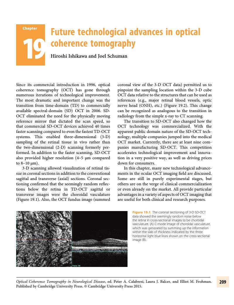
Summary
A summary is not available for this content so a preview has been provided. Please use the Get access link above for information on how to access this content.
- Type
- Chapter
- Information
- Optical Coherence Tomography in Neurologic Diseases , pp. 209 - 217Publisher: Cambridge University PressPrint publication year: 2015
References
Sander, , , B., Larsen, M., Thrane, L., et al., Enhanced optical coherence tomography imaging by multiple scan averaging. Br J Ophthalmol 2005; 89(2): 207–12.CrossRefGoogle ScholarPubMed
Sakamoto, , , A., Hangai, M., and Yoshimura, N., Spectral-domain optical coherence tomography with multiple B-scan averaging for enhanced imaging of retinal diseases. Ophthalmology, 2008;115(6): 1071–1078 e7.CrossRefGoogle ScholarPubMed
Pappuru, , , R.R., Briceno, C., Ouyang, Y., et al., Clinical significance of B-scan averaging with SD-OCT. Ophthalmic Surg Lasers Imaging, 2012;43(1): 63–8.CrossRefGoogle ScholarPubMed
Jorgensen, , , T.M., Thomadsen, J., Christensen, U., et al., Enhancing the signal-to-noise ratio in ophthalmic optical coherence tomography by image registration–method and clinical examples. J Biomed Opt, 2007;12(4): p. 041208.CrossRefGoogle ScholarPubMed
Wu, , , W., Tan, O., Pappuru, R.R., et al., Assessment of frame-averaging algorithms in OCT image analysis. Ophthalmic Surg Lasers Imaging Retina, 2013;44(2): 168–75.CrossRefGoogle ScholarPubMed
Chin, , , E.K., Sedeek, R.W., Li, Y., et al., Reproducibility of macular thickness measurement among five OCT instruments: effects of image resolution, image registration, and eye tracking. Ophthalmic Surg Lasers Imaging, 2012; 43(2) 97–108.CrossRefGoogle ScholarPubMed
Drexler, , , W. and Fujimoto, J.G., Optical Coherence Tomography: Technology and Applications. 2008. New York:Springer.CrossRefGoogle Scholar
Blatter, , , C., Klein, T., Grajciar, B., et al., Ultrahigh-speed non-invasive widefield angiography. J Biomed Opt, 2012. 17(7): 070505.CrossRefGoogle ScholarPubMed
Goda, , , K., Fard, A., Malik, O., et al., High-throughput optical coherence tomography at 800 nm. Opt Express, 2012. 20(18): 19612–7.CrossRefGoogle Scholar
Klein, , , T., Wieser, W., Eigenwillig, C.M., et al., Megahertz OCT for ultrawide-field retinal imaging with a 1050 nm Fourier domain mode-locked laser. Opt Express, 2011. 19(4): 3044–62.CrossRefGoogle ScholarPubMed
Potsaid, , , B., Baumann, B., Huang, D., et al., Ultrahigh speed 1050nm swept source/Fourier domain OCT retinal and anterior segment imaging at 100,000 to 400,000 axial scans per second. Opt Express, 2010. 18(19): 20029–48.CrossRefGoogle Scholar
Zawadzki, , , R.J., Choi, S.S., Jones, S.M., et al., Adaptive optics-optical coherence tomography: optimizing visualization of microscopic retinal structures in three dimensions. J Opt Soc Am A Opt Image Sci Vis, 2007. 24(5): 1373–83.CrossRefGoogle ScholarPubMed
Zawadzki, , , R.J., Choi, S.S., Fuller, A.R., et al., Cellular resolution volumetric in vivo retinal imaging with adaptive optics-optical coherence tomography. Opt Express, 2009. 17(5): 4084–94.CrossRefGoogle ScholarPubMed
Ragazzoni, , , R., Marchetti, E., and Valente, G., Adaptive-optics corrections available for the whole sky. Nature, 2000. 403(6765): 54–6.CrossRefGoogle ScholarPubMed
Miller, , , D.T., Kocaoglu, O.P., Wang, Q., et al., Adaptive optics and the eye (super resolution OCT). Eye (Lond), 2011. 25(3): 321–30.CrossRefGoogle ScholarPubMed
Kocaoglu, , , O.P., Lee, S., Jonnal, R.S., et al., Imaging cone photoreceptors in three dimensions and in time using ultrahigh resolution optical coherence tomography with adaptive optics. Biomed Opt Express, 2011. 2(4): 748–63.CrossRefGoogle ScholarPubMed
Yannuzzi, , , L.A., Rohrer, K.T., Tindel, L.J., et al., Fluorescein angiography complication survey. Ophthalmology, 1986. 93(5): 611–7.CrossRefGoogle ScholarPubMed
Hope-Ross, , , M., Yannuzzi, L.A., Gragoudas, E.S., et al., Adverse reactions due to indocyanine green. Ophthalmology, 1994. 101(3): 529–33.CrossRefGoogle ScholarPubMed
Drexler, , , W. and Fujimoto, J.G., State-of-the-art retinal optical coherence tomography. Prog Retin Eye Res, 2008. 27(1):45–88.CrossRefGoogle ScholarPubMed
Wehbe, , , H., Ruggeri, M., Jiao, S., et al., Automatic retinal blood flow calculation using spectral domain optical coherence tomography. Opt Express, 2007. 15(23): 15193–206.CrossRefGoogle ScholarPubMed
Makita, , , S., Fabritius, T., and Yasuno, Y., Quantitative retinal-blood flow measurement with three-dimensional vessel geometry determination using ultrahigh-resolution Doppler optical coherence angiography. Opt Lett, 2008. 33(8): 836–8.CrossRefGoogle ScholarPubMed
Wang, , , Y., Bower, B.A., Izatt, J.A., et al., Retinal blood flow measurement by circumpapillary Fourier domain Doppler optical coherence tomography. J Biomed Opt, 2008. 13(6): 064003.CrossRefGoogle ScholarPubMed
Szkulmowska, , , A., Szkulmowski, M., Szlag, D., et al., Three-dimensional quantitative imaging of retinal and choroidal blood flow velocity using joint Spectral and Time domain Optical Coherence Tomography. Opt Express, 2009. 17(13): 10584–98.CrossRefGoogle ScholarPubMed
Makita, , , S., Hong, Y., Yamanari, M., et al., Optical coherence angiography. Opt Express, 2006. 14(17): 7821–40.CrossRefGoogle ScholarPubMed
An, , , L. and Wang, R.K., In vivo volumetric imaging of vascular perfusion within human retina and choroids with optical micro-angiography. Opt Express, 2008. 16(15): 11438–52.CrossRefGoogle ScholarPubMed
Kim, , , D.Y., Fingler, J., Werner, J.S., et al., In vivo volumetric imaging of human retinal circulation with phase-variance optical coherence tomography. Biomed Opt Express, 2011. 2(6): 1504–13.CrossRefGoogle ScholarPubMed
Makita, , , S., Jaillon, F., Yamanari, M., et al., Comprehensive in vivo micro-vascular imaging of the human eye by dual-beam-scan Doppler optical coherence angiography. Opt Express, 2011. 19(2): 1271–83.CrossRefGoogle ScholarPubMed
Miura, , , M., Makita, S., Iwasaki, T., et al., Three-dimensional visualization of ocular vascular pathology by optical coherence angiography in vivo. Invest Ophthalmol Vis Sci, 2011. 52(5): 2689–95.CrossRefGoogle ScholarPubMed
Hwang, , , J.C., Konduru, R., Zhang, X., et al., Relationship among visual field, blood flow, and neural structure measurements in glaucoma. Invest Ophthalmol Vis Sci, 2012. 53(6): 3020–6.CrossRefGoogle ScholarPubMed
Adhi, , , M. and Duker, J.S., Optical coherence tomography–current and future applications. Curr Opin Ophthalmol, 2013. 24(3): 213–21.CrossRefGoogle ScholarPubMed
Pircher, , , M., Goetzinger, E., Leitgeb, R., et al., Transversal phase resolved polarization sensitive optical coherence tomography. Phys Med Biol, 2004. 49(7): 1257–63.CrossRefGoogle ScholarPubMed
Pircher, , , M., Gotzinger, E., Leitgeb, R., et al., Imaging of polarization properties of human retina in vivo with phase resolved transversal PS-OCT. Opt Express, 2004. 12(24): 5940–51.CrossRefGoogle ScholarPubMed
Gotzinger, , , E., Pircher, M., and Hitzenberger, C.K., High speed spectral domain polarization sensitive optical coherence tomography of the human retina. Opt Express, 2005. 13(25): 10217–29.Google ScholarPubMed
Zotter, , , S., Pircher, M., Gotzinger, E., et al., Measuring retinal nerve fiber layer birefringence, retardation, and thickness using wide-field, high-speed polarization sensitive spectral domain OCT. Invest Ophthalmol Vis Sci, 2013. 54(1): 72–84.CrossRefGoogle ScholarPubMed
Gotzinger, , , E., Pircher, M., Baumann, B., et al., Three-dimensional polarization sensitive OCT imaging and interactive display of the human retina. Opt Express, 2009. 17(5): 4151–65.CrossRefGoogle ScholarPubMed
Windisch, , , B.K., Harasymowycz, P.J., See, J.L., et al., Comparison between confocal scanning laser tomography, scanning laser polarimetry and optical coherence tomography on the ability to detect localised retinal nerve fibre layer defects in glaucoma patients. Br J Ophthalmol, 2009. 93(2): 225–30.CrossRefGoogle ScholarPubMed
Ferreras, , , A., Pablo, L.E., Pajarin, A.B., et al., Scanning laser polarimetry: logistic regression analysis for perimetric glaucoma diagnosis. Eye (Lond), 2009. 23(3): 593–600.CrossRefGoogle ScholarPubMed
Yamanari, , , M., Miura, M., Makita, S., et al., Phase retardation measurement of retinal nerve fiber layer by polarization-sensitive spectral-domain optical coherence tomography and scanning laser polarimetry. J Biomed Opt, 2008. 13(1): 014013.CrossRefGoogle ScholarPubMed
Moon, , , B.G., Sung, K.R., Cho, J.W., et al., Glaucoma progression detection by retinal nerve fiber layer measurement using scanning laser polarimetry: event and trend analysis. Korean J Ophthalmol, 2012. 26(3): 174–81.CrossRefGoogle ScholarPubMed
Hoffmann, , , E.M. and Schulze, A., [Glaucoma diagnosis using scanning laser polarimetry]. Ophthalmologe, 2009. 106(8): 696–8, 700–1.Google ScholarPubMed
Gotzinger, , , E., Pircher, M., Baumann, B., et al., Retinal nerve fiber layer birefringence evaluated with polarization sensitive spectral domain OCT and scanning laser polarimetry: a comparison. J Biophotonics, 2008. 1(2): 129–39.CrossRefGoogle ScholarPubMed
Fortune, , , B., Wang, L., Cull, G., et al., Intravitreal colchicine causes decreased RNFL birefringence without altering RNFL thickness. Invest Ophthalmol Vis Sci, 2008. 49(1): 255–61.CrossRefGoogle ScholarPubMed
Zaveri, , , M.S., Conger, A., Salter, A., et al., Retinal imaging by laser polarimetry and optical coherence tomography evidence of axonal degeneration in multiple sclerosis. Arch Neurol, 2008. 65(7): 924–8.CrossRefGoogle ScholarPubMed
Bagga, , , H., Greenfield, D.S., Feuer, W., et al., Scanning laser polarimetry with variable corneal compensation and optical coherence tomography in normal and glaucomatous eyes. Am J Ophthalmol, 2003. 135(4): 521–9.CrossRefGoogle ScholarPubMed
Sehi, , , M., Ume, S., and Greenfield, D.S., Scanning laser polarimetry with enhanced corneal compensation and optical coherence tomography in normal and glaucomatous eyes. Invest Ophthalmol Vis Sci, 2007. 48(5): 2099–104.CrossRefGoogle ScholarPubMed
Michels, , , S., Pircher, M., Geitzenauer, W., et al., Value of polarisation-sensitive optical coherence tomography in diseases affecting the retinal pigment epithelium. Br J Ophthalmol, 2008. 92(2): 204–9.CrossRefGoogle ScholarPubMed
Merchant, , , K.Y., Su, D., Park, S.C., et al., Enhanced depth imaging optical coherence tomography of optic nerve head Drusen. Ophthalmology, 2013.CrossRefGoogle Scholar
Hedels, , , C. and Krohn, J., Enhanced depth imaging optical coherence tomography of optic disc maculopathy without a visible optic pit. Clin Experiment Ophthalmol, 2013.CrossRefGoogle Scholar
Skondra, , , D., Papakostas, T., and Vavvas, D.G., Enhanced depth imaging optical coherence tomography in age-related macular degeneration. Semin Ophthalmol, 2012. 27(5–6): 209–12.CrossRefGoogle ScholarPubMed
Rahman, , , W., Chen, F.K., Yeoh, J., et al., Enhanced depth imaging of the choroid in patients with neovascular age-related macular degeneration treated with anti-VEGF therapy versus untreated patients. Graefes Arch Clin Exp Ophthalmol, 2013. 251(6): 1483–8.CrossRefGoogle ScholarPubMed
da Silva, , , F.T., Sakata, V.M., Nakashima, A., et al., Enhanced depth imaging optical coherence tomography in long-standing Vogt-Koyanagi-Harada disease. Br J Ophthalmol, 2013. 97(1): 70–4.CrossRefGoogle ScholarPubMed
Nakayama, , , M., Keino, H., Okada, A.A., et al., Enhanced depth imaging optical coherence tomography of the choroid in Vogt-Koyanagi-Harada disease. Retina, 2012. 32(10): 2061–9.CrossRefGoogle ScholarPubMed
Querques, , , G., Lattanzio, R., Querques, L., et al., Enhanced depth imaging optical coherence tomography in type 2 diabetes. Invest Ophthalmol Vis Sci, 2012. 53(10): 6017–24.CrossRefGoogle ScholarPubMed
Shields, , , C.L., Kaliki, S., Rojanaporn, D., et al., Enhanced depth imaging optical coherence tomography of small choroidal melanoma: comparison with choroidal nevus. Arch Ophthalmol, 2012. 130(7): 850–6.CrossRefGoogle ScholarPubMed
Shah, , , S.U., Kaliki, S., Shields, C.L., et al., Enhanced depth imaging optical coherence tomography of choroidal nevus in 104 cases. Ophthalmology, 2012. 119(5): 1066–72.CrossRefGoogle ScholarPubMed
Park, , , H.Y., Jeon, S.H., and Park, C.K., Enhanced depth imaging detects lamina cribrosa thickness differences in normal tension glaucoma and primary open-angle glaucoma. Ophthalmology, 2012. 119(1): 10–20.CrossRefGoogle ScholarPubMed
Park, , , S.C., De Moraes, C.G., Teng, C.C., et al., Enhanced depth imaging optical coherence tomography of deep optic nerve complex structures in glaucoma. Ophthalmology, 2012. 119(1): 3–9.CrossRefGoogle ScholarPubMed
Wong, , , I.Y., Koizumi, H., and Lai, W.W., Enhanced depth imaging optical coherence tomography. Ophthalmic Surg Lasers Imaging, 2011. 42 Suppl: S75–84.CrossRefGoogle ScholarPubMed
Ikuno, , , Y., Maruko, I., Yasuno, Y., et al., Reproducibility of retinal and choroidal thickness measurements in enhanced depth imaging and high-penetration optical coherence tomography. Invest Ophthalmol Vis Sci, 2011. 52(8): 5536–40.CrossRefGoogle ScholarPubMed
Imamura, , , Y., Iida, T., Maruko, I., et al., Enhanced depth imaging optical coherence tomography of the sclera in dome-shaped macula. Am J Ophthalmol, 2011. 151(2): 297–302.CrossRefGoogle ScholarPubMed
Margolis, , , R. and Spaide, R.F., A pilot study of enhanced depth imaging optical coherence tomography of the choroid in normal eyes. Am J Ophthalmol, 2009. 147(5): 811–5.CrossRefGoogle ScholarPubMed
Spaide, , , R.F., Enhanced depth imaging optical coherence tomography of retinal pigment epithelial detachment in age-related macular degeneration. Am J Ophthalmol, 2009. 147(4): 644–52.CrossRefGoogle ScholarPubMed
Spaide, , , R.F., Koizumi, H., and Pozzoni, M.C., Enhanced depth imaging spectral-domain optical coherence tomography. Am J Ophthalmol, 2008. 146(4): 496–500.CrossRefGoogle ScholarPubMed
Hale, , , G.M. and Querry, M.R., Optical Constants of Water in the 200-nm to 200-microm Wavelength Region. Appl Opt, 1973. 12(3): 555–63.CrossRefGoogle ScholarPubMed
de Bruin, , , D.M., Burnes, D.L., Loewenstein, J., et al., In vivo three-dimensional imaging of neovascular age-related macular degeneration using optical frequency domain imaging at 1050 nm. Invest Ophthalmol Vis Sci, 2008. 49(10): 4545–52.CrossRefGoogle Scholar
Lee, , , E.C., de Boer, J.F., Mujat, M., et al., In vivo optical frequency domain imaging of human retina and choroid. Opt Express, 2006. 14(10): 4403–11.CrossRefGoogle ScholarPubMed
Yasuno, , , Y., Miura, M., Kawana, K., et al., Visualization of sub-retinal pigment epithelium morphologies of exudative macular diseases by high-penetration optical coherence tomography. Invest Ophthalmol Vis Sci, 2009. 50(1): 405–13.CrossRefGoogle ScholarPubMed
Maruko, , , I., Iida, T., Sugano, Y., et al., Morphologic analysis in pathologic myopia using high-penetration optical coherence tomography. Invest Ophthalmol Vis Sci, 2012. 53(7): 3834–8.CrossRefGoogle ScholarPubMed
Nagase, , , S., Miura, M., Makita, S., et al., High-penetration optical coherence tomography with enhanced depth imaging of polypoidal choroidal vasculopathy. Ophthalmic Surg Lasers Imaging, 2012. 43 Online: p. e5–9.CrossRefGoogle Scholar
Usui, , , S., Ikuno, Y., Miki, A., et al., Evaluation of the choroidal thickness using high-penetration optical coherence tomography with long wavelength in highly myopic normal-tension glaucoma. Am J Ophthalmol, 2012. 153(1): 10–6 e1.CrossRefGoogle ScholarPubMed
Jaillon, , , F., Makita, S., Min, E.J., et al., Enhanced imaging of choroidal vasculature by high-penetration and dual-velocity optical coherence angiography. Biomed Opt Express, 2011. 2(5): 1147–58.CrossRefGoogle ScholarPubMed
Nakai, , , K., Gomi, F., Ikuno, Y., et al., Choroidal observations in Vogt-Koyanagi-Harada disease using high-penetration optical coherence tomography. Graefes Arch Clin Exp Ophthalmol, 2012. 250(7): 1089–95.CrossRefGoogle ScholarPubMed
Chung, , , S.E., Kang, S.W., Lee, J.H., et al., Choroidal thickness in polypoidal choroidal vasculopathy and exudative age-related macular degeneration. Ophthalmology, 2011. 118(5): p. 840–5.CrossRefGoogle ScholarPubMed
Hong, , , Y.J., Miura, M., Makita, S., et al., Noninvasive investigation of deep vascular pathologies of exudative macular diseases by high-penetration optical coherence angiography. Invest Ophthalmol Vis Sci, 2013. 54(5): 3621–31.CrossRefGoogle ScholarPubMed
Koizumi, , , H., Yamagishi, T., Yamazaki, T., et al., Subfoveal choroidal thickness in typical age-related macular degeneration and polypoidal choroidal vasculopathy. Graefes Arch Clin Exp Ophthalmol, 2011. 249(8): 1123–8.CrossRefGoogle ScholarPubMed
Rishi, , , P., Rishi, E., Mathur, G., et al., Ocular perfusion pressure and choroidal thickness in eyes with polypoidal choroidal vasculopathy, wet-age-related macular degeneration, and normals. Eye (Lond), 2013.CrossRefGoogle Scholar
Klein, , , T., Andre, R., Wieser, W., et al., Joint aperture detection for speckle reduction and increased collection efficiency in ophthalmic MHz OCT. Biomed Opt Express, 2013. 4(4): 619–34.CrossRefGoogle ScholarPubMed
Wax, , , A., Yang, C., and Izatt, J.A., Fourier-domain low-coherence interferometry for light-scattering spectroscopy. Opt Lett, 2003. 28(14): 1230–2.CrossRefGoogle ScholarPubMed