Skip to main content Accessibility help
×
Hostname: page-component-76fb5796d-25wd4 Total loading time: 0 Render date: 2024-04-27T22:21:30.756Z Has data issue: false hasContentIssue false

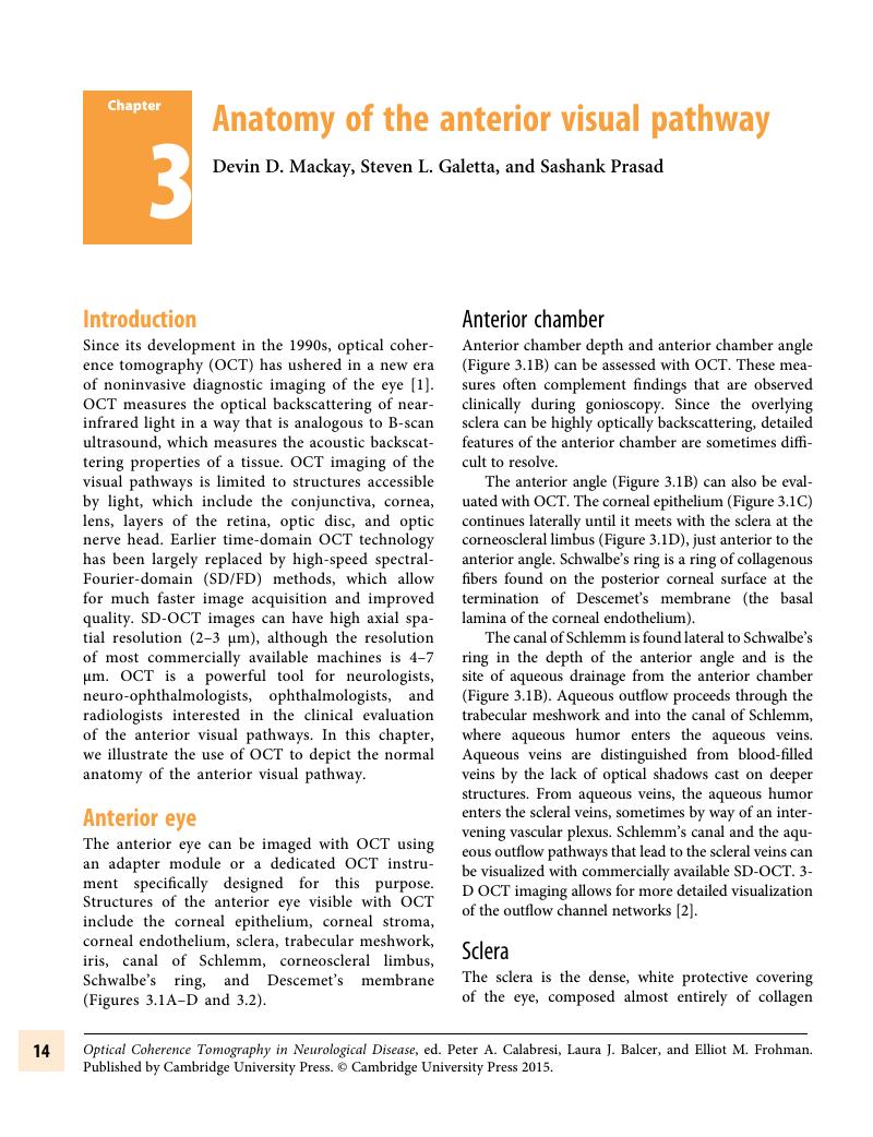
Chapter 3 - Anatomy of the anterior visual pathway

Published online by Cambridge University Press:  05 May 2015

Peter A. Calabresi
Affiliation:
Department of Neurology, Johns Hopkins University Hospital, Baltimore
Laura J. Balcer
Affiliation:
Department of Neurology, NYU Langone Medical Center, New York
Elliot M. Frohman
Affiliation:
Department of Neurology, UT Southwestern Medical Center, Dallas
Get access

Summary

Image of the first page of this content. For PDF version, please use the ‘Save PDF’ preceeding this image.'
Type
Chapter
Information
Publisher: Cambridge University Press
Print publication year: 2015

Access options

Get access to the full version of this content by using one of the access options below. (Log in options will check for institutional or personal access. Content may require purchase if you do not have access.)

References

Huang, D, Swanson, EA, Lin, CP, et al. Optical coherence tomography. Science 1991; 254: 1178–81.CrossRefGoogle ScholarPubMed
Kagemann, L, Wollstein, G, Ishikawa, H. Visualization of the conventional outflow pathway in the living human eye. Ophthalmology 2012; 119: 1563–8.CrossRefGoogle ScholarPubMed
Riordan-Eva, P. Chapter 1. Anatomy & embryology of the eye. In: Riordan-Eva, P, Cunningham, Jr. ET, eds. Vaughan & Asbury’s General Ophthalmology. 18th ed. New York: McGraw-Hill; 2011. http://www.accessmedicine.com/content.aspx?aID=55780501. Accessed May 22, 2013.Google Scholar
Wollstein, G, Folio, LS, Nevins, JE, et al. Chapter 2. Interpretation of the optical coherence tomography image. In: Schuman, JS, Puliafito, CA, Fujimoto, JG, Duker, JS, eds. Optical Coherence Tomography of Ocular Diseases. 3rd ed. Thorofare, NJ: SLACK Incorporated; 2013.Google Scholar
Považay, B, Herman, B, Unterhuber, A, et al. Three-dimensional optical coherence tomography at 1050 nm versus 800 nm in retinal pathologies: enhanced performance and choroidal penetration in cataract patients. J Biomed Optics 2007; 12: 041211.CrossRefGoogle ScholarPubMed
Drexler, W, Morgner, U, Ghanta, RK, et al. Ultrahigh-resolution ophthalmic optical coherence tomography. Nat Med 2001; 7: 502–7 (Erratum in: Nat Med 2001; 7: 636).CrossRefGoogle ScholarPubMed
Booij, JC, Baas, DC, Beisekeeva, J, et al. The dynamic nature of Bruch’s membrane. Prog Retin Eye Res 2010; 29: 118.CrossRefGoogle ScholarPubMed
Srinivasan, VJ, Monson, BK, Wojtkowski, M, et al. Characterization of outer retinal morphology with high-speed, ultra-high resolution optical coherence tomography. Invest Ophthalmol Vis Sci 2008; 49: 1571–9.CrossRefGoogle Scholar
Drexler, W, Fujimoto, JG. State-of-the-art retinal optical coherence tomography. Prog Ret Eye Res 2008; 27: 4588.CrossRefGoogle ScholarPubMed
Hee, MR, Izatt, JA, Swanson, EA, et al. Optical coherence tomography of the human retina. Arch Ophthalmol 1995; 113: 325–32.CrossRefGoogle ScholarPubMed
Anger, EM, Unterhuber, A, Hermann, B, et al. Ultrahigh resolution optical coherence tomography of the monkey fovea. Identification of retinal sublayers by correlation with semithin histology sections. Exp Eye Res 2004; 78: 1117–25.CrossRefGoogle ScholarPubMed
Spraul, CW, Lang, GE, Grossniklaus, HE. Morphometric analysis of the choroid, Bruch’s membrane, and retinal pigment epithelium in eyes with age-related macular degeneration. Inv Ophth & Vis Sci 1996; 37: 2724–35.Google ScholarPubMed
Hirsch, J,d Curcio, CA. The spatial resolution capacity of human foveal retina. Vision Res 29: 1095–101.Google Scholar
Rosenzweig, MR, Breedlove, SM, Leiman, AL. Biological Psychology 3rd Edition, Sunderland, MA: Sinauer Associates, Inc. 2002; Chapter 10, pp. 288–93.Google Scholar
Prasad, S, Galetta, SL. Anatomy and physiology of the afferent visual system. Handb Clin Neurol 2011; 102: 319.CrossRefGoogle ScholarPubMed
Osterberg, G. Topography of the layer of rods and cones in the human retina. Acta Ophthalmol 1935; 13: 1103.Google Scholar
Flores-Herr, N, Protti, DA, Wassle, H. Synaptic currents generating the inhibitory surround of ganglion cells in the mammalian retina. J Neurosci 2001; 21: 4852–63.CrossRefGoogle ScholarPubMed
Schuman, JS, Pedut-Kloizman, T, Hertzmark, E, et al. Reproducibility of nerve fiber layer thickness measurements using optical coherence tomography. Ophthalmology 1996; 103: 1889–98.CrossRefGoogle ScholarPubMed
Costa, RA, Skaf, M, Melo, LAS, et al. Retinal assessment using optical coherence tomography. Prog Ret Eye Res 2006; 25: 325–53.CrossRefGoogle ScholarPubMed
Savini, G, Zanini, M, Carelli, V, et al. Correlation between retinal nerve fibre layer thickness and optic nerve head size: an optical coherence tomography study. Br J Ophthalmol 2005; 89: 489–92.CrossRefGoogle ScholarPubMed
Bruesch, SR, Arey, LB. The number of myelinated and unmyelinated fibers in the optic nerve of vertebrates. J Comp Neurol 1942; 77: 631655.CrossRefGoogle Scholar
Jonas, JB, Gusek, GC, Naumann, GO. Optic disc, cup and neuroretinal rim size, configuration and correlations in normal eyes. Invest Ophthalmol Vis Sci 1988; 29: 1151–8.Google ScholarPubMed
Mariotte, LA. A new discovery touching vision. Phil Trans R Soc Lond 1668; 3: 668–71.Google Scholar
Strouthidis, NG, Grimm, J, Williams, GA, Cull, GA, Wilson, DJ, Burgoyne, CF. A Comparison of optic nerve head morphology viewed by spectral domain optical coherence tomography and by serial histology. Invest Ophthalmol Vis Sci 2010; 51: 1464–74.CrossRefGoogle ScholarPubMed
Kim, TW, Kagemann, L, Girard, MJ, et al. Imaging of the lamina cribrosa in glaucoma: perspectives of pathogenesis and clinical Applications. Curr Eye Res 2013; 38: 903909.CrossRefGoogle ScholarPubMed
Spaide, RF, Koizumi, H, Pozonni, MC. Enhanced depth imaging spectral-domain optical coherence tomography. Am J Ophthalmol 2008; 146: 496500.CrossRefGoogle ScholarPubMed
Park, SC, De Moraes, CG, Teng, CC, Tello, C, Liebmann, JM, Ritch, R. Enhanced depth imaging optical coherence tomography of deep optic nerve complex structures in glaucoma. Ophthalmology 2012; 119: 39.CrossRefGoogle ScholarPubMed
Wojtkowski, M, Srinivasan, VJ, Fugimoto, JG, et al. Three-dimension retinal imaging with high-speed ultrahigh-resolution optical coherence tomography. Ophthalmology 2005; 112: 1734–46.CrossRefGoogle Scholar
Walter, SD, Ishikawa, H, Galetta, KM, et al. Ganglion cell loss in relation to visual disability in multiple sclerosis. Ophthalmology 2012; 119: 1250–7.CrossRefGoogle ScholarPubMed
Makita, S, Hong, Y, Yamanari, M, et al. Optical coherence angiography. Opt Express 2006; 14: 7821–40.CrossRefGoogle ScholarPubMed

Save book to Kindle

To save this book to your Kindle, first ensure coreplatform@cambridge.org is added to your Approved Personal Document E-mail List under your Personal Document Settings on the Manage Your Content and Devices page of your Amazon account. Then enter the ‘name’ part of your Kindle email address below. Find out more about saving to your Kindle.

Note you can select to save to either the @free.kindle.com or @kindle.com variations. ‘@free.kindle.com’ emails are free but can only be saved to your device when it is connected to wi-fi. ‘@kindle.com’ emails can be delivered even when you are not connected to wi-fi, but note that service fees apply.

Find out more about the Kindle Personal Document Service.

Available formats
×

Save book to Dropbox

To save content items to your account, please confirm that you agree to abide by our usage policies. If this is the first time you use this feature, you will be asked to authorise Cambridge Core to connect with your account. Find out more about saving content to Dropbox.

Available formats
×

Save book to Google Drive

To save content items to your account, please confirm that you agree to abide by our usage policies. If this is the first time you use this feature, you will be asked to authorise Cambridge Core to connect with your account. Find out more about saving content to Google Drive.

Available formats
×