Skip to main content Accessibility help
×
Hostname: page-component-76fb5796d-qxdb6 Total loading time: 0 Render date: 2024-04-26T15:56:37.959Z Has data issue: false hasContentIssue false

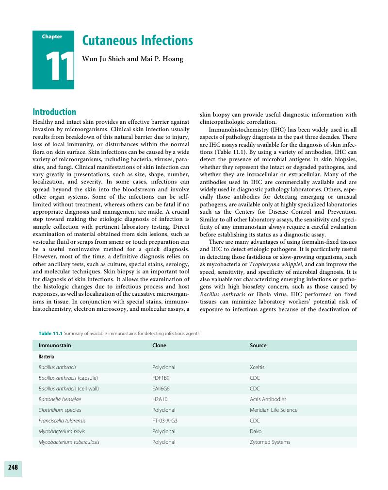
Chapter 11 - Cutaneous Infections

Published online by Cambridge University Press:  04 November 2017

Mai P. Hoang
Affiliation:
Harvard Medical School, Boston
Get access

Summary

Image of the first page of this content. For PDF version, please use the ‘Save PDF’ preceeding this image.'
Type
Chapter
Information
Publisher: Cambridge University Press
Print publication year: 2017

Access options

Get access to the full version of this content by using one of the access options below. (Log in options will check for institutional or personal access. Content may require purchase if you do not have access.)

References

Godyn, JJ, Siderits, R, Dzaman, J. Cutaneous anthrax. Arch Pathol Lab Med 2004;128(6):709–10.Google Scholar
Shieh, W-J, Guarner, J, Paddock, C, et al. The critical role of pathology in the investigation of bioterrorism-related cutaneous anthrax. Am J Pathol 2003;163(5):1901–10.Google Scholar
Quinn, C, Dull, P, Semenova, V, et al. Immune responses to Bacillus anthracis protective antigen in patients with bioterrorism-related cutaneous or inhalation anthrax. J Infect Dis 2004;190(7):1228–36.CrossRefGoogle ScholarPubMed
Dauphin, LA, Marston, CK, Bhullar, V, et al. Swab protocol for rapid laboratory diagnosis of cutaneous anthrax. J Clin Microbiol 2012;50(12):3960–67.Google Scholar
Tatti, KM, Greer, P, White, E, et al. Morphologic, immunologic, and molecular methods to detect bacillus anthracis in formalin-fixed tissues. Appl Immunohistochem Mol Morphol 2006;14(2):234–43.CrossRefGoogle ScholarPubMed
Murakawa, GJ. American Academy of Dermatology 1997 awards for young investigators in Dermatology. Pathogenesis of Bartonella henselae in cutaneous and systemic disease. J Am Acad Dermatol 1997;37(5 Pt 1):775–76.Google ScholarPubMed
Pierard-Franchimont, C, Quatresooz, P, Pierard, GE. Skin diseases associated with Bartonella infection: Facts and controversies. Clin Dermatol 2010;28(5):483–88.Google Scholar
Maass, M, Schreiber, M, Knobloch, J. Detection of Bartonella bacilliformis in cultures, blood, and formalin preserved skin biopsies by use of the polymerase chain reaction. Trop Med Parasitol 1992;43(3):191–94.Google Scholar
Hansmann, Y, DeMartino, S, Piemont, Y, et al. Diagnosis of cat scratch disease with detection of Bartonella henselae by PCR: A study of patients with lymph node enlargement. J Clin Microbiol 2005;43(8):3800–6.Google Scholar
Czarnetzki, BM, Pomeranz, JR, Khandekar, PK, Wolinsky, E, Belcher, RW. Cat-scratch disease skin test. Studies of specificity and histopathologic features. Arch Dermatol 1975;111(6):736–39.CrossRefGoogle ScholarPubMed
Brazier, JS, Duerden, BI, Hall, V, et al. Isolation and identification of Clostridium spp. from infections associated with the injection of drugs: Experiences of a microbiological investigation team. J Med Microbiol 2002,51(11):985–89.Google Scholar
Bhatnagar, J, Deleon-Carnes, M, Kellar, KL, et al. Rapid, simultaneous detection of Clostridium sordellii and Clostridium perfringens in archived tissues by a novel PCR-based microsphere assay: Diagnostic implications for pregnancy-associated toxic shock syndrome cases. Infect Dis Obstet Gynecol 2012, 2012:972845.Google Scholar
Tsokos, M, Schalinski, S, Paulsen, F, et al. Pathology of fatal traumatic and nontraumatic clostridial gas gangrene: A histopathological, immunohistochemical, and ultrastructural study of six autopsy cases. Int J Legal Med 2008;122(1):3541.Google Scholar
Guarner, J, Bartlett, J, Reagan, S, et al. Immunohistochemical evidence of Clostridium sp, Staphylococcus aureus, and group A Streptococcus in severe soft tissue infections related to injection drug use. Hum Pathol 2006;37(11):1482–88.CrossRefGoogle ScholarPubMed
Eliasson, H, Broman, T, Forsman, M, Bäck, E. Tularemia: Current epidemiology and disease management. Infect Dis Clin North Am 2006;20(2):289311.Google Scholar
Syrjala, H, Karvonen, J, Salminen, A. Skin manifestations of tularemia: A study of 88 cases in northern Finland during 16 years (1967–1983). Acta Derm Venereol 1984;64(6):513–16.Google Scholar
Splettstoesser, WD, Tomaso, H, Dahouk Al, S, Neubauer, H, Schuff-Werner, P. Diagnostic procedures in tularaemia with special focus on molecular and immunological techniques. J Vet Med B Infect Dis Vet Public Health 2005;52(6):249–61.Google Scholar
Byington, CL, Bender, JM, Ampofo, K, et al. Tularemia with vesicular skin lesions may be mistaken for infection with herpes viruses. Clin Infect Dis 2008;47(1):e46. doi: 10.1086/588843.CrossRefGoogle ScholarPubMed
Asano, S, Mori, K, Yamazaki, K, et al. Temporal differences of onset between primary skin lesions and regional lymph node lesions for tularemia in Japan: A clinicopathologic and immunohistochemical study of 19 skin cases and 54 lymph node cases. Virchows Arch 2012;460(6):651–58.CrossRefGoogle Scholar
Degitz, K. Detection of mycobacterial DNA in the skin. Etiologic insights and diagnostic perspectives. Arch Dermatol 1996;132(1):7175.Google Scholar
Beyt, BEJ, Ortbals, DW, Santa Cruz, DJ, et al. Cutaneous mycobacteriosis: Analysis of 34 cases with a new classification of the disease. Medicine 1981;60(2):95109.CrossRefGoogle ScholarPubMed
Farina, MC, Gegundez, MI, Pique, E, et al. Cutaneous tuberculosis: A clinical, histopathologic, and bacteriologic study. J Am Acad Dermatol 1995;33(3):433–40.Google Scholar
Jordaan, HF, Van Niekerk, DJ, Louw, M. Papulonecrotic tuberculid. A clinical, histopathological, and immunohistochemical study of 15 patients. Am J Dermatopathol 1994;16(5):474–85.Google Scholar
Min, K-W, Ko, JY, Park, CK. Histopathological spectrum of cutaneous tuberculosis and non-tuberculous mycobacterial infections. J Cutan Pathol 2012;39(6):582–95.Google Scholar
Dodiuk-Gad, R, Dyachenko, P, Ziv, M, et al. Nontuberculous mycobacterial infections of the skin: A retrospective study of 25 cases. J Am Acad Dermatol 2007;57(3):413–20.Google Scholar
Sia, TY, Taimur, S, Blau, DM, et al. Clinical and pathological evaluation of mycobacterium marinum group skin infections associated with fish markets in New York City. Clin Infect Dis 2016;62(5):590–95.Google Scholar
Mahaisavariya, P, Chaiprasert, A, Khemngern, S, et al. Nontuberculous mycobacterial skin infections: Clinical and bacteriological studies. J Med Assoc Thai 2003;86(1):5260.Google ScholarPubMed
Lee, WJ, Kang, SM, Sung, H, et al. Non-tuberculous mycobacterial infections of the skin: A retrospective study of 29 cases. J Dermatol 2010;37(11):965–72.Google Scholar
Cook, SM, Bartos, RE, Pierson, CL, Frank, TS. Detection and characterization of atypical mycobacteria by the polymerase chain reaction. Diagn Mol Pathol 1994;3(1):5358.Google Scholar
Mahaisavariya, P, Manonukul, J, Khemngern, S, Chaiprasert, A. Mycobacterial skin infections: Comparison between histopathologic features and detection of acid fast bacilli in pathologic section. J Med Assoc Thai 2004;87(6):709–12.Google Scholar
Noordeen, SK. Eliminating leprosy as a public health problem; why the optimism is justified. Int J Lepr Other Mycobact Dis 1995;63(4):559–66.Google ScholarPubMed
Declercq, E. Guide to eliminating leprosy as a public health problem. Lepr Rev 2001;72(1):106–7.Google Scholar
Ridley, DS, Jopling, WH. Classification of leprosy according to immunity. A five-group system. Int J Lepr Other Mycobact Dis 1966;34(3):255–73.Google Scholar
Skinsnes, OK. Immuno-pathology of leprosy: The century in review. Pathology, pathogenesis, and the development of classification. Int J Lepr Other Mycobact Dis 1973;41(3):329–60.Google Scholar
Ridley, DS. Histological classification and the immunological spectrum of leprosy. Bull World Health Organ 1974;51(5):451–65.Google Scholar
Fine, PE, Job, CK, Lucas, SB, et al. Extent, origin, and implications of observer variation in the histopathological diagnosis of suspected leprosy. Int J Lepr Other Mycobact Dis 1993;61(2):270–82.Google Scholar
Fleury, RN, Bacchi, CE. S-100 protein and immunoperoxidase technique as an aid in the histopathologic diagnosis of leprosy. Int J Lepr Other Mycobact Dis 1987;55(2):338–44.Google ScholarPubMed
de Wit, MY, Faber, WR, Krieg, SR, et al. Application of a polymerase chain reaction for the detection of Mycobacterium leprae in skin tissues. J Clin Microbiol 1991;29(5):906–10.Google Scholar
Nishimura, M, Kwon, KS, Shibuta, K, et al. Methods in pathology. An improved method for DNA diagnosis of leprosy using formaldehyde-fixed, paraffin-embedded skin biopsies. Mod Pathol 1994;7(2):253–56.Google Scholar
van Deuren, M, Brandtzaeg, P, van der Meer, JW. Update on meningococcal disease with emphasis on pathogenesis and clinical management. Clin Microbiol Rev 2000;13(1):144–66.Google Scholar
Stephens, DS, Greenwood, B, Brandtzaeg, P. Epidemic meningitis, meningococcaemia, and Neisseria meningitides. Lancet 2007;369(9580):21962210.CrossRefGoogle Scholar
Guarner, J, Greer, PW, Whitney, A, et al. Pathogenesis and diagnosis of human meningococcal disease using immunohistochemical and PCR assays. Am J Clin Pathol 2004;122(5):754–64.Google Scholar
Arend, SM, Lavrijsen, AP, Kuijken, I, van der Plas, RN, Kuijper, EJ. Prospective controlled study of the diagnostic value of skin biopsy in patients with presumed meningococcal disease. Eur J Clin Microbiol Infect Dis 2006;25(10):643–49.CrossRefGoogle ScholarPubMed
O’Brien, JP, Goldenberg, DL, Rice, PA. Disseminated gonococcal infection: A prospective analysis of 49 patients and a review of pathophysiology and immune mechanisms. Medicine 1983;62(6):395406.Google Scholar
Jain, S, Win, HN, Chalam, V, Yee, L. Disseminated gonococcal infection presenting as vasculitis: A case report. J Clin Pathol 2007;60(1):9091.Google Scholar
Walker, DH. Rocky Mountain spotted fever: A seasonal alert. Clin Infect Dis 1995;20(5):1111–17.Google Scholar
Chapman, AS, Murphy, SM, Demma, LJ, et al. Rocky Mountain spotted fever in the United States, 1997–2002. Vector Borne Zoonotic Dis 2006;6(2):170–78.Google Scholar
Paddock, CD, Finley, RW, Wright, CS, et al. Rickettsia parkeri rickettsiosis and its clinical distinction from Rocky Mountain spotted fever. Clin Infect Dis 2008;47(9):1188–96.Google Scholar
Koss, T, Carter, EL, Grossman, ME, et al. Increased detection of rickettsialpox in a New York City hospital following the anthrax outbreak of 2001: Use of immunohistochemistry for the rapid confirmation of cases in an era of bioterrorism. Arch Dermatol 2003;139(12):1545–52.Google Scholar
Sexton, DJ, Kaye, KS. Rocky mountain spotted fever. Med Clin North Am 2002;86(2):351–60viiviii.CrossRefGoogle ScholarPubMed
Sexton, DJ, Corey, GR. Rocky Mountain “spotless” and “almost spotless” fever: A wolf in sheep’s clothing. Clin Infect Dis 1992;15(3):439–48.CrossRefGoogle Scholar
Dumler, JS, Walker, DH. Diagnostic tests for Rocky Mountain spotted fever and other rickettsial diseases. Dermatol Clin 1994;12(1):2536.Google Scholar
Walker, DH, Herrero-Herrero, JI, Ruiz-Beltran, R, Bullon-Sopelana, A, Ramos-Hidalgo, A. The pathology of fatal Mediterranean spotted fever. Am J Clin Pathol 1987;87(5):669–72.Google Scholar
Kao, GF, Evancho, CD, Ioffe, O, Lowitt, MH, Dumler, JS. Cutaneous histopathology of Rocky Mountain spotted fever. J Cutan Pathol 1997;24(10):604–10.Google Scholar
Dumler, JS, Gage, WR, Pettis, GL, Azad, AF, Kuhadja, FP. Rapid immunoperoxidase demonstration of Rickettsia rickettsii in fixed cutaneous specimens from patients with Rocky Mountain spotted fever. Am J Clin Pathol 1990;93(3):410–14.Google Scholar
Paddock, CD, Greer, PW, Ferebee, TL, et al. Hidden mortality attributable to Rocky Mountain spotted fever: Immunohistochemical detection of fatal, serologically unconfirmed disease. J Infect Dis 1999;179(6):1469–76.Google Scholar
Seong, SY, Choi, MS, Kim, IS. Orientia tsutsugamushi infection: Overview and immune responses. Microbes Infect 2001;3(1):1121.CrossRefGoogle ScholarPubMed
Kim, D-M, Won, KJ, Park, CY, et al. Distribution of eschars on the body of scrub typhus patients: A prospective study. Am J Trop Med Hyg 2007;76(5):806–9.Google Scholar
Lee, J-H, Lee, J-H, Chung, KM, et al. Dynamics of clinical symptoms in patients with scrub typhus. Jpn J Infect Dis 2013;66(2):155–57.Google Scholar
Koraluru, M, Bairy, I, Varma, M, Vidyasagar, S. Diagnostic validation of selected serological tests for detecting scrub typhus. Microbiol Immunol 2015;59(7):371–74.CrossRefGoogle ScholarPubMed
Kim, CM, Cho, MK, Kim, DM, et al. Accuracy of conventional PCR targeting the 16S rRNA gene with the Ot-16sRF1 and Ot-16sRR1 primers for diagnosis of scrub typhus: A case-control study. J Clin Microbiol 2016,54(1):178–79.Google Scholar
Park, JH, Hart MS: The pathology of scrub typhus. Am J Clin Pathol 1946;16:139–49.CrossRefGoogle ScholarPubMed
Kim, D-M, Lim, S-C, Won, KJ, et al. Severe scrub typhus confirmed early via immunohistochemical staining. Am J Trop Med Hyg 2007;77(4):719–22.CrossRefGoogle ScholarPubMed
Carapetis, JR, Steer, AC, Mulholland, EK, Weber, M. The global burden of group A streptococcal diseases. Lancet Infect Dis 2005;5(11):685–94.Google Scholar
Nelson, GE, Pondo, T, Toews, K-A, et al. Epidemiology of Invasive Group A Streptococcal Infections in the United States, 2005–2012. Clin Infect Dis 2016,63(4):478–86.Google Scholar
Hoge, CW, Schwartz, B, Talkington, DF, et al. The changing epidemiology of invasive group A streptococcal infections and the emergence of streptococcal toxic shock-like syndrome. A retrospective population-based study. JAMA 1993;269(3):384–89.Google Scholar
Low, DE, McGeer, A. Skin and soft tissue infection: Necrotizing fasciitis. Curr Opin Infect Dis 1998;11(2):119–23.Google Scholar
Parks, T, Smeesters, PR, Steer AC: Streptococcal skin infection and rheumatic heart disease. Curr Opin Infect Dis 2012;25(2):145–53.Google Scholar
Shulman, ST, Bisno, AL, Clegg, HW, et al. Clinical practice guideline for the diagnosis and management of group A streptococcal pharyngitis: 2012 update by the Infectious Diseases Society of America. Clin Infect Dis 2012;55(10):e86e102.Google Scholar
Cohen, JF, Chalumeau, M, Levy, C, et al. Effect of clinical spectrum, inoculum size and physician characteristics on sensitivity of a rapid antigen detection test for group A streptococcal pharyngitis. Eur J Clin Microbiol Infect Dis 2013;32(6):787–93.Google Scholar
Hartman-Adams, H, Banvard, C, Juckett, G. Impetigo: Diagnosis and treatment. Am Fam Physician 2014;90(4):229–35.Google Scholar
Guarner, J, Sumner, J, Paddock, CD, et al. Diagnosis of invasive group A streptococcal infections by using immunohistochemical and molecular assays. Am J Clin Pathol 2006;126(1):148–55.Google Scholar
Cohen, PR. Community-acquired methicillin-resistant Staphylococcus aureus skin infections: Implications for patients and practitioners. Am J Clin Dermatol 2007;8(5):259–70.CrossRefGoogle ScholarPubMed
Lyell, A. The staphylococcal scalded skin syndrome in historical perspective: Emergence of dermopathic strains of Staphylococcus aureus and discovery of the epidermolytic toxin. A review of events up to 1970. J Am Acad Dermatol 1983;9(2):285–94.Google Scholar
Rieger-Fackeldey, E, Plano, LRW, Kramer, A, Schulze, A. Staphylococcal scalded skin syndrome related to an exfoliative toxin A- and B-producing strain in preterm infants. Eur J Pediatr 2002;161(12):649–52.CrossRefGoogle Scholar
Ladhani, S. Understanding the mechanism of action of the exfoliative toxin of **Staphylococcus aureus.** FEMS Immunol Med Microbiol 2003;39(2):181–89.CrossRefGoogle ScholarPubMed
Singh, G. Pathogenesis of staphylococcal infections of the skin. Int J Dermatol 1975;14(10):755–60.Google Scholar
Peeling, RW, Hook, EW. The pathogenesis of syphilis: The Great Mimicker, revisited. J Pathol 2006;208(2):224–32.Google Scholar
Hoang, MP, High, WA, Molberg, KH. Secondary syphilis: A histologic and immunohistochemical evaluation. J Cutan Pathol 2004;31(9):595–99.Google Scholar
Martin-Ezquerra, G, Fernandez-Casado, A, Barco, D, et al. Treponema pallidum distribution patterns in mucocutaneous lesions of primary and secondary syphilis: An immunohistochemical and ultrastructural study. Hum Pathol 2009;40(5):624–30.Google Scholar
Buffet, M, Grange, PA, Gerhardt, P, et al. Diagnosing Treponema pallidum in secondary syphilis by PCR and immunohistochemistry. J Invest Dermatol 2007;127(10):2345–50.Google Scholar
Stenseth, NC, Atshabar, BB, Begon, M, et al. Plague: Past, present, and future. PLoS Med 2008;5(1):e3. doi: 10.1371/journal.pmed.0050003.Google Scholar
Inglesby, TV, Dennis, DT, Henderson, DA, et al. Plague as a biological weapon: Medical and public health management. Working Group on Civilian Biodefense. JAMA 2000;283(17):2281–90.Google Scholar
Perry, RD, Fetherston, JD. **Yersinia pestis** – etiologic agent of plague. Clin Microbiol Rev 1997;10(1):3566.Google Scholar
Cieslak, TJ, Talbot, TB, Hartstein, BH. Biological warfare and the skin I: Bacteria and toxins. Clin Dermatol 2002;20(4):346–54.Google Scholar
Butler, T, Hudson, BW. The serological response to Yersinia pestis infection. Bull World Health Organ 1977;55(1):3942.Google Scholar
Radnedge, L, Gamez-Chin, S, McCready, PM, et al. Identification of nucleotide sequences for the specific and rapid detection of **Yersinia pestis.** Appl Environ Microbiol 2001;678):3759–62.Google Scholar
Guarner, J, Shieh, W-J, Greer, PW, et al. Immunohistochemical detection of Yersinia pestis in formalin-fixed, paraffin-embedded tissue. Am J Clin Pathol 2002;117(2):205–9.Google Scholar
Frank, KM, Schneewind, O, Shieh, WJ. Investigation of a researcher’s death due to septicemic plague. N Engl J Med 2011;364(26):2563–64.Google Scholar
Grist, NR, Bell, EJ, Assad, F. Enteroviruses in human disease. Prog Med Virol 1978;24:114–57.Google Scholar
Strikas, RA, Anderson, LJ, Parker, RA. Temporal and geographic patterns of isolates of nonpolio enteroviruses in the United States. J Infect Dis 1986;153(2):346–51.Google Scholar
Ooi, MH, Wong, SC, Lewthwaite, P, Cardosa, MJ, Solomon, T. Clinical features, diagnosis, and management of enterovirus 71. Lancet Neurol 2010;9(11):10971105.Google Scholar
Yan, JJ, Wang, JR, Liu, CC, Yang, HB, Su, IJ. An outbreak of enterovirus 71 infection in Taiwan 1998: A comprehensive pathological, virological, and molecular study on a case of fulminant encephalitis. J Clin Virol 2000;17(1):1322.CrossRefGoogle Scholar
Aswathyraj, S, Arunkumar, G, Alidjinou, EK, Hober, D. Hand, foot, and mouth disease (HFMD): Emerging epidemiology and the need for a vaccine strategy. Med Microbiol Immunol 2016;205(5):397407.Google Scholar
Bubba, L, Pellegrinelli, L, Pariani, E, et al. A novel multiplex one-step real-time RT-PCR assay for the simultaneous identification of enterovirus and parechovirus in clinical fecal samples. J Prev Med Hyg 2015;56(2):E5760.Google Scholar
Shieh, WJ, Jung, SM, Hsueh, C, et al. Pathologic studies of fatal cases in outbreak of hand, foot, and mouth disease, Taiwan. Emerg Infect Dis 2001;7(1):146–48.Google Scholar
Guerrant, RL, Walker, DH, Weller, PF. Tropical Infectious Diseases: Principles, Pathogens and Practice. Elsevier Health Sciences; 2011.Google Scholar
Singh, SK, Ruzek, D. Viral Hemorrhagic Fevers. CRC Press; 2013.Google Scholar
Zaki, SR, Shieh, WJ, Greer, PW, et al. A novel immunohistochemical assay for the detection of Ebola virus in skin: Implications for diagnosis, spread, and surveillance of Ebola hemorrhagic fever. Commission de Lutte contre les Epidémies à Kikwit. J Infect Dis 1999;179(Suppl 1):S3647.Google Scholar
Saadiah, S, Sharifah, BI, Robson, A, Greaves, MW. Skin histopathology and immunopathology are not of prognostic value in dengue haemorrhagic fever. Br J Dermatol 2008;158(4):836–37.Google Scholar
Bernstein, DI, Bellamy, AR, Hook, EW, et al. Epidemiology, clinical presentation, and antibody response to primary infection with herpes simplex virus type 1 and type 2 in young women. Clin Infect Dis 2013;56(3):344–51.Google Scholar
Hofstetter, AM, Rosenthal, SL, Stanberry, LR. Current thinking on genital herpes. Curr Opin Infect Dis 2014;27(1):7583.Google Scholar
Benedetti, J, Corey, L, Ashley, R. Recurrence rates in genital herpes after symptomatic first-episode infection. Ann Intern Med 1994;121(11):847–54.Google Scholar
Leinweber, B, Kerl, H, Cerroni, L. Histopathologic features of cutaneous herpes virus infections (herpes simplex, herpes varicella/zoster): A broad spectrum of presentations with common pseudolymphomatous aspects. Am J Surg Pathol 2006;30(1):5058.Google Scholar
Hoyt, B, Bhawan, J. Histological spectrum of cutaneous herpes infections. Am J Dermatopathol 2014;36(8):609–19.CrossRefGoogle ScholarPubMed
Weinberg, JM. Herpes zoster: Epidemiology, natural history, and common complications. J Am Acad Dermatol 2007;57(6 Suppl):S130–35.Google Scholar
Oxman, MN, Levin, MJ, Johnson, GR, et al, Shingles Prevention Study Group. A vaccine to prevent herpes zoster and postherpetic neuralgia in older adults. N Engl J Med 2005;352(22)2271–84.Google Scholar
el Hayderi, L, Bontems, S, Nikkels-Tassoudji, N, et al. Satellite lesions accompanying herpes zoster: A new prognostic sign for high-risk zoster. Br J Dermatol 2015;172(6):1530–34.Google Scholar
Wilson, DA, Yen-Lieberman, B, Schindler, S, et al. Should varicella-zoster virus culture be eliminated? A comparison of direct immunofluorescence antigen detection, culture, and PCR, with a historical review. J Clin Microbiol 2012;50(12):4120–22.Google Scholar
Boer, A, Herder, N, Winter, K, Falk, T. Herpes folliculitis: Clinical, histopathological, and molecular pathologic observations. Br J Dermatol 2006;154(4):743–46.Google Scholar
Muraki, R, Baba, T, Iwasaki, T, Sata, T, Kurata, T. Immunohistochemical study of skin lesions in herpes zoster. Virchows Arch A Pathol Anat Histopathol 1992;420(1):7176.Google Scholar
Horie, C, Mizukawa, Y, Yamazaki, Y, Shiohara, T. Varicella-zoster virus antigen expression of eccrine gland and duct epithelium in herpes zoster lesions. Br J Dermatol 2011;165(4):802–7.Google Scholar
Pisapia, DJ, Lavi, E. VZV, temporal arteritis, and clinical practice: False positive immunohistochemical detection due to antibody cross-reactivity. Exp Mol Pathol 2016;100(1):114–15.Google Scholar
Fernandez Flores, A. Epstein-Barr virus in cutaneous pathology. Am J Dermatopathol 2013;35(8):763–86.Google Scholar
Tan, HH, Goh, CL. Viral infections affecting the skin in organ transplant recipients: Epidemiology and current management strategies. Am J Clin Dermatol 2006;7(1):1329.Google Scholar
Chisholm, C, Lopez, L. Cutaneous infections caused by Herpesviridae: A review. Arch Pathol Lab Med 2011;135(10):1357–62.CrossRefGoogle ScholarPubMed
Hennard, C, Pfuhl, T, Buettner, M, et al. The antibody 2B4 directed against the Epstein-Barr virus (EBV)-encoded nuclear antigen 1 (EBNA1) detects MAGE-4: Applications for studies on the EBV association of human cancers. J Pathol 2006;209(4):430–35.Google Scholar
Kempf, W, Flaig, MJ, Kutzner, H. Molecular diagnostics in infectious skin diseases. J Dtsch Dermatol Ges 2013;11(Suppl 4):5058.Google ScholarPubMed
Drago, F, Aragone, MG, Lugani, C, Rebora, A. Cytomegalovirus infection in normal and immunocompromised humans. A review. Dermatology 2000;200(3):189–95.Google Scholar
Cohen, JI, Corey, GR. Cytomegalovirus infection in the normal host. Medicine 1985;64(2):100–14.Google Scholar
Fine, JD, Arndt, KA. The TORCH syndrome: A clinical review. J Am Acad Dermatol 1985;12(4):697706.Google Scholar
Pariser, RJ. Histologically specific skin lesions in disseminated cytomegalovirus infection. J Am Acad Dermatol 1983;9(6):937–46.Google Scholar
Resnik, KS, DiLeonardo, M, Maillet, M. Histopathologic findings in cutaneous cytomegalovirus infection. Am J Dermatopathol 2000;22(5):397407.Google Scholar
Choi, YL, Kim, JA, Jang, KT, et al. Characteristics of cutaneous cytomegalovirus infection in non-acquired immune deficiency syndrome, immunocompromised patients. Br J Dermatol 2006;155(5):977–82.Google Scholar
Boutolleau, D, Fernandez, C, Andre, E, et al. Human herpesvirus (HHV)-6 and HHV-7: Two closely related viruses with different infection profiles in stem cell transplantation recipients. J Infect Dis 2003;187(2):179–86.Google Scholar
Sumiyoshi, Y, Akashi, K, Kikuchi, M. Detection of human herpes virus 6 (HHV6) in the skin of a patient with primary HHV 6 infection and erythroderma. J Clin Pathol 1994;47(8):762–63.Google Scholar
Broccolo, F, Drago, F, Careddu, AM, et al. Additional evidence that pityriasis rosea is associated with reactivation of human herpes virus-6 and -7. J Invest Dermatol 2005;124(6):1234–40.Google Scholar
Levine, PH, Jahan, N, Murari, P, Manak, M, Jaffe, ES. Detection of human herpes virus 6 in tissues involved by sinus histiocytosis with massive lymphadenopathy (Rosai-Dorfman disease). J Infect Dis 1992;166(2):291–95.Google Scholar
Ortonne, N, Fillet, AM, Kosuge, H, et al. Cutaneous Destombes-Rosai-Dorfman disease: Absence of detection of HHV-6 and HHV-8 in skin. J Cutan Pathol 2002;29(2):113–18.Google Scholar
Hiatt, KM, Nelson, AM, Lichy, JH, Fandburg-Smith, JC. Classic Kaposi sarcoma in the United States over the last two decades: A clinicopathologic and molecular study of 438 non-HIV-related Kaposi sarcoma patients with comparison to HIV-related Kaposi sarcoma. Mod Pathol 2008;21(5):572–82.Google Scholar
Hong, A, Davies, S, Lee, CS. Immunohistochemical detection of the human herpes virus 8 (HHV8) latent nuclear antigen-1 in Kaposi’s sarcoma. Pathology 2003;35(5):448450.Google Scholar
Cheuk, W, Wong, KO, Wong, CS, et al. Immunostaining for human herpes virus 8 latent nuclear antigen-1 helps distinguish Kaposi sarcoma from its mimickers. Am J Clin Pathol 2004;121(3):335–42.Google Scholar
McDonagh, DP, Liu, J, Gaffey, MJ, et al. Detection of Kaposi’s sarcoma-associated herpes virus-like DNA sequence in angiosarcoma. Am J Pathol 1996;149(4):1363–68.Google Scholar
Nuovo, GJ, Friedman, D, Richart, DM. In situ hybridization analysis of human papillomavirus DNA segregation patterns in lesions of the female genital tract. Gynecol Oncol 1990;36(2):256–62.Google Scholar
Marshburn, PB, Trofatter, KF. Recurrent condyloma acuminatum in women over age 40: Association with immunosuppression and malignant disease. Am J Obstet Gynecol 1988;159(2):429–33.Google Scholar
Mahajan, A. Practical issues in the application of p16 immunohistochemistry in diagnostic pathology. Hum Pathol 2016;51(5):6474.Google Scholar
Kostopoulou, E, Samara, M, Kollia, P, et al. Different patterns of p16 immunoreactivity in cervical biopsies: Correlation to lesion grade and HPV detection, with a review of the literature. Eur J Gynaecol Oncol 2011;32(1):54–61.Google Scholar
O’Neill, CJ, McCluggage, WG. p16 expression in the female genital tract and its value in diagnosis. Adv Anat Pathol 2006;13(1):815.Google Scholar
Lenhoff, A. Five FDA-approved HPV assays. MLO Med Lab Obs 2012;44(7):14, 16, 18.Google Scholar
Bishop, JA, Ma, XJ, Wang, H, et al. Detection of transcriptionally active high-risk HPV in patients with head and neck squamous cell carcinoma as visualized by a novel E6/E7 mRNA in situ hybridization method. Am J Surg Pathol 2012;36(12):1874–82.Google Scholar
La Grenade, L, Manns, A, Fletcher, V, et al. Clinical, pathologic, and immunologic features of human T-lymphotropic virus type I-associated infective dermatitis in children. Arch Dermatol 1998;134(4):439–44.Google Scholar
Amano, M, Kurokawa, M, Ogata, K, et al. New entity, definition and diagnostic criteria of cutaneous adult T-cell leukemia/lymphoma: Human T-lymphotropic virus type 1 proviral DNA load can distinguish between cutaneous and smoldering types. J Dermatol 2008;35(5):270–75.Google Scholar
Hasui, K, Wang, J, Tanaka, Y, et al. Development of ultra-super sensitive immunohistochemistry and its application to the etiological study of adult T-cell leukemia/lymphoma. Acta Histochem Cytochem 2012;45(2):83106.Google Scholar
Santonja, C, Nieto-Gonzalez, G, Santos-Briz, A, et al. Immunohistochemical detection of parvovirus B19 in “gloves and socks” papular purpuric syndrome: Direct evidence for viral endothelial involvement. Report of three cases and review of the literature. Am J Dermatopathol 2011;33(8):790–95.Google Scholar
Bonvicini, F, La Placa, M, Manaresi, E, et al. Parvovirus B19 DNA is commonly harboured in human skin. Dermatology 2010;220(2):138–42.Google Scholar
Schwarz, TF, Wiersbitzky, S, Pambor, M. Case report: Detection of parvovirus B19 in a skin biopsy of a patient with erythema infectiosum. J Med Virol 1994;43(2):171–74.Google Scholar
Takahashi, M, Ito, M, Sakamoto, F, et al. Human parvovirus B19 infection: Immunohistochemical and electron microscopic studies of skin lesions. J Cutan Pathol 1995;22(2):168–72.Google Scholar
Feng, H, Shuda, M, Chang, Y, et al. Clonal integration of a polyomavirus in human Merkel cell carcinoma. Science 2008;319(5866):10961100.Google Scholar
Andres, C, Belloni, B, Puchta, U, Sander, CA, Flaig, MJ. Prevalence of MCPyV in Merkel cell carcinoma and non-MCC tumors. J Cutan Pathol 2010;37(1):2834.Google Scholar
Mertz, KD, Pfaltz, M, Junt, T, et al. Merkel cell polyomavirus is present in common warts and carcinoma in situ of the skin. Hum Pathol 2010;41(10):1369–79.Google Scholar
Shuda, M, Arora, R, Kwun, HJ, et al. Human Merkel cell polyomavirus infection I. MCV T antigen expression in Merkel cell carcinoma, lymphoid tissues and lymphoid tumors. Int J Cancer 2009;125(6):1243–49.Google Scholar
Shuda, M, Kwun, HJ, Feng, H, et al. Human Merkel cell polyomavirus small T antigen is an oncoprotein targeting the 4E-BP1 translation regulator. J Clin Invest 2011;121(9):3623–34.Google Scholar
Rodig, SJ, Cheng, J, Wardzala, J, et al. Improved detection suggests all Merkel cell carcinomas harbor Merkel polyomavirus. J Clin Invest 2012;122(2):4645–53.Google Scholar
Busam, JK, Jungbluth, AA, Rekthman, N, et al. Merkel cell polyomavirus expression in Merkel cell carcinomas and its absence in combined tumors and pulmonary neuroendocrine carcinomas. Am J Surg Pathol 2009;33(9):1378–85.Google Scholar
Levine, RS, Peterson, AT, Yorita, KL, et al. Ecological niche and geographic distribution of human monkeypox in Africa. PLoS One 2007;2(1):e176.Google Scholar
Fleischauer, AT, Kile, JC, Davidson, M, et al. Evaluation of human-to-human transmission of monkeypox from infected patients to health care workers. Clin Infect Dis 2005;40(5):689–94.Google Scholar
Centers for Disease Control and Prevention CDC. Multistate outbreak of monkeypox–Illinois, Indiana, and Wisconsin, 2003. MMWR Morb Mortal Wkly Rep 2003;52(23):537–40.Google Scholar
Reed, KD, Melski, JW, Graham, MB, et al. The detection of monkeypox in humans in the Western Hemisphere. N Engl J Med 2004;350(4):342–50.Google Scholar
McCollum, AM, Damon, IK. Human Monkeypox. Clin Infect Dis 2014;58(2):260–67.Google Scholar
Li, Y, Olson, VA, Laue, T, Laker, MT, Damon, IK. Detection of monkeypox virus with real-time PCR assays. J Clin Virol 2006,36(3):194203.Google Scholar
Goldsmith, CS, Ksiazek, TG, Rollin, PE, et al. Cell culture and electron microscopy for identifying viruses in diseases of unknown cause. Emerg Infect Dis 2013;19(6):864–69.Google Scholar
Guarner, J, Johnson, BJ, Paddock, CD, et al. Veterinary Monkeypox Virus Working Group: Monkeypox transmission and pathogenesis in prairie dogs. Emerg Infect Dis 2004;10(3):426–31.Google Scholar
Hessami, M, Keney, DA, Pearson, LD, Storz, J. Isolation of parapox viruses from man and animals: Cultivation and cellular changes in bovine fetal spleen cells. Comp Immunol Microbiol Infect Dis 1979;2(1):17.Google Scholar
Tondury, B, Kuhne, A, Kutzner, H, et al. Molecular diagnostics of parapox virus infections. J Dtsch Dermatol Ges 2010;8(9):681–84.Google Scholar
Leavell, UWJ, McNamara, MJ, Muelling, R, et al. Orf. Report of 19 human cases with clinical and pathological observations. JAMA 1968;204(8):657–64.Google Scholar
Johannessen, JV, Krogh, HK, Solberg, I, et al. Human orf. J Cutan Pathol 1975;2(6):265–83.Google Scholar
Kuokkanen, K, Launis, J, Mörttinen, A. Erythema nodosum and erythema multiforme associated with milker’s nodules. Acta Derm Venereol 1976;56(1):6972.Google Scholar
Groves, RW, Wilson-Jones, E, MacDonald, DM. Human orf and milkers’ nodule: A clinicopathologic study. J Am Acad Dermatol 1991;25(4):706–11.Google Scholar
Evins, S, Leavell, UWJ, Phillips, IA. Intranuclear inclusions in milker’s nodules. Arch Dermatol 1971;103(1):9193.Google Scholar
Sanchez, RL, Hebert, A, Lucia, H, Swedo, J. Orf. A case report with histologic, electron microscopic, and immunoperoxidase studies. Arch Pathol Lab Med 1985;109(2):166–70.Google Scholar
Helton, J, Loveless, M, White, CRJ. Cutaneous acanthamoeba infection associated with leukocytoclastic vasculitis in an AIDS patient. Am J Dermatopathol 1993;15(2):146–49.Google Scholar
Tan, B, Weldon-Linne, CM, Rhone, DP, Penning, CL, Visvesvara, GS. Acanthamoeba infection presenting as skin lesions in patients with the acquired immunodeficiency syndrome. Arch Pathol Lab Med 1993;117(10):1043–46.Google Scholar
Gullett, J, Mills, J, Hadley, K, et al. Disseminated granulomatous acanthamoeba infection presenting as an unusual skin lesion. Am J Med 1979;67(5):891–96.Google Scholar
Wortman, PD. Acanthamoeba infection. Int J Dermatol 1996;35(1):4851.Google Scholar
Galarza, C, Ramos, W, Gutierrez, EL, et al. Cutaneous acanthamebiasis infection in immunocompetent and immunocompromised patients. Int J Dermatol 2009;48(12):1324–29.Google Scholar
Rosenberg, AS, Morgan, MB. Disseminated acanthamoebiasis presenting as lobular panniculitis with necrotizing vasculitis in a patient with AIDS. J Cutan Pathol 2001;28(6):307–13.Google Scholar
Guarner, J, Bartlett, J, Shieh, W-J, et al. Histopathologic spectrum and immunohistochemical diagnosis of amebic meningoencephalitis. Mod Pathol 2007;20(12):1230–37.Google Scholar
Lobo, SA, Patil, K, Jain, S, et al. Diagnostic challenges in Balamuthia mandrillaris infections. Parasitol Res 2013;112(12):4015–19.Google Scholar
Martinez, DY, Seas, C, Bravo, F, et al. Successful treatment of Balamuthia mandrillaris amoebic infection with extensive neurological and cutaneous involvement. Clin Infect Dis 2010;51(2):e711.CrossRefGoogle ScholarPubMed
Qvarnstrom, Y, Visvesvara, GS, Sriram, R, da Silva, AJ. Multiplex real-time PCR assay for simultaneous detection of Acanthamoeba spp., Balamuthia mandrillaris, and Naegleria fowleri. J Clin Microbiol 2006;44(10):3589–95.Google Scholar
Pritzker, AS, Kim, BK, Agrawal, D, Southern, PMJ, Pandya, AG. Fatal granulomatous amebic encephalitis caused by Balamuthia mandrillaris presenting as a skin lesion. J Am Acad Dermatol 2004;50(2 Suppl):S3841.Google Scholar
Farnon, EC, Kokko, KE, Budge, PJ, et al. Balamuthia Transplant Investigation Teams: Transmission of Balamuthia mandrillaris by organ transplantation. Clin Infect Dis 2016;63(7):878–88.Google Scholar
Grevelink, SA, Lerner, EA. Leishmaniasis. J Am Acad Dermatol 1996;34(2 Pt 1):257–72.Google Scholar
Pavli, A, Maltezou, HC. Leishmaniasis, an emerging infection in travelers. Int J Infect Dis 2010;14(12):e1032–39.Google Scholar
Ryan, ET, Maguire, JH, Strickland, GT, Solomon, T, Hill, DR. Hunter’s Tropical Medicine and Emerging Infectious Disease. W B Saunders Company; 2012.Google Scholar
Sangueza, OP, Sangueza, JM, Stiller, MJ, Sangueza, P. Mucocutaneous leishmaniasis: A clinicopathologic classification. J Am Acad Dermatol 1993;28(6):927–32.Google Scholar
Goihman-Yahr, M. American mucocutaneous leishmaniasis. Dermatol Clin 1994;12(4):703–12.Google Scholar
Salman, SM, Rubeiz, NG, Kibbi, AG. Cutaneous leishmaniasis: Clinical features and diagnosis. Clin Dermatol 1999;17(3):291–96.Google Scholar
Gazozai, S, Iqbal, J, Bukhari, I, Bashir, S. Comparison of diagnostic methods in cutaneous Leishmaniasis (histopathology compared to skin smears). Pak J Pharm Sci 2010;23(4):363–66.Google Scholar
de Almeida, ME, Steurer, FJ, Koru, O, et al. Identification of Leishmania spp. by molecular amplification and DNA sequencing analysis of a fragment of rRNA internal transcribed spacer 2. J Clin Microbiol 2011;49(9):3143–49.Google Scholar
Kurban, AK, Malak, JA, Farah, FS, Chaglassian, HT. Histopathology of cutaneous leishmaniasis. Arch Dermatol 1966;93(4):396401.Google Scholar
Gutierrez, Y, Salinas, GH, Palma, G, et al. Correlation between histopathology, immune response, clinical presentation, and evolution in Leishmania braziliensis infection. Am J Trop Med Hyg 1991;45(3):281–89.Google Scholar
Mehregan, DR, Mehregan, AH, Mehregan, DA. Histologic diagnosis of cutaneous leishmaniasis. Clin Dermatol 1999;17(3):297304.Google Scholar
Kenner, JR, Aronson, NE, Bratthauer, GL, et al. Immunohistochemistry to identify Leishmania parasites in fixed tissues. J Cutan Pathol 1999;26(3):130–36.Google Scholar
Neblett Fanfair, R, Benedict, K, Bos, J, et al. Necrotizing cutaneous mucormycosis after a tornado in Joplin, Missouri, in 2011. N Engl J Med 2012;367(23):2214–25.Google Scholar
Ritter, JM, Muehlenbachs, A, Blau, DM, et al; Exserohilum Infections Working Group. Exserohilum infections associated with contaminated steroid injections: A clinicopathologic review of 40 cases. Am J Pathol 2013;183(3):881–92.Google Scholar
Bialek, R, Konrad, F, Kern, J, et al. PCR based identification and discrimination of agents of mucormycosis and aspergillosis in paraffin wax embedded tissue. J Clin Pathol 2005;58(11):1180–84.Google Scholar
Lau, A, Chen, S, Sorrell, T, et al. Development and clinical application of a panfungal PCR assay to detect and identify fungal DNA in tissue specimens. J Clin Microbiol 2007;45(2):380–85.Google Scholar
Muñoz-Cadavid, C, Rudd, S, Zaki, SR, et al. Improving molecular detection of fungal DNA in formalin-fixed paraffin-embedded tissues: Comparison of five tissue DNA extraction methods using panfungal PCR. J Clin Microbiol 2010;48(6):2147–53.CrossRefGoogle ScholarPubMed

Save book to Kindle

To save this book to your Kindle, first ensure coreplatform@cambridge.org is added to your Approved Personal Document E-mail List under your Personal Document Settings on the Manage Your Content and Devices page of your Amazon account. Then enter the ‘name’ part of your Kindle email address below. Find out more about saving to your Kindle.

Note you can select to save to either the @free.kindle.com or @kindle.com variations. ‘@free.kindle.com’ emails are free but can only be saved to your device when it is connected to wi-fi. ‘@kindle.com’ emails can be delivered even when you are not connected to wi-fi, but note that service fees apply.

Find out more about the Kindle Personal Document Service.

Available formats
×

Save book to Dropbox

To save content items to your account, please confirm that you agree to abide by our usage policies. If this is the first time you use this feature, you will be asked to authorise Cambridge Core to connect with your account. Find out more about saving content to Dropbox.

Available formats
×

Save book to Google Drive

To save content items to your account, please confirm that you agree to abide by our usage policies. If this is the first time you use this feature, you will be asked to authorise Cambridge Core to connect with your account. Find out more about saving content to Google Drive.

Available formats
×