

Book contents
- Immunohistochemistry in Diagnostic Dermatopathology
- Immunohistochemistry in Diagnostic Dermatopathology
- Copyright page
- Dedication
- Contents
- Contributors
- Preface and Acknowledgments
- Chapter 1 Introduction to Immunohistochemistry
- Chapter 2 Epithelial or Squamous Neoplasms
- Chapter 3 Neoplasms of Cutaneous Appendages
- Chapter 4 Inflammatory Dermatoses Mimicking Lymphomas
- Chapter 5 Cutaneous Lymphoid Neoplasms
- Chapter 6 Melanocytic neoplasms
- Chapter 7 Soft Tissue Neoplasms
- Chapter 8 Miscellaneous Tumors
- Chapter 9 Detection of Genetic Syndromes
- Chapter 10 Immunobullous Disorders
- Chapter 11 Cutaneous Infections
- Chapter 12 Therapeutic and Prognostic Applications
- Index
- References
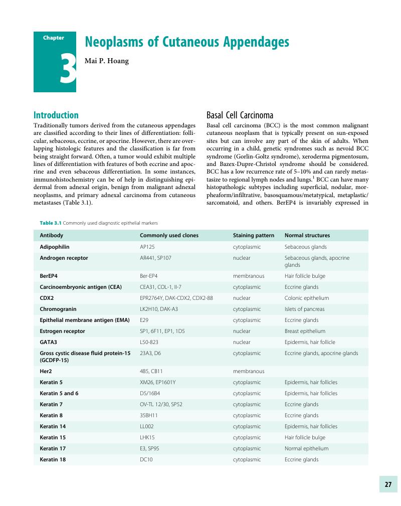
Chapter 3 - Neoplasms of Cutaneous Appendages
Published online by Cambridge University Press: 04 November 2017
By
Edited by

Book contents
- Immunohistochemistry in Diagnostic Dermatopathology
- Immunohistochemistry in Diagnostic Dermatopathology
- Copyright page
- Dedication
- Contents
- Contributors
- Preface and Acknowledgments
- Chapter 1 Introduction to Immunohistochemistry
- Chapter 2 Epithelial or Squamous Neoplasms
- Chapter 3 Neoplasms of Cutaneous Appendages
- Chapter 4 Inflammatory Dermatoses Mimicking Lymphomas
- Chapter 5 Cutaneous Lymphoid Neoplasms
- Chapter 6 Melanocytic neoplasms
- Chapter 7 Soft Tissue Neoplasms
- Chapter 8 Miscellaneous Tumors
- Chapter 9 Detection of Genetic Syndromes
- Chapter 10 Immunobullous Disorders
- Chapter 11 Cutaneous Infections
- Chapter 12 Therapeutic and Prognostic Applications
- Index
- References
Summary
A summary is not available for this content so a preview has been provided. Please use the Get access link above for information on how to access this content.
- Type
- Chapter
- Information
- Immunohistochemistry in Diagnostic Dermatopathology , pp. 27 - 54Publisher: Cambridge University PressPrint publication year: 2017
References
Wysong, A, Aasi, SZ, Tang, JY. Update on metastatic basal cell carcinoma: A summary of published cases from 1981 through 2011. JAMA Dermatol 2013;149(5):615–16.Google Scholar
Beer, TW, Shepherd, P, Theaker, JM. BerEP4 and epithelial membrane antigen aid distinction of basal cell, squamous cell and basosquamous carcinomas of the skin. Histopathology 2000;37(3):218–23.Google Scholar
LeBoit, PE, Burg, G, Weedon, D, et al. World Health Organization Classification of Tumours. Pathology and Genetics of Skin Tumours. Lyon, France: IARC Press, 2006.Google Scholar
Kurzen, H, Esposito, L, Langbein, L, Hartschuh, W. Cytokeratins as markers of follicular differentiation: An immunohistochemical study of trichoblastoma and basal cell carcinoma. Am J Dermatopathol 2001;23(6):501–9.Google Scholar
Onishi, T, Watanabe, S. Immunohistochemical analysis of cytokeratin expression in various trichogenic tumors. Am J Dermatopathol 1999;21(4):337–43.Google Scholar
Evangelista, MT, North, JP. Comparative analysis of cytokeratin 15, TDAG51, cytokeratin 20 and androgen receptor in sclerosing adnexal neoplasms and variants of basal cell carcinoma. J Cutan Pathol 2015;42(11):824–31.Google Scholar
Costache, M, Bresch, M, Boer, A. Desmoplastic trichoepithelioma versus morphoeic basal cell carcinoma: A critical reappraisal of histomorphological and immunohistochemical criteria for differentiation. Histopathology 2008;52(7):865–76.Google Scholar
Katona, TM, Perkins, SM, Billings, SD. Does the panel of cytokeratin 20 and androgen receptor antibodies differentiate desmoplastic trichoepithelioma from morpheaform/infiltrative basal cell carcinoma? J Cutan Pathol 2008;35(2):174–79.Google Scholar
Krahl, D, Sellheyer, K. p75 neurotrophin receptor differentiates between morphoeic basal cell carcinoma and desmoplastic trichoepithelioma: Insights into the histogenesis of adnexal tumours based on embryology and hair follicle biology. Br J Dermatol 2010;163(1):138–45.Google Scholar
Yeh, I, McCalmont, TH, LeBoit, PE. Differential expression of PHLDA1 (TDAG51) in basal cell carcinoma and trichoepithelioma. Br J Dermatol 2012;167(5):1106–10.Google Scholar
Pham, TTN, Selim, MA, Burchette, JL, et al. CD10 expression in trichoepithelioma and basal cell carcinoma. J Cutan Pathol 2006;33(2):123–28.Google Scholar
Chiller, K, Passaro, D, Scheuller, M, et al. Microcystic adnexal carcinoma: Forty-eight cases, their treatment, and their outcome. Arch Dermatol 2000;136(11):1355–59.Google Scholar
Tse, JY, Nguyen, AT, Le, LP, Hoang, MP. Microcystic adnexal carcinoma versus desmoplastic trichoepithelioma: A comparative study. Am J Dermatopathol 2013;35(1):50–55.Google Scholar
Sellheyer, K, Nelson, P, Kutzner, H, Patel, RM. The immunohistochemical differential diagnosis of microcystic adnexal carcinoma, desmoplastic trichoepithelioma and morpheaform basal cell carcinoma using BerEP4 and stem cell markers. J Cutan Pathol 2013;40(4):363–70.Google Scholar
Hoang, MP, Dresser, KA, Kapur, P, High, WA, Mahalingam, M. Microcystic adnexal carcinoma: An immunohistochemical reappraisal. Mod Pathol 2008;21(2):178–85.Google Scholar
Smith, KJ, Williams, J, Corbett, D, Skelton, H. Microcystic adnexal carcinoma: An immunohistochemical study including markers of proliferation and apoptosis. Am J Surg Pathol 2001;25(4):464–71.Google Scholar
Carvalho, J, Fullen, D, Lowe, L, Su, L, Ma, L. The expression of CD23 in cutaneous non-lymphoid neoplasms. J Cutan Pathol 2007;34(9):693–98.Google Scholar
Bogner, PN, Su, LD, Fullen, DR. Cluster designation 5 staining of normal and non-lymphoid neoplastic skin. J Cutan Pathol 2005;32(1):50–54.Google Scholar
Vidal, CI, Goldberg, M, Burstein, DE, Emanuel, HJ, Emanuel, PO. P63 immunohistochemistry is a useful adjunct in distinguishing sclerosing cutaneous tumors. Am J Dermatopathol 2010;32(3):257–61.Google Scholar
Jedrych, J, McNiff, JM. Expression of p75 neurotrophin receptor in desmoplastic trichoepithelioma, infiltrative basal cell carcinoma, and microcystic adnexal carcinoma. Am J Dermatopathol 2013;35(3):308–15.Google Scholar
Kurokawa, I, Nishijima, S, Kusumoto, K, et al. Trichilemmoma: An immunohistochemical study of cytokeratins. Br J Dermatol 2003;149(1):99–104.Google Scholar
Illueca, C, Monteagudo, C, Revert, A, et al. Diagnostic value of CD34 immunostaining in desmoplastic trichilemmoma. J Cutan Pathol 1998;25(8):435–39.Google Scholar
Dalton, SRLeBoit, PE. Squamous cell carcinoma with clear cells: How often is there evidence of tricholemmal differentiation? Am J Dermatopathol 2008;30(4):333–39.Google Scholar
Fan, YS, Carr, RA, Sanders, DS, et al. Characteristic BerEP4 and EMA expression in sebaceoma is immunohistochemically distinct from basal cell carcinoma. Histopathology 2007;31(1):80–86.Google Scholar
Shields, JA, Demirici, H, Marr, BP, Eagle, RC Jr, Shields, CL. Sebaceous carcinoma of the eyelids: Personal experience with 60 cases. Ophthalmology 2004;111(12):2151–57.Google Scholar
Asadi-Amoli, F, Khoshnevis, F, Haeri, H, et al. Comparative examination of androgen receptor reactivity for differential diagnosis of sebaceous carcinoma from squamous cell and basal cell carcinoma. Am J Clin Pathol 2010;134(1):22–26.Google Scholar
Ansai, S, Koseki, S, Hozumi, Y, Kondo, S. An immunohistochemical study of lysozyme, CD-15 (Leu M1), and gross cystic disease fluid protein-15 in various skin tumors. Assessment of the specificity and sensitivity of markers of apocrine differentiation. Am J Dermatopathol 1995;17(3):249–55.Google Scholar
Ostler, DA, Prieto, VG, Reed, JA, et al. Adipophilin expression in sebaceous tumors and other cutaneous lesions with clear cell histology: An immunohistochemical study of 117 cases. Mod Pathol 2010;23(4):567–73.CrossRefGoogle ScholarPubMed
Sramek, B, Lisle, A, Loy, T. Immunohistochemistry in ocular carcinomas. J Cutan Pathol 2008;35(7):641–46.Google Scholar
Sinard, JH. Immunohistochemical distinction of ocular sebaceous carcinoma from basal cell and squamous cell carcinoma. Arch Ophthalmol 1999;117(6):775–83.Google Scholar
Boussahmain, C, Mochel, MC, Hoang, MP. Perilipin and adipophilin expression in sebaceous carcinoma and mimics. Hum Pathol 2013;44(9):1811–16.Google Scholar
Shikata, N, Kurokawa, I, Andachi, H, et al. Expression of androgen receptors in skin appendage tumors: An immunohistochemical study. J Cutan Pathol 1995;22(2):149–53.Google Scholar
Ansai, S, Arase, S, Kawana, S, Kimura, T. Immunohistochemical findings of sebaceous carcinoma and sebaceoma: Retrieval of cytokeratin expression by a panel of anti-cytokeratin monoclonal antibodies. J Dermatol 2011;38(10):951–58.Google Scholar
Robson, A, Greene, J, Ansari, N, et al. Eccrine porocarcinoma (malignant eccrine poroma): A clinicopathologic study of 69 cases. Am J Surg Pathol 2001;25(6):710–20.Google Scholar
Mahalingam, M, Richards, JE, Selim, MA, Muzikansky, A, Hoang, MP. An immunohistochemical comparison of cytokeratin 7, cytokeratin 15, cytokeratin 19, CAM5.2, carcinoembryonic antigen, and nestin in differentiating porocarcinoma from squamous cell carcinoma. Hum Pathol 2012;43(8):1265–72.Google Scholar
Mahomed, F, Blok, J, Grayson, W. The squamous variant of eccrine porocarcinoma: A clinicopathological study of 21 cases. J Clin Pathol 2008;61():361–65.Google Scholar
Dotto, JE, Glusac, EJ. P63 is a useful marker for cutaneous spindle cell squamous cell carcinoma. J Cutan Pathol 2006;33(6):413–17.Google Scholar
Alomari, AK, Glusac, EJ, McNiff, JM. p40 is a more specific marker than p63 for cutaneous poorly differentiated squamous cell carcinoma. J Cutan Pathol 2014;41(11):839–45.Google Scholar
Sigel, JE, Skacel, M, Bergfeld, WF, et al. The utility of cytokeratin 5/6 in the recognition of cutaneous spindle cell squamous cell carcinoma. J Cutan Pathol 2001;28(10):520–24.Google Scholar
Heyderman, E, Graham, RM, Chapman, DV, Richardson, TC, McKee, PH. Epithelial markers in primary skin cancer: An immunoperoxidase study of the distribution of epithelial membrane antigen (EMA) and carcinoembryonic antigen (CEA) in 65 primary skin carcinomas. Histopathology 1984;8(3):423–34.CrossRefGoogle ScholarPubMed
Beer, TW, Shepherd, P, Theaker, JM. Ber-EP4 and epithelial membrane antigen aid distinction of basal cell, squamous cell and basosquamous carcinomas of the skin. Histopathology 2000;37(3):218–23.Google Scholar
Ansai, S, Koseki, S, Hozumi, Y, Kondo, S. An immunohistochemical study of lysozyme, CD-15 (Leu M1), and gross cystic disease fluid protein-15 in various skin tumors. Assessment of the specificity and sensitivity of markers of apocrine differentiation. Am J Dermatopathol 1995;17(3):249–55.Google Scholar
Goh, SGN, Dayrit, JF, Calonje, E. Sarcomatoid eccrine porocarcinoma: Report of two cases and a review of the literature. J Cutan Pathol 2007;34(1):55–60.Google Scholar
Qureshi, HS, Ormsby, A, Lee, MW, Zarbo, RJ, Ma, CK. The diagnostic utility of p63, CK5/6, CK7, and CK20 in distinguishing primary cutaneous adnexal neoplasms from metastatic carcinomas. J Cutan Pathol 2004;31(2):145–52.Google Scholar
Pulitzer, M, Desman, G, Busam, KJ. CK7 expression in primary cutaneous squamous cell carcinoma. J Cutan Pathol 2010;37(9):966–72.Google Scholar
Mahalingam, M, Nguyen, LP, Richards, JE, Muzikansky, A, Hoang, MP. The diagnostic utility of immunohistochemistry in distinguishing primary skin adnexal carcinomas from metastatic adenocarcinoma to skin: An immunohistochemical reappraisal using cytokeratin 15, nestin, p63, D2-40, and calretinin. Mod Pathol 2010;23(5):713–19.CrossRefGoogle ScholarPubMed
Kurokawa, I, Urakawa, Y, Senba, Y, et al. Keratin profiles may differ between intraepidermal and intradermal invasive eccrine porocarcinoma. Oncol Rep 2006;16(3):473–77.Google Scholar
Chen, S, Takahara, M, Kido, M, et al. Increased expression of an epidermal stem cell marker, cytokeratin 19, in cutaneous squamous cell carcinoma. Br J Dermatol 2008;159(4):952–55.Google Scholar
Yada, K, Kashima, K, Daa, T, et al. Expression of CD10 in basal cell carcinoma. Am J Dermatopathol 2004;26(6):463–71.Google Scholar
Lee, JJ, Mochel, MC, Piris, A, et al. p40 exhibits better specificity than p63 in distinguishing primary skin adnexal carcinomas from cutaneous metastases. Hum Pathol 2014;45(5):1078–83.Google Scholar
Gu, LH, Ichiki, Y, Kitajima, Y. Aberrant expression of p16 and RB protein in eccrine porocarcinoma. J Cutan Pathol 2002;29(8):473–79.Google Scholar
Hernandez-Parez, E, Cestoni-Parducci, R. Nodular hidradenoma and hidradenocarcinoma. A 10-year review. J Am Acad Dermatol 1985;12(1 Pt 1):15–20.Google Scholar
Nazarian, RM, Kapur, P, Rakheja, D, et al. Atypical and malignant hidradenomas: A histologic and immunohistochemical study. Mod Pathol 2009;22(4):600–10.Google Scholar
Mambo, NC. The significance of atypical nuclear changes in benign eccrine acrospiromas: A clinical and pathological study of 18 cases. J Cutan Pathol 1984;11(1):35–44.Google Scholar
Young, AW Jr, Herman, EW, Tovell, HM. Syringoma of the vulva: Incidence, diagnosis, and cause of pruritus. Obstet Gynecol 1980;55(4):515–18.Google Scholar
Kazakov, DV, Bouda, J Jr, Kacerovska, D, Michal, M. Vulvar syringomas with deep extension: A potential histopathologic mimic of microcystic adnexal carcinoma. Int J Gynecol Pathol 2011;30(1):92–94.Google Scholar
Missall, TA, Burkemper, NM, Jensen, SL, Hurley, MY. Immunohistochemical differentiation of four benign eccrine tumors. J Cutan Pathol 2009;36(2):190–96.Google Scholar
Wallace, ML, Smoller, BR. Progesterone receptor positivity supports hormonal control of syringomas. J Cutan Pathol 1995;22(5):442–45.Google Scholar
Kazakov, DV, Suster, S, LeBoit, PE, et al. Mucinous carcinoma of the skin, primary, and secondary: A clinicopathologic study of 63 cases with emphasis on the morphologic spectrum of primary cutaneous forms: Homologies with mucinous lesions in the breast. Am J Surg Pathol 2005;29(6):764–82.Google Scholar
Qureshi, HS, Salama, ME, Chitale, D, et al. Primary cutaneous mucinous carcinoma: Presence of myoepithelial cells as a clue to the cutaneous origin. Am J Dermatopathol 2004;26(5):353–58.Google Scholar
Levy, G, Finkelstein, A, McNiff, JM. Immunohistochemical techniques to compare primary vs. metastatic mucinous carcinoma of the skin. J Cutan Pathol 2010;37(4):411–15.Google Scholar
Zembowicz, A, Garcia, CF, Tannous, ZS, et al. Endocrine mucin-producing sweat gland carcinoma. Twelve new cases suggest that it is a precursor of some invasive mucinous carcinomas. Am J Surg Pathol 2005;29(10):1330–39.Google Scholar
Mortensen, AL, Heegaard, S, Clemmensen, O, Prause, JU. Signet ring cell carcinoma of the eyelid – the monocle tumour. APMIS 2008;116(4):326–32.Google Scholar
Requena, L, Prieto, VG, Requena, C, et al. Primary signet-ring cell/histiocytoid carcinoma of the eyelid: A clinicopathologic study of 5 cases and review of the literature. Am J Surg Pathol 2011;35(3):378–91.Google Scholar
Robson, A, Lazar, AJ, Ben Nagi, J, et al. Primary cutaneous apocrine carcinoma: A clinicopathologic analysis of 24 cases. Am J Surg Pathol 2008;32(5):682–90.Google Scholar
Japaze, H, Imina, J, Diaz, C, et al. “Pure” invasive apocrine carcinoma of the breast: A new clinicopathological entity? Breast 2005;14(1):3–10.Google Scholar
Celis, JE, Cabezon, T, Moreira, JMA, et al. Molecular characterization of apocrine carcinoma of the breast: Validation of an apocrine protein signature in a well-defined cohort. Mol Oncol 2009;3(3):220–37.Google Scholar
Niemeier, LA, Dabbs, DJ, Beriwal, S, Striebel, JM, Bhargava, R. Androgen receptor in breast cancer: Expression in estrogen receptor-positive tumors and in estrogen receptor-negative tumors with apocrine differentiation. Mod Pathol 2010;23(2):205–12.Google Scholar
Le, LP, Dias-Santagata, D, Pawlak, AC, et al. Apocrine-eccrine carcinomas: Molecular and immunohistochemical analyses. PLoS One 2012;7(10):e47290. doi: 10.1371/journal.pone.0047290.Google Scholar
Piris, A, Peng, Y, Boussahmain, C, et al. Cutaneous and mammary apocrine carcinomas have different immunoprofiles. Hum Pathol 2014;45(2):320–26.Google Scholar
Plumb, SJ, Argenyi, ZB, Stone, MS, De Young, BR. Cytokeratin 5/6 immunostaining in cutaneous adnexal neoplasms and metastatic adenocarcinoma. Am J Dermatopathol 2004;26(6):447–51.Google Scholar
Chanda, JJ. Extramammary Paget’s disease: Prognosis and relationship to internal malignancy. J Am Acad Dermatol 1985;13(6):1009–14.Google Scholar
Shepherd, V, Davidson, EJ, Davies-Humphreys, J. Extramammary Paget’s disease. BJOG 2005;112(3):273–79.Google Scholar
Lam, C, Funaro, D. Extramammary Paget’s disease: Summary of current knowledge. Dermatol Clin 2010;28(4):807–26.Google Scholar
Goldblum, JR, Hart, WR. Perianal Paget’s disease: A histologic and immunohistochemical study of 11 cases with and without associated rectal adenocarcinoma. Am J Surg Pathol 1998;22(2):170–79.Google Scholar
De Nisi, MC, D’Amuri, A Toscano, M, Lalinga, AV, Pirtoli, L, Miracco, C. Usefulness of CDX2 in the diagnosis of extramammary Paget disease associated with malignancies of intestinal type. Br J Dermatol 2005;153(3):677–79.Google Scholar
Ohnishi, T, Watanabe, S. The use of cytokeratins 7 and 20 in the diagnosis of primary and secondary extramammary Paget’s disease. Br J Dermatol 2000;142(2):243–47.Google Scholar
Perrotto, J, Abbott, JJ, Ceilley, RI, Ahmed, I. The role of immunohistochemistry in discriminating primary from secondary extramammary Paget’s disease. Am J Dermatopathol 2010;32(2):137–43.Google Scholar
Brown, HM, Wilkinson, EJ. Uroplakin-III to distinguish primary vulvar Paget disease from Paget disease secondary to urothelial carcinoma. Hum Pathol 2002;33(5):545–48.Google Scholar
Plaza, JA, Torres-Cabala, C, Ivan, D, Prieto, VG. HER-2/neu expression in extramammary Paget disease: A clinicopathologic and immunohistochemistry study of 47 cases with and without underlying malignancy. J Cutan Pathol 2009;36(7):729–33.Google Scholar
Liegl, B, Horn, HC. Moinfar, F. Androgen receptors are frequently expressed in mammary and extramammary Paget’s disease. Mod Pathol 2005;18(10):1283–88.Google Scholar
Hikita, T, Ohtsuki, Y, Maeda, T, Furihata, M. Immunohistochemical and fluorescence studies on noninvasive and invasive extramammary Paget’s disease. Int J Surg Pathol 2012;20(5):441–48.Google Scholar
Kuan, S-F, Montag, AG, Hart, J, Krausz, T, Recant, W. Differential expression of mucin genes in mammary and extramammary Paget’s disease. Am J Surg Pathol 2001;25(12):1469–77.Google Scholar
Shaco-Levy, R, Bean, SM, Vollmer, RT, et al. Paget disease of the vulva: A histologic study of 56 cases correlating pathologic features and disease course. Int J Gynecol Pathol 2010;29(1):69–78.Google Scholar
Chang, J, Prieto, VG, Sangueza, M, Plaza, JA. Diagnostic utility of p63 expression in the differential diagnosis of pagetoid squamous cell carcinoma in situ and extramammary Paget disease: A histopathologic study of 70 cases. Am J Dermatopathol 2014;36(1):49–55.Google Scholar
Wollina, U, Graefe, T, Konrad, H, Schönlebe, J. Cutaneous metastases of internal cancer. Acta Dermatoven APA 2004;13(3):79–84.Google Scholar
Reis-Filho, JS, Torio, B, Albergaria, A, Schmitt, FC. p63 expression in normal skin and usual cutaneous carcinomas. J Cutan Pathol 2002;29(9):517–23.Google Scholar
Ivan, D, Diwan, A, Prieto, VG. Expression of p63 in primary cutaneous adnexal neoplasms and adenocarcinoma metastatic to the skin. Mod Pathol 2005;18(1):137–42.Google Scholar
Rollins-Raval, M, Chivukula, M, Tseng, GC, Jukic, D, Dabbs, DJ. An immunohistochemical panel to differentiate metastatic breast carcinoma to skin from primary sweat gland carcinomas with a review of the literature. Arch Pathol Lab Med 2011;135(8):975–83.Google Scholar
Chang, A, Amin, A, Gabrielson, E, et al. Utility of GATA3 immunohistochemistry in differentiating urothelial carcinoma from prostate adenocarcinoma and squamous cell carcinomas of the uterine cervix, anus, and lung. Am J Surg Pathol 2012;36(10):1472–76.Google Scholar
North, JP, McCalmont, TH, Fehr, A, et al. Detection of MYB alterations and other immunohistochemical markers in primary cutaneous adenoid cystic carcinoma. Am J Surg Pathol 2015;39(10):1347–56.Google Scholar
Dessauvagie, BF, Wood, BA. CD117 and CD43 are useful adjuncts in the distinction of adenoid cystic carcinoma from adenoid basal cell carcinoma. Pathology 2015;47(2):130–33.Google Scholar
Emanuel, P, Wang, B, Wu, M, Burstein, DE. p63 immunohistochemistry in the distinction of adenoid cystic carcinoma from basaloid squamous cell carcinoma. Mod Pathol 2005;18(5):645–50.Google Scholar
Persson, M, Andren, Y, Mark, J, et al. Recurrent fusion of MYB and NFIB transcription factor genes in carcinomas of the breast and head and neck. Proc Nalt Acad Sci USA 2009;106(44):18740–44.Google Scholar
Von Holstein, SL, Fehr, A, Persson, M, et al. Adenoid cystic carcinoma of the lacrimal gland: MYB gene activation, genomic imbalances, and clinical characteristics. Ophthalmology 2013;210(10):2130–38.Google Scholar
Mitani, Y, Li, J, Rao, PH, Zhao, YJ, et al. Comprehensive analysis of the MYB-NFIB gene fusion in salivary adenoid cystic carcinoma: Incidence, variability and clinicopathologic significance. Clin Cancer Res 2010;16(19):4722–31.Google Scholar
West, RB, Kong, C, Clarke, N, et al. MYB expression and translocation in adenoid cystic carcinomas and other salivary gland tumors with clinicopathologic correlation. Am J Surg Pathol 2011;35(1):92–9.Google Scholar
Bell, D, Roberts, D, Karpowicz, M, et al. Clinical significance of Myb protein and downstream target genes in salivary adenoid cystic carcinoma. Cancer Biol Ther 2011;12(7):569–73.Google Scholar
Brill, LB, Kanner, WA, Fehr, A, et al. Analysis of MYB expression and MYB-NFIB gene fusions in adenoid cystic carcinoma and other salivary neoplasms. Mod Pathol 2011;24(9):1169–76.Google Scholar
Bullerdiek, J, Wobst, G, Meyer-Bolte, K, et al. Cytogenetic subtyping of 220 salivary gland pleomorphic adenomas: Correlation to occurrence, histological subtype, and in vitro cellular behavior. Cancer Genet Cytogenet 1993;65(1):27–32.Google Scholar
Matsuyama, A, Hisaoka, M, Nagao, Y, Hashimoto, H. Aberrant PLAG1 expression in pleomorphic adenomas of the salivary gland: A molecular genetic and immunohistochemical study. Virchows Arch 2011;458(5):583–92.Google Scholar
Matsuyama, A, Hisaoka, M, Hashimoto, H. PLAG1 expression in cutaneous mixed tumors: An immunohistochemical and molecular genetic study. Virchows Arch 2011;459(5):539–45.Google Scholar
Matsuyama, A, Hisaoka, M, Hashimoto, H. PLAG1 expression in mesenchymal tumors: An immunohistochemical study with special emphasis on the pathogenetical distinction between soft tissue myoepithelioma and pleomorphic adenoma of the salivary gland. Pathol Int 2012;62(1):1–7.Google Scholar
Bahrami, A, Dalton, JD, Shivakumar, B, Krane, JF. PLAG1 alteration in carcinoma ex pleomorphic adenoma: Immunohistochemical and fluorescence in situ hybridization studies of 22 cases. Head Neck Pathol 2012;6(3):328–35.Google Scholar
Mendoza, PR, Jakobiec, FA, Krane, JF. Immunohistochemical features of lacrimal gland epithelial tumors. Am J Ophthalmol 2013;156(6):1147–58.Google Scholar
Snow, SN, Reizner, GT. Mucinous eccrine carcinoma of the eyelid. Cancer 1992;70(8):2099–104.Google Scholar
Hanby, AM, McKee, P, Jeffery, M, et al. Primary mucinous carcinoma of the skin express AFF1, TFF3, estrogen receptor, and progresterone receptors. Am J Surg Pathol 1998;22(9):1125–31.Google Scholar
Rahilly, MA, Beattie, GJ, Lessels, AM. Mucinous eccrine carcinoma of the vulva with neuroendocrine differentiation. Histopathology 1995;27(1):82–86.Google Scholar
Pardal, J, Sundram, U, Selim, MA, Hoang, MP. GATA3 and MYB expression in cutaneous adnexal neoplasms. Am J Dermatopathol 2017;39(4):279–86.Google Scholar
Bigby, SM, Charlton, A, Miller, MV, et al. Biphasic sarcomatoid basal cell carcinoma (carcinosarcoma): Four cases with immunohistochemistry and review of the literature. J Cutan Pathol 2005;32(2):141–47.Google Scholar
McKee, PH, Fletcher, CDM, Stavrinos, P, Pambakian, H. Carcinosarcoma arising in eccrine spiradenoma. A clinicopathological and immunohistochemical study of two cases. Am J Dermatopathol 1990;12(4):335–43.Google Scholar
Patel, NK, McKee, PH, Smith, NP, Fletcher, CD. Primary metaplastic carcinoma (carcinosarcoma) of the skin. A clinicopathologic study of four cases and review of the literature. Am J Dermatopathol 1997;19(4):363–72.Google Scholar
Brasanac, D, Boricic, I, Todorovic, V, Tomanovic, N. Primary cutaneous carcinosarcoma: Case report with expanded immunohistochemical analysis. Int J Dermatol 2008;47(5):496–501.Google Scholar
Tran, TA, Muller, S, Chaudahri, PJ, Carlson, JA. Cutaneous carcinosarcoma: Adnexal vs. epidermal types define high- and low-risk tumors. Results of a meta-analysis. J Cutan Pathol 2005;32(1):2–11.Google Scholar
Paniz-Mondolfi, A, Singh, R, Jour, G, et al. Cutaneous carcinosarcoma: Further insights into its mutational landscape through massive parallel genome sequencing. Virchows Arch 2014;465(3):339–50.Google Scholar
Thompson, L, Chang, B, Barsky, SH. Monoclonal origins of malignant mixed tumors (carcinosarcoma): Evidence for a divergent histogenesis. Am J Surg Pathol 1996;20(3):277–85.Google Scholar
Boyd, AS, Rapini, RP. Cutaneous collision tumors: An analysis of 69 cases and review of the literature. Am J Dermatopathol 1994;16(3):253–57.Google Scholar
Burkhalter, A, White, W. Malignant melanoma in situ colonizing basal cell carcinoma: A simulator of invasive melanoma. Am J Dermatopathol 1997;19(3):303–7.Google Scholar
Rodriguez, J, Nonaka, D, Kuhn, E, et al. Combined high-grade basal cell carcinoma and malignant melanoma of the skin (“malignant basomelanocytic tumor”): Report of two cases and review of the literature. Am J Dermatopathol 2005;27(4):314–18.Google Scholar
Goeser, M, DiMaio, DJ. A colonization of basal cell carcinoma by malignant melanoma in situ resembling a malignant basomelanocytic tumor. Am J Dermatopathol 2014;36(11):e179–e182.Google Scholar
Busam, KJ, Halpern, A, Marghoob, AA. Malignant melanoma metastatic to a basal cell carcinoma simulating the pattern of a basomelanocytic tumor. Am J Surg Pathol 2006;30(1):133–36.Google Scholar
Erickson, LA, Myers, JL, Mihm, MC, Markovic, SN, Pittelkow, MR. Malignant basomelanocytic tumor manifesting as metastatic melanoma. Am J Surg Pathol 2004;28(10):1393–96.Google Scholar
Florell, SR, Zone, JJ, Gerwels, JW. Basal cell carcinomas are populated by melanocytes and Langerhans cells. Am J Dermatopathol 2001;23(1):24–28.Google Scholar