

Book contents
- Immunohistochemistry in Diagnostic Dermatopathology
- Immunohistochemistry in Diagnostic Dermatopathology
- Copyright page
- Dedication
- Contents
- Contributors
- Preface and Acknowledgments
- Chapter 1 Introduction to Immunohistochemistry
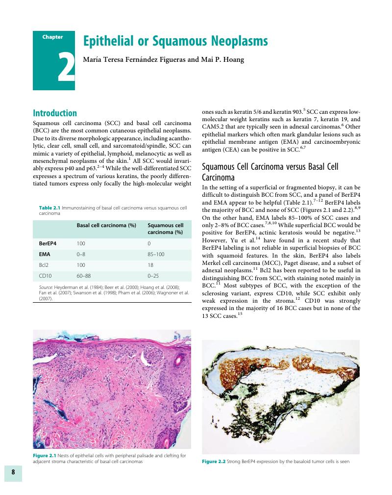
- Chapter 2 Epithelial or Squamous Neoplasms
- Chapter 3 Neoplasms of Cutaneous Appendages
- Chapter 4 Inflammatory Dermatoses Mimicking Lymphomas
- Chapter 5 Cutaneous Lymphoid Neoplasms
- Chapter 6 Melanocytic neoplasms
- Chapter 7 Soft Tissue Neoplasms
- Chapter 8 Miscellaneous Tumors
- Chapter 9 Detection of Genetic Syndromes
- Chapter 10 Immunobullous Disorders
- Chapter 11 Cutaneous Infections
- Chapter 12 Therapeutic and Prognostic Applications
- Index
- References
Chapter 2 - Epithelial or Squamous Neoplasms
Published online by Cambridge University Press: 04 November 2017
Edited by

Book contents
- Immunohistochemistry in Diagnostic Dermatopathology
- Immunohistochemistry in Diagnostic Dermatopathology
- Copyright page
- Dedication
- Contents
- Contributors
- Preface and Acknowledgments
- Chapter 1 Introduction to Immunohistochemistry
- Chapter 2 Epithelial or Squamous Neoplasms
- Chapter 3 Neoplasms of Cutaneous Appendages
- Chapter 4 Inflammatory Dermatoses Mimicking Lymphomas
- Chapter 5 Cutaneous Lymphoid Neoplasms
- Chapter 6 Melanocytic neoplasms
- Chapter 7 Soft Tissue Neoplasms
- Chapter 8 Miscellaneous Tumors
- Chapter 9 Detection of Genetic Syndromes
- Chapter 10 Immunobullous Disorders
- Chapter 11 Cutaneous Infections
- Chapter 12 Therapeutic and Prognostic Applications
- Index
- References
Summary
A summary is not available for this content so a preview has been provided. Please use the Get access link above for information on how to access this content.
- Type
- Chapter
- Information
- Immunohistochemistry in Diagnostic Dermatopathology , pp. 8 - 26Publisher: Cambridge University PressPrint publication year: 2017
References
Lohmann, CM, Solomon, AR. Clinicopathologic variants of cutaneous squamous cell carcinoma. Adv Anat Pathol 2001;8(1):27–36.Google Scholar
Dotto, JE, Glusac, EJ. P63 is a useful marker for cutaneous spindle cell squamous cell carcinoma. J Cutan Pathol 2006;33(6):413–17.CrossRefGoogle ScholarPubMed
Henderson, SA, Torres-Cabala, CA, Curry, JL, et al. p40 is more specific than p63 for the distinction of atypical fibroxanthoma from other cutaneous spindle cell malignancies. Am J Surg Pathol 2014;38(8):1102–10.Google Scholar
Alomari, AK, Glusac, EJ, McNiff, JM. p40 is a more specific marker than p63 for cutaneous poorly differentiated squamous cell carcinoma. J Cutan Pathol 2014;41(11):839–45.Google Scholar
Sigel, JE, Skacel, M, Bergfeld, WF, et al. The utility of cytokeratin 5/6 in the recognition of cutaneous spindle cell squamous cell carcinoma. J Cutan Pathol 2001;28(10):520–24.CrossRefGoogle ScholarPubMed
Mahalingam, M, Richards, JE, Selim, MA, Muzikansky, A, Hoang, MP. An immunohistochemical comparison of cytokeratin 7, cytokeratin 15, cytokeratin 19, CAM5.2, carcinoembryonic antigen, and nestin in differentiating porocarcinoma from squamous cell carcinoma. Hum Pathol 2012;43(8):1265–72.Google Scholar
Heyderman, E, Graham, RM, Chapman, DV, Richardson, TC, McKee, PH. Epithelial markers in primary skin cancer: An immunoperoxidase study of the distribution of epithelial membrane antigen (EMA) and carcinoembryonic antigen (CEA) in 65 primary skin carcinomas. Histopathology 1984;8(3):423–34.Google Scholar
Beer, TW, Shepherd, P, Theaker, JM. Ber-EP4 and epithelial membrane antigen aid distinction of basal cell, squamous cell and basosquamous carcinomas of the skin. Histopathology 2000;37(3):218–23.CrossRefGoogle ScholarPubMed
Hoang, MP, Dresser, KA, Kapur, P, High, WA, Mahalingam, M. Microcystic adnexal carcinoma: An immunohistochemical reappraisal. Mod Pathol 2008;21(2):178–85.Google Scholar
Fan, YS, Carr, RA, Sanders, DS, et al. Characteristic Ber-EP4 and EMA expression in sebaceoma is immunohistochemically distinct from basal cell carcinoma. Histopathology 2007;51(1):80–86.Google Scholar
Swanson, PE, Fitzpatrick, MM, Ritter, JH, Glusac, EJ, Wick, MR. Immunohistologic differential diagnosis of basal cell carcinoma, squamous cell carcinoma, and trichoepithelioma in small cutaneous biopsy specimens. J Cutan Pathol 1998;25(3):153–59.Google Scholar
Pham, TT, Selim, MA, Burchette, JL Jr, et al. CD10 expression in trichoepithelioma and basal cell carcinoma. J Cutan Pathol 2006;33(2):123–28.Google Scholar
Tope, WD, Nowfar-Rad, M, Kist, DA. Ber-EP4-positive phenotype differentiates actinic keratosis from superficial basal cell carcinoma. Dermatol Surg 2000;26(5):415–18.Google Scholar
Yu, L, Galan, A, McNiff, JM. Caveats in BerEP4 staining to differentiate basal and squamous cell carcinoma. J Cutan Pathol 2009;36(10):1074–76.Google Scholar
Wagnoner, J, Keehn, C, Morgan, MB. CD-10 immunostaining differentiates superficial basal cell carcinoma from cutaneous squamous cell carcinoma. Am J Dermatopathol 2007;29(6):555–58.Google Scholar
Shields, JA, Demirici, H, Marr, BP, Eagle, RC Jr, Shields, CL. Sebaceous carcinoma of the ocular region: A review. Surv Ophthalmol 2005;50(2):103–22.CrossRefGoogle ScholarPubMed
Asadi-Amoli, F, Khoshnevis, F, Haeri, H, et al. Comparative examination of androgen receptor reactivity for differential diagnosis of sebaceous carcinoma from squamous cell and basal cell carcinoma. Am J Clin Pathol 2010;134(1):22–26.Google Scholar
Ansai, S, Koseki, S, Hozumi, Y, Kondo, S. An immunohistochemical study of lysozyme, CD-15 (Leu M1), and gross cystic disease fluid protein-15 in various skin tumors. Assessment of the specificity and sensitivity of markers of apocrine differentiation. Am J Dermatopathol 1995;17(3):249–55.CrossRefGoogle ScholarPubMed
Ostler, DA, Prieto, VG, Reed, JA, et al. Adipophilin expression in sebaceous tumors and other cutaneous lesions with clear cell histology: An immunohistochemical study of 117 cases. Mod Pathol 2010;23(4):567–73.Google Scholar
Sramek, B, Lisle, A, Loy, T. Immunohistochemistry in ocular carcinomas. J Cutan Pathol 2008;35(7):641–46.Google Scholar
Sinard, JH. Immunohistochemical distinction of ocular sebaceous carcinoma from basal cell and squamous cell carcinoma. Arch Ophthalmol 1999;117(6):775–83.CrossRefGoogle ScholarPubMed
Boussahmain, C, Mochel, MC, Hoang, MP. Perilipin and adipophilin expression in sebaceous carcinoma and mimics. Hum Pathol 2013;44(9):1811–16.Google Scholar
Shikata, N, Kurokawa, I, Andachi, H, et al. Expression of androgen receptors in skin appendage tumors: An immunohistochemical study. J Cutan Pathol 1995;22(2):149–53.CrossRefGoogle ScholarPubMed
Ansai, S, Arase, S, Kawana, S, Kimura, T. Immunohistochemical findings of sebaceous carcinoma and sebaceoma: Retrieval of cytokeratin expression by a panel of anti-cytokeratin monoclonal antibodies. J Dermatol 2011;38(10):951–58.Google Scholar
Ansai, S, Takeichi, H, Arase, S, Kawana, S, Kimura, T. Sebaceous carcinoma: An immunohistochemical reappraisal. Am J Dermatopathol 2011;33(6):579–87.CrossRefGoogle ScholarPubMed
Mahomed, F, Blok, J, Grayson, W. The squamous variant of eccrine porocarcinoma: A clinicopathological study of 21 cases. J Clin Pathol 2008;61(3):361–65.Google ScholarPubMed
Robson, A, Greene, J, Ansari, N, et al. Eccrine porocarcinoma (malignant eccrine poroma): A clinicopathologic study of 69 cases. Am J Surg Pathol 2001;25(6):710–20.Google Scholar
Goh, SGN, Dayrit, JF, Calonje, E. Sarcomatoid eccrine porocarcinoma: Report of two cases and a review of the literature. J Cutan Pathol 2007;34(1):55–60.CrossRefGoogle Scholar
Qureshi, HS, Ormsby, A, Lee, MW, Zarbo, RJ, Ma, CK. The diagnostic utility of p63, CK5/6, CK7, and CK20 in distinguishing primary cutaneous adnexal neoplasms from metastatic carcinomas. J Cutan Pathol 2004;31(2):145–52.CrossRefGoogle Scholar
Pulitzer, M, Desman, G, Busam, KJ. CK7 expression in primary cutaneous squamous cell carcinoma. J Cutan Pathol 2010;37(9):966–72.Google Scholar
Mahalingam, M, Nguyen, LP, Richards, JE, Muzikansky, A, Hoang, MP. The diagnostic utility of immunohistochemistry in distinguishing primary skin adnexal carcinomas from metastatic adenocarcinoma to skin: An immunohistochemical reappraisal using cytokeratin 15, nestin, p63, D2–40, and calretinin. Mod Pathol 2010;23(5):713–19.Google Scholar
Kurokawa, I, Urakawa, Y, Senba, Y, et al. Keratin profiles may differ between intraepidermal and intradermal invasive eccrine porocarcinoma. Oncol Rep 2006;16(3):473–77.Google Scholar
Chen, S, Takahara, M, Kido, M, et al. Increased expression of an epidermal stem cell marker, cytokeratin 19, in cutaneous squamous cell carcinoma. Br J Dermatol 2008;159(4):952–55.Google Scholar
Yada, K, Kashima, K, Daa, T, et al. Expression of CD10 in basal cell carcinoma. Am J Dermatopathol 2004;26(6):463–71.Google Scholar
Lee, JJ, Mochel, MC, Piris, A, et al. p40 exhibits better specificity than p63 in distinguishing primary skin adnexal carcinomas from cutaneous metastases. Hum Pathol 2014;45(5):1078–83.Google Scholar
Afshar, M, Deroide, F, Robson, A. BerEP4 is widely expressed in tumors of the sweat apparatus: A source of potential diagnostic error. J Cutan Pathol. 2013;40(2):259–64.CrossRefGoogle ScholarPubMed
Folpe, AL, Cooper, K. Best practices in diagnostic immunohistochemistry: Pleomorphic cutaneous spindle cell tumors. Arch Pathol Lab Med 2007;131(10):1517–24.Google Scholar
Ko, CJ, McNiff, JM, Glusac, EJ. Squamous cell carcinoma with single cell infiltration: A potential diagnostic pitfall and the utility of MNF116 and p63. J Cutan Pathol 2008;35(4):353–57.CrossRefGoogle ScholarPubMed
Bishop, JA, Montgomery, EA, Westra, WH. Use of p40 and p63 immunohistochemistry and human papillomavirus testing as ancillary tools for the recognition of head and neck sarcomatoid carcinoma and its distinction from benign and malignant mesenchymal processes. Am J Surg Pathol 2014;38(2):257–64.Google Scholar
Miettinen, M, McCue, PA, Sariomo-Rikala, M, et al. Sox-10 – a marker for not only schwannian and melanocytic neoplasms but also myoepithelial cell tumors of soft tissue: A systematic analysis of 5134 tumors. Am J Surg Pathol 2015;39(6):826–35.Google Scholar
Hultgren, TL, DiMaio, DJ. Immunohistochemical staining of CD10 in atypical fibroxanthomas. J Cutan Pathol 2007;34(5):415–19.Google Scholar
Mirza, B, Weedon, D. Atypical fibroxanthoma: A clinicopathological study of 89 cases. Australas J Dermatol 2005;46(4):235–38.CrossRefGoogle ScholarPubMed
Luzar, B, Calonje, E. Morphological and immunohistochemical characteristics of atypical fibroxanthoma with a special emphasis on potential diagnostic pitfalls: A review. J Cutan Pathol 2010;37(3):301–9.Google Scholar
Chokoeva, AA, Tchernev, G, Cardoso, JC, et al. Vulvar sarcomas: Short guideline for histopathological recognition and clinical management. Part 1. Int J Immunopathol Pharmacol 2015;28(2):168–77.Google Scholar
Costa, LC, Leite, C, Cardoso, S, et al. Expression of epithelial-mesenchymal transition markers at the invasive front of oral squamous cell carcinoma. J Appl Oral Sci 2015; 23(2):169–78.Google Scholar
Mentzel, T, Requena, L, Kaddu, S, et al. Cutaneous myoepithelial neoplasms: Clinicopathologic and immunohistochemical study of 20 cases suggesting a continuous spectrum ranging from benign mixed tumor of the skin to cutaneous myoepithelioma and myoepithelial carcinoma. J Cutan Pathol 2003;30(5):294–302.Google Scholar
Hornick, JL, Fletcher, CD. Myoepithelial tumors of soft tissue: A clinicopathologic and immunohistochemical study of 101 cases with evaluation of prognostic parameters. Am J Surg Pathol 2003;27(9):1183–96.Google Scholar
Folpe, AL, Schoolmeester, JK, McCluggage, WG, et al. SMARCB1-deficient vulvar neoplasms: A clinicopathologic, immunohistochemical, and molecular genetic study of 14 cases. Am J Surg Pathol. 2015;39(6):836–49.Google Scholar
Watanabe, S, Ichikawa, E, Takahashi, H, Otsuka, F. Changes of cytokeratin and involucrin expression in squamous cell carcinomas of the skin during progression to malignancy. Br J Dermatol 1995;132(5):730–39.Google Scholar
Calder, KB, Coplowitz, S, Schlauder, S, Morgan, MB. A case series and immunophenotypic analysis of CK20-/CK7+ primary neuroendocrine carcinoma of the skin. J Cutan Pathol 2007;34(12):918–23.Google Scholar
Reddi, DM, Puri, PK. Expression of focal TTF-1 expression in a case of CK7/CK20-positive Merkel cell carcinoma. J Cutan Pathol 2013;40(4):431–33.Google Scholar
Kurokawa, M, Nabeshima, K, Akiyama, Y, et al. CD56: A useful marker for diagnosing Merkel cell carcinoma. J Dermatol Sci 2003;31(3):219–24.Google Scholar
Fernández-Figueras, MT, Puig, L, Musulén, E, et al. Expression profiles associated with aggressive behavior in Merkel cell carcinoma. Mod Pathol 2007;20(1):90–101.Google Scholar
McCalmont, TH. Paranuclear dots of neurofilament reliably identify Merkel cell carcinoma. J Cutan Pathol 2010;37(8):821–23.Google Scholar
Wong, SQ, Waldeck, K, Vergara, IA, et al. UV-associated mutations underlie the etiology of MCV-negative Merkel cell carcinomas. Cancer Res 2015;75(24):5228–34.Google Scholar
Molina-Ruiz, AM, Santonja, C, Rütten, A, et al. Immunohistochemistry in the diagnosis of cutaneous viral infections – part II: Cutaneous viral infections by parvoviruses, poxviruses, paramyxoviridae, picornaviridae, retroviruses and filoviruses. Am J Dermatopathol 2015;37(2):93–106.Google Scholar
Requena, L, Kutzner, H, Palmedo, G, et al. Cutaneous involvement in multiple myeloma: A clinicopathologic, immunohistochemical, and cytogenetic study of 8 cases. Arch Dermatol. 2003;139(4):475–86.Google Scholar
Robson, A, Shukur, Z, Ally, M, et al. Immunocytochemical p63 expression discriminates between primary cutaneous follicle centre cell and diffuse large B-cell lymphoma-leg type, and is of the TAp63 isoform. Histopathology 2016;69(1):11–19.Google Scholar
Reyes, MC, Cooper, K. An update on vulvar intraepithelial neoplasia: Terminology and a practical approach to diagnosis. J Clin Pathol 2014;67(4):290–94.Google Scholar
Wasserman, JK, Bateman, J, Mai, KT. Differentiated squamous intraepithelial neoplasia associated with squamous cell carcinoma of the anal canal. Histopathology 2016;68(6):834–42.Google Scholar
Downes, MR. Review of in situ and invasive penile squamous cell carcinoma and associated non-neoplastic dermatological conditions. J Clin Pathol 2015;68(5):333–40.Google Scholar
Darragh, TM, Colgan, TJ, Thomas Cox, J, et al. The Lower Anogenital Squamous Terminology Standardization project for HPV-associated lesions: Background and consensus recommendations from the College of American Pathologists and the American Society for Colposcopy and Cervical Pathology. Int J Gynecol Pathol 2013;32(1):76–115.Google Scholar
Bornstein, J, Bogliatto, F, Haefner, H, et al. The 2015 International Society for the Study of Vulvovaginal Disease (ISSVD) terminology of vulvar squamous intraepithelial lesions. J Low Genit Tract Dis 2016;20(1):11–14.Google Scholar
Ordi, J1, Alejo, M, Fusté, V, et al. HPV-negative vulvar intraepithelial neoplasia (VIN) with basaloid histologic pattern: An unrecognized variant of simplex (differentiated) VIN. Am J Surg Pathol 2009;33(11):1659–65.Google Scholar
Maniar, KP, Sanchez, B, Paintal, A, Gursel, DB, Nayar, R. Role of the biomarker p16 in downgrading – IN 2 diagnoses and predicting higher-grade lesions. Am J Surg Pathol 2015;39(12):1708–18.Google Scholar
Velazquez, EF, Chaux, A, Cubilla, AL. Histologic classification of penile intraepithelial neoplasia. Semin Diagn Pathol 2012;29(2):96–102.CrossRefGoogle ScholarPubMed
Roberts, JM, Jin, F, Thurloe, JK, Biro, C, et al. High reproducibility of histological diagnosis of human papillomavirus-related intraepithelial lesions of the anal canal. Pathology 2015;47(4):308–13.Google Scholar
Maniar, KP, Nayar, R. HPV-related squamous neoplasia of the lower anogenital tract: An update and review of recent guidelines. Adv Anat Pathol 2014;21(5):341–58.Google Scholar
Stewart, CJ, Crook, ML. Fascin and cyclin D1 immunoreactivity in non-neoplastic vulvar squamous epithelium, vulvar intraepithelial neoplasia and invasive squamous carcinoma: Correlation with Ki67 and p16 protein expression. J Clin Pathol 2014;67(4):319–25.Google Scholar
Pirog, EC. Immunohistochemistry and in situ hybridization for the diagnosis and classification of squamous lesions of the anogenital region. Semin Diagn Pathol 2015;32(5):409–18.Google Scholar
Patil, DT, Yang, B. Utility of human papillomavirus capsid protein L1 and p16 in the assessment and accurate classification of anal squamous intraepithelial lesions. Am J Clin Pathol 2015;144(1):113–21.Google Scholar
Chaux, A, Pfannl, R, Lloveras, B, et al. Distinctive association of p16INK4a overexpression with penile intraepithelial neoplasia depicting warty and/or basaloid features: A study of 141 cases evaluating a new nomenclature. Am J Surg Pathol 2010;34(3):385–92.Google Scholar
Ferrándiz-Pulido, C, Masferrer, E, de Torres, I, et al. Identification and genotyping of human papillomavirus in a Spanish cohort of penile squamous cell carcinomas: Correlation with pathologic subtypes, p16(INK4a) expression, and prognosis. J Am Acad Dermatol 2013;68(1):73–82.CrossRefGoogle Scholar
Dong, F, Kojiro, S, Borger, DR, Growdon, WB, Oliva, E. Squamous cell carcinoma of the vulva: A subclassification of 97 cases by clinicopathologic, immunohistochemical, and molecular features (p16, p53, and EGFR). Am J Surg Pathol 2015;39(8):1045–53.Google Scholar
Ramirez, N, Guerra, F, Camporeale, G, et al. Expressions of E2 and E7-HPV16 proteins in pre-malignant and malignant lesions of the uterine cervix. Biotech Histochem 2015;90(8):573–80.Google Scholar
Sagasta, A, Castillo, P, Saco, A, et al. p16 staining has limited value in predicting the outcome of histological low-grade squamous intraepithelial lesions of the cervix. Mod Pathol 2016;29(1):51–59.Google Scholar
Bala, R, Pinsky, BA, Beck, AH, et al. p16 is superior to ProEx C in identifying high-grade squamous intraepithelial lesions (HSIL) of the anal canal. Am J Surg Pathol 2013;37(5):659–68.CrossRefGoogle ScholarPubMed
Kreuter, A, Jesse, M, Potthoff, A, et al. Expression of proliferative biomarkers in anal intraepithelial neoplasia of HIV-positive men. J Am Acad Dermatol 2010;63(3):490–98.Google Scholar
Walts, AE, Lechago, J, Hu, B, et al. P16 and Ki67 immunostains decrease intra- and interobserver variability in the diagnosis and grading of anal intraepithelial neoplasia (AIN) Clin Med Pathol 2008;1:7–13.Google Scholar
Kim, SA, Hong, R. Significance of intracellular localization of surviving in cervical squamous cell lesions: Correlation with disease progression. Oncol Lett 2014;7(5):1589–93.Google Scholar
Paquette, C, Mills, AM, Stoler, MH. Predictive value of cytokeratin 7 immunohistochemistry in cervical low-grade squamous intraepithelial lesion as a marker for risk of progression to a high-grade lesion. Am J Surg Pathol 2016;40(2):236–43.Google Scholar
Nazarian, RM, Primiani, A, Doyle, LA, et al. Cytokeratin 17: An adjunctive marker of invasion in squamous neoplastic lesions of the anus. Am J Surg Pathol 2014;38(1):78–85.Google Scholar
Regauer, S, Reich, O, Eberz, B. Vulvar cancers in women with vulvar lichen planus: A clinicopathological study. J Am Acad Dermatol 2014;71(4):698–707.Google Scholar
Mentrikoski, MJ, Stelow, EB, Culp, S, Frierson, HF Jr, Cathro, HP. Histologic and immunohistochemical assessment of penile carcinomas in a North American population. Am J Surg Pathol 2014;38(10):1340–48.Google Scholar
Singh, N, Leen, SL, Han, G, et al. Expanding the morphologic spectrum of differentiated VIN (dVIN) through detailed mapping of cases with p53 loss. Am J Surg Pathol 2015;39(1):52–60.Google Scholar
Liegl, B, Regauer, S. p53 immunostaining in lichen sclerosus is related to ischaemic stress and is not a marker of differentiated vulvar intraepithelial neoplasia (d-VIN). Histopathology 2006;48(3):268–74.Google Scholar
Holway, AH, Rieger-Christ, KM, Miner, WR, et al. Somatic mutation of PTEN in vulvar cancer. Clin Cancer Res 2000;6(8):3228–35.Google ScholarPubMed
Growdon, WB, Boisvert, SL, Akhavanfard, S, et al. Decreased survival in EGFR gene amplified vulvar carcinoma. Gynecol Oncol 2008;111(2):289–97.Google Scholar
Carlson, BC, Hofer, MD, Ballek, N, et al. Protein markers of malignant potential in penile and vulvar lichen sclerosus. J Urol 2013;190(2):399–406.Google Scholar
Graham, RP, Arnold, CA, Naini, BV, Lam-Himlin, DM. Basaloid squamous cell carcinoma of the anus revisited. Am J Surg Pathol 2016;40(3):354–60.Google Scholar
Kazakov, DV, Belousova, IE, Sima, R, Michal, M. Mammary type tubulolobular carcinoma of the anogenital area: Report of a case of a unique tumor presumably originating in anogenital mammary-like glands. Am J Surg Pathol 2006;30(9):1193–96.Google Scholar
Roberts, JM, Cornall, AM, Ekman, D, et al. Papillary immature metaplasia of the anal canal: A low-grade lesion that can mimic a high-grade lesion. Am J Surg Pathol 2016;40(3):348–53.Google Scholar
Emanuel, P, Wang, B, Wu, M, Burstein, DE. p63 immunohistochemistry in the distinction of adenoid cystic carcinoma from basaloid squamous cell carcinoma. Mod Pathol 2005;18(5):645–50.Google Scholar