
Book contents
- Immunohistochemistry in Diagnostic Dermatopathology
- Immunohistochemistry in Diagnostic Dermatopathology
- Copyright page
- Dedication
- Contents
- Contributors
- Preface and Acknowledgments
- Chapter 1 Introduction to Immunohistochemistry
- Chapter 2 Epithelial or Squamous Neoplasms
- Chapter 3 Neoplasms of Cutaneous Appendages
- Chapter 4 Inflammatory Dermatoses Mimicking Lymphomas
- Chapter 5 Cutaneous Lymphoid Neoplasms
- Chapter 6 Melanocytic neoplasms
- Chapter 7 Soft Tissue Neoplasms
- Chapter 8 Miscellaneous Tumors
- Chapter 9 Detection of Genetic Syndromes
- Chapter 10 Immunobullous Disorders
- Chapter 11 Cutaneous Infections
- Chapter 12 Therapeutic and Prognostic Applications
- Index
- References
Chapter 7 - Soft Tissue Neoplasms
Published online by Cambridge University Press: 04 November 2017
Edited by

Book contents
- Immunohistochemistry in Diagnostic Dermatopathology
- Immunohistochemistry in Diagnostic Dermatopathology
- Copyright page
- Dedication
- Contents
- Contributors
- Preface and Acknowledgments
- Chapter 1 Introduction to Immunohistochemistry
- Chapter 2 Epithelial or Squamous Neoplasms
- Chapter 3 Neoplasms of Cutaneous Appendages
- Chapter 4 Inflammatory Dermatoses Mimicking Lymphomas
- Chapter 5 Cutaneous Lymphoid Neoplasms
- Chapter 6 Melanocytic neoplasms
- Chapter 7 Soft Tissue Neoplasms
- Chapter 8 Miscellaneous Tumors
- Chapter 9 Detection of Genetic Syndromes
- Chapter 10 Immunobullous Disorders
- Chapter 11 Cutaneous Infections
- Chapter 12 Therapeutic and Prognostic Applications
- Index
- References
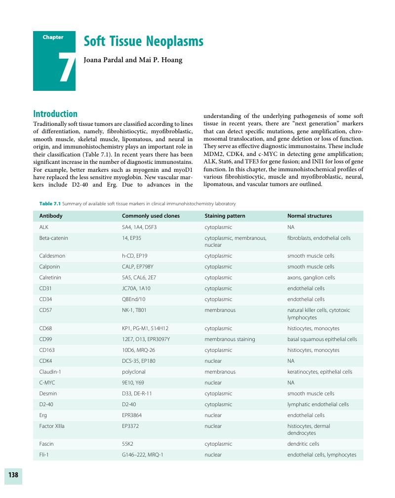
Summary
A summary is not available for this content so a preview has been provided. Please use the Get access link above for information on how to access this content.
- Type
- Chapter
- Information
- Immunohistochemistry in Diagnostic Dermatopathology , pp. 138 - 185Publisher: Cambridge University PressPrint publication year: 2017
References
Wambacher-Gasser, B, Zelger, B, Zelger, BG, Steiner, H. Clear cell dermatofibroma. Histopathology 1997;30(1):64–69.Google Scholar
Zelger, BG, Steiner, H, Kutzner, H, Rutten, A, Zelger, B. Granular cell dermatofibroma. Histopathology 1997;31(3):258–62.Google Scholar
Gleason, BC, Fletcher, CD. Deep “benign” fibrous histiocytoma: Clinicopathologic analysis of 69 cases of a rare tumor indicating occasional metastatic potential. Am J Surg Pathol 2008;32(3):354–62.Google Scholar
Calonje, E, Fletcher, CD. Aneurysmal benign fibrous histiocytoma: Clinicopathological analysis of 40 cases of a tumour frequently misdiagnosed as a vascular neoplasm. Histopathology 1995;26(4):323–31.Google Scholar
Kaddu, S, McMenamin, ME, Fletcher, CD. Atypical fibrous histiocytoma of the skin: Clinicopathologic analysis of 59 cases with evidence of infrequent metastasis. Am J Surg Pathol 2002;26(1):35–46.Google Scholar
Sachdev, R, Sundram, U. Expression of CD163 in dermatofibroma, cellular fibrous histiocytoma, and dermatofibrosarcoma protuberans: Comparison with CD68, CD34 and factor 13a. J Cutan Pathol 2006;33(5):353–60.Google Scholar
Horenstein, MG, Prieto, VG, Nuckols, JD, Burchette, JL, Shea, CR. Indeterminate fibrohistioctyic lesions of the skin: Is there a spectrum between dermatofibroma and dermatofibrosarcoma protuberans. Am J Surg Pathol 2000;24(7):996–1003.Google Scholar
West, RB, Harwell, J, Linn, SC, et al. Apo D in soft tissue tumors: A novel marker for dermatofibrosarcoma protuberans. Am J Surg Pathol 2004;28(8):1063–69.Google Scholar
Mori, T, Misago, N, Yamamoto, O, Toda, S, Narisawa, Y. Expression of nestin in dermatofibrosarcoma protuberans in comparison to dermatofibroma. J Dermatol 2008;35(7):419–25.Google Scholar
Labonte, S, Hanna, W, Bandarchi-Chamhaleh, B. A study of CD117 expression in dermatofibrosarcoma and cellular dermatofibroma. J Cutan Pathol 2007;34(11):856–60.Google Scholar
Bandarchi, B, Ma, L, Marginean, C, et al. D2-40, a novel immunohistochemical marker in differentiating dermatofibroma from dermatofibrosarcoma protuberans. Mod Pathol 2010;23(3):434–38.Google Scholar
Kamino, H, Lee, JY, Berke, A. Pleomorphic fibroma of the skin: A benign neoplasm with cytologic atypia. A clinicopathologic study of eight cases. Am J Surg Pathol 1989;13(2):107–13.Google Scholar
Rudolph, P, Schubert, C, Zelger, BG, et al. Different expression of CD34 and Ki-M1p in pleomorphic fibroma and dermatofibroma with monster cells. Am J Dermatopathol 1999;21(5):414–19.CrossRefGoogle ScholarPubMed
Mahmood, MN, Salama, ME, Chaffiins, M, et al. Solitary sclerotic fibroma of the skin: A possible link with pleomorphic fibroma with immunophenotypic expression for O13 (CD99) and CD34. J Cutan Pathol 2003;30(10):631–36.Google Scholar
Puig, L, Fernandez Figueras, MT, Bielsa, I, et al. Multinucleate cell angiohistiocytoma: A fibrohistiocytic proliferation with increased mast cell numbers and vascular hyperplasia. J Cutan Pathol 2002;29(4):232–37.Google Scholar
Hanft, VN, Shea, CR, McNutt, NS, et al. Expression of CD34 in sclerotic (“plywood”) fibromas. Am J Dermatopathol 2000;22(1):17–21.Google Scholar
Shitabata, PK, Crouch, EC, Fitzgibbon, JF, et al. Cutaneous sclerotic fibroma. Immunohistochemical evidence of a fibroblastic neoplasm with ongoing type I collagen synthesis. Am J Dermatopathol 1995;17(4):339–43.Google Scholar
Kutzner, H, Mentzel, T, Palmedo, G, et al. Plaque-like CD34-positive dermal fibroma (“medalion-like dermal dendrocyte hamartoma”): Clinicopathologic, immunohistochemical, and molecular analysis of 5 cases emphasizing its distinction from superficial, plaque-like dermatofibrosarcoma protuberans. Am J Surg Pathol 2010;34(2):190–201.Google Scholar
De Feraudy, S, Fletcher, CDM. Fibroblastic connective tissue nevus. A rare cutaneous lesion analyzed in a series of 25 cases. Am J Surg Pathol 2012;36(10):1509–15.Google Scholar
Erdag, G, Qureshi, HS, Patterson, JW, Wick, MR. Solitary fibrous tumors of the skin: A clinicopathologic study of 10 cases and review of the literature. J Cutan Pathol 2007;34(11):844–50.Google Scholar
Chmielecki, J, Crago, AM, Rosenberg, M, et al. Whole-exome sequencing identifies a recurrent NAB2-STAT6 fusion in solitary fibrous tumors. Nat Genet 2013;45(2):131–32.Google Scholar
Doyle, LA, Vivero, M, Fletcher, CD, Mertens, F, Hornick, JL. Nuclear expression of STAT6 distinguishes solitary fibrous tumor from histologic mimics. Mod Pathol 2014;27(3):290–95.Google Scholar
Frew, JW. Multinucleate cell angiohistiocytoma: Clinicopathological correlation of 142 cases with insights into etiology and pathogenesis. Am J Dermatopathol 2015;37(3):222–28.Google Scholar
Cesinaro, AM, Roncati, L, Maiorana, A. Estrogen receptor alpha overexpression in multinucleate cell angiohistiocytoma: New insights into the pathogenesis of a reactive process. Am J Dermatopathol 2010;32(7):655–59.Google Scholar
Saab, ST, McClain, CM, Coffin, CM. Fibrous hamartoma of infancy: A clinicopathologic analysis of 60 cases. Am J Surg Pathol 2014;38(3):394–401.Google Scholar
Fetsch, JF, Miettinen, M. Calcifying aponeurotic fibroma: A clinicopathologic study of 22 cases arising in uncommon sites. Hum Pathol 1998;29(12):1504–10.Google Scholar
Maluf, HM, DeYoung, BR, Swanson, PE, Wick, MR. Fibroma and giant cell tumor of tendon sheath: A comparative histological and immunohistological study. Mod Pathol 1995;8(2):155–59.Google Scholar
Hoang, MP, Rogers, BB, Albores Saavedra, J. Giant cell tumor of the skin: A morphologic and immunohistochemical study of five cases. Ann Diagn Pathol 2002;6(5):288–93.Google Scholar
Folpe, AL, Fanburg-Smith, JC, Billings, SD, et al. Most osteomalacia-associated mesenchymal tumors are a single histopathologic entity: An analysis of 32 cases and a comprehensive review of the literature. Am J Surg Pathol 2004;28(1):1–30.Google Scholar
Carlson, JW, Fletcher, CD. Immunohistochemistry for beta-catenin in the differential diagnosis of spindle cell lesions: Analysis of a series and review of the literature. Histopathology 2007;51(4):509–14.Google Scholar
Bhattacharya, B, Dilworth, HP, Iacobuzio-Donahue, C, et al. Nuclear beta-catenin expression distinguishes deep fibromatosis from other benign and malignant fibroblastic and myofibroblastic lesions. Am J Surg Pathol 2005;29(5):653–59.Google Scholar
De Feraudy, S, Fletcher, CD. Intradermal nodular fasciitis: A rare lesion analyzed in a series of 24 cases. Am J Surg Pathol 2010;34(9):1377–81.CrossRefGoogle Scholar
Perez-Monteil, MD, Plaza, JA, Dominguez Malagon, H, Suster, S. Differential expression of smooth muscle myosin, smooth muscle actin, h-caldesmon, and calponin in the diagnosis of myofibroblastic and smooth muscle lesions of skin and soft tissue. Am J Dermatopathol 2006;28(2):105–11.Google Scholar
Coffin, CM, Watterson, J, Priest, JR, Dehner, LP. Extrapulmonary inflammatory myofibroblastic tumor (inflammatory pseudotumor). A clinicopathologic and immunohistochemical study of 84 cases. Am J Surg Pathol 1995;19(8):859–72.Google Scholar
Antonescu, CR, Suurmeijer, AJ, Zhang, L, et al. Molecular characterization of inflammatory myofibroblastic tumors with frequent ALK and ROS1 gene fusions and rare novel RET rearrangement. Am J Surg Pathol 2015;39(7):957–67.Google Scholar
Yamamoto, H, Yoshida, A, Taguchi, K, et al. ALK, ROS1 and NTRK3 gene rearrangements in inflammatory myofibroblastic tumors. Histopathology 2016;69(1):72–83.Google Scholar
Laskin, WB, Fetsch, JF, Miettinen, M. Myxoinflammatory fibroblastic sarcoma: A clinicopathologic analysis of 104 cases, with emphasis on predictors of outcome. Am J Surg Pathol 2014;38(1):1–12.Google Scholar
Kovarik, CL, Barrett, T, Auerbach, A, Cassarino, DS. Acral myxoinflammatory sarcoma: Case series and immunohistochemical analysis. J Cutan Pathol 2008;35(2):192–96.Google Scholar
Hassaneim, AM, Atkinson, SP, Al-Guran, SZ, Jain, SM, Reith, JD. Acral myxoinflammatory fibroblastic sarcoma: Are they all low-grade neoplasms? J Cutan Pathol 2008;35(2):186–91.Google Scholar
Hollmann, TJ, Bovee, JVMG, Fletcher, CDM. Digital fibromyxoma (superficial acral fibromyxoma). A detailed characterization of 124 cases. Am J Surg Pathol 2012;36(6):789–98.Google Scholar
Misago, N, Ohkawa, T, Yanai, T, Narisawa, Y. Superficial acral fibromyxoma on the tip of the big toe: Expression of CD10 and nestin. J Eur Acad Dermatol Venereol 2008;22(2):255–57.Google Scholar
McNiff, JM, Subtil, A, Cowper, SE, Lazova, R, Glusac, EJ. Cellular digital fibromas. Distinctive CD34-positive lesions that may mimic dermatofibrosarcoma protuberans. J Cutan Pathol 2005;32(6):413–18.Google Scholar
Doyle, LA, Moller, E, Dal Cin, P, et al. MUC4 is a highly sensitive and specific marker for low-grade fibromyxoid sarcoma. Am J Surg Pathol 2011;35(5):733–41.Google Scholar
Möller, E, Hornick, JL, Magnusson, L, et al. **FUS-CREB3L2/L1**-Positive Sarcomas Show a Specific Gene Expression Profile with Upregulation of **CD24** and **FOXL1.** Clin Cancer Res 2011;17(9):2646–56.Google Scholar
Calonje, E, Fletcher, CD. Myoid differentiation in dermatofibrosarcoma protuberans and its fibrosarcomatous variant: Clinicopathologic analysis of 5 cases. J Cutan Pathol 1996;23(1):30–36.Google Scholar
Segura, S, Salgado, R, Toll, A, et al. Identification of t(17;22)(q22q13)(COL1A1/PDGFB) in dermatofibrosarcoma protuberans by fluorescence in situ hybridization in paraffin-embedded tissue microarrays. Hum Pathol 2011;42(2):176–84.Google Scholar
Li, N, McNiff, J, Hui, P, Manfioletti, G, Tallini, G. Differential expression of HMGA1 and HMGA2 in dermatofibroma and dermatofibrosarcoma protuberans: Potential diagnostic applications and comparison with histologic findings, CD34, and factor XIIIa immunoreactivity. Am J Dermatopathol 2004;26(4):267–72.Google Scholar
Thway, K, Fisher, C. Angiomatoid fibrous histiocytoma: The current status of pathology and genetics. Arch Pathol Lab Med 2015;139(5):674–82.Google Scholar
Moosavi, C, Jha, P, Fanburg-Smith, JC. An update on plexiform fibrohistiocytic tumor and addition of 66 new cases from the Armed Forces Institute of Pathology, in honor of Frank M. Enzinger, MD. Ann Diagn Pathol 2007;11(5):313–19.Google Scholar
Tallon, B, Beer, TW. MiTF positivity in atypical fibroxanthoma: A diagnostic pitfall. Am J Dermatopathol 2014;36(11):888–91.Google Scholar
Thum, C, Hollowood, K, Birch, J, Goodlad, JR, Brenn, T. Aberrant Melan-A expression in atypical fibroxanthoma and undifferentiated pleomorphic sarcoma of the skin. J Cutan Pathol 2011;38(12):954–60.Google Scholar
Henderson, SA, Torres-Cabala, CA, Curry, JL, et al. p40 is more specific than p63 for the distinction of atypical fibroxanthoma from other cutaneous spindle cell malignancies. Am J Surg Pathol 2014;38(8):1102–10.Google Scholar
Rangdaeng, S, Truong, LD. Comparative immunohistochemical staining for desmin and muscle-specific actin. A study of 576 cases. Am J Clin Pathol 1991;96(1):32–45.Google Scholar
Tardio, JC, Azorin, D, Hernandez-Nunez, A, et al. Dermatomyofibromas presenting in pediatric patients: Clinicopathologic characteristics and differential diagnosis. J Cutan Pathol 2011;38(12):967–72.Google Scholar
Matsuyama, A, Hisaoka, M, Hashimoto, H. Angioleiomyoma: A clinicopathologic and immunohistochemical reappraisal with special reference to the correlation with myopericytoma. Hum Pathol 2007;38(4):645–51.Google Scholar
Chaudhary, KS, Shousha, S. Leiomyoma of the nipple, and normal subareola muscle fibers, are oestrogen and progesterone receptor positive. Histopathology 2004;44(6):626–28.Google Scholar
Idriss, MH, Kazlouskaya, V, Malhotta, S, Andres, C, Elston, DM. Phosphohistone-H3 and Ki-67 immunostaining in cutaneous pilar leiomyoma and leiomyosarcoma (atypical intradermal smooth muscle neoplasm). J Cutan Pathol 2013;40(6):557–63.Google Scholar
Deyrup, AT, Lee, VK, Hill, CE, et al. Epstein-Barr virus-associated smooth muscle tumors are distinctive mesenchymal tumors reflecting multiple infection events: A clinicopathologic and molecular analysis of 29 tumors from 19 patients. Am J Surg Pathol 2006;30(1):75–82.Google Scholar
Laskin, WB, Miettinen, M, Fetsch, JF. Infantile digital fibroma/fibromatosis. A clinicopathological and immunohistochemical study of 69 tumors from 57 patients with long-term follow-up. Am J Surg Pathol 2009;33(1):1–3.Google Scholar
Fletcher, CDM, Achu, P, Van Noorden, S, McKee, PH. Infantile myofibromatosis: A light microscopic, histochemical and immunohistochemical study suggesting true muscle differentiation. Histopathology 1987;11(3):245–58.Google Scholar
Calonje, E, Guerin, D, McCormick, D, Fletcher, CD. Superficial angiomyxoma: Clinicopathologic analysis of a series of distinctive but poorly recognized cutaneous tumors with tendency for recurrence. Am J Surg Pathol 1999;23(8):910–17.Google Scholar
Fetsch, JF, Laskin, WB, Lefkowitz, M, Kindblom, LG, Meis-Kindblom, JM. Aggressive angiomyxoma: A clinicopathologic study of 29 female patients. Cancer 1996;78(1):79–90.Google Scholar
Bigby, SM, Symmans, PJ, Miller, MV, Dray, MS, Jones, RW. Aggressive angiomyxoma of the female genital tract and pelvis: Clinicopathologic features with immunohistochemical analysis. Int J Gynecol Pathol 2011;30(5):505–13.Google Scholar
Dreux, N, Marty, M, Chibon, F, et al. Value and limitation of immunohistochemical expression of HMBA2 in mesenchymal tumors: About a series of 1052 cases. Mod Pathol 2010;23(12):1657–66.Google Scholar
Flucke, U, van Krieken, JH, Mentzel, T. Cellular angiofibroma: Analysis of 25 cases emphasizing its relationship to spindle cell lipoma and mammary-type myofibroblastoma. Mod Pathol 2011;24(1):82–89.Google Scholar
Chen, E, Fletcher, CD. Cellular angiofibroma with atypical or sarcomatous transformation: Clinicopathologic analysis of 13 cases. Am J Surg Pathol 2010;34(5):707–14.Google Scholar
Iwasa, Y, Fletcher, CD. Cellular angiofibroma: Clinicopathologic and immunohistochemical analysis of 51 cases. Am J Surg Pathol 2004;28(11):1426–35.Google Scholar
Laskin, WB, Fetsch, JF, Tavassoli, FA. Angiofibroblastoma of the female genital tract: Analysis of 17 cases including a lipomatous variant. Hum Pathol 1997;28(9):1046–55.Google Scholar
Nielsen, GP, Rosenberg, AF, Young, RH, et al. Angiomyofibroblastoma of the vulva and vagina. Mod Pathol 1996;9(3):284–91.Google Scholar
Qiu, X, Montgomery, E, Sun, B. Inflammatory myofibroblastic tumor and low-grade myofibroblastic sarcoma: A comparative study of clinicopathologic features and further observations on the immunohistochemical profile of myofibroblasts. Hum Pathol 2008;39(6):846–56.Google Scholar
Wang, NP, Marx, J, McNutt, MA, Rutledge, JC, Gown, AM. Expression of myogenic regulatory proteins (myogenin and MyoD1) in small blue round cell tumors of childhood. Am J Pathol 1995;147(8):1799–810.Google Scholar
Tsokos, M, Linnoila, RI, Chandra, RS, Triche, TJ. Neuron specific enolase in the diagnosis of neuroblastoma and other small round cell tumors in children. Hum Pathol 1984;15(6):575–84.Google Scholar
Fetsch, JF, Laskin, WB, Miettinen, M. Nerve sheath myxoma: A clinicopathologic and immunohistochemical analysis of 57 morphologically distinctive, S-100 protein- and GFAP-positive, myxoid peripheral nerve sheath tumors with a predilection for the extremities and a high local recurrence rate. Am J Surg Pathol 2005;29(12):1615–24.Google Scholar
Laskin, WB, Fetsch, JF, Miettinen, M. The “neurothekeoma”: Immunohistochemical analysis distinguishes the true nerve sheath myxoma from its mimics. Hum Pathol 2000;31(10):1230–41.Google Scholar
Fetsch, JF, Laskin, WB, Hallman, JR, Lupton, GP, Miettinen, M. Neurothekeoma: An analysis of 178 tumors with detailed immunohistochemical data and long-term patient follow-up information. Am J Surg Pathol 2007;31(7):1103–14.Google Scholar
Calonje, E, Guerin, D, McCormick, D, Fletcher, CD. Superficial angiomyxoma: Clinicopathologic analysis of a series of distinctive but poorly recognized cutaneous tumors with tendency for recurrence. Am J Surg Pathol 1999;23(8):910–17.Google Scholar
Fetsch, JF, Miettinen, M. Sclerosing perineurioma: A clinicopathologic study of 19 cases of a distinctive soft tissue lesion with a predilection for the fingers and palms of young adults. Am J Surg Pathol 1997;21(12):1433–42.Google Scholar
Fetsch, JF, Laskin, WB, Miettinen, M. Superficial acral fibromyxoma: A clinicopathologic and immunohistochemical analysis of 37 cases of a distinctive soft tissue tumor with a predilection for the fingers and toes. Hum Pathol 2001;32(7):704–14.Google Scholar
Hornick, JL, Fletcher, CD. Cellular neurothekeoma: Detailed characterization in a series of 133 cases. Am J Surg Pathol 2007;31(3):329–40.Google Scholar
Page, RN, King, R, Mihm, MC Jr, Googe, PB. Microphthalmia transcription factor and NKI/C3 expression in cellular neurothekeoma. Mod Pathol 2004;17(2):230–34.Google Scholar
Suarez, A, High, WA. Immunohistochemical analysis of KBA.62 in 18 neurothekeomas: A potential marker for differentiating neurothekeoma, but a marker that may lead to confusion with melanocytic tumors. J Cutan Pathol 2014;41(1):36–41.Google Scholar
Plaza, JA, Torres-Cabala, C, Evans, H, Diwan, AH, Prieto, VG. Immunohistochemical expression of S100A6 in cellular neurothekeoma: Clinicopathologic and immunohistochemical analysis of 31 cases. Am J Dermatopathol 2009;31(5):419–22.Google Scholar
Wang, AR, May, D, Bourne, P, et al. PGP9.5: A marker for cellular neurothekeoma. Am J Surg Pathol 1999;23(11):1401–07.Google Scholar
Antonescu, CR, Scheithauer, BW, Woodruff, JM. Tumors of the peripheral nervous system. Atlas of tumor pathology.4th series. Fascicle 19. Washington, D.C.: Armed Forces Institute of Pathology; 2013.Google Scholar
Johnson, MD, Glick, AD, Davis, BW. Immunohistochemical evaluation of Leu-7, myelin basic protein, S-100 protein, glial-fibrillary acidic-protein, and LN3 immunoreactivity in nerve sheath tumors and sarcomas. Arch Pathol Lab Med 1988;112(2):155–60.Google Scholar
Nonaka, D, Chiriboga, L, Rubin, DP. Sox10: A pan-schwannian and melanocytic marker. Am J Surg Pathol 2008;32(9):1291–98.Google Scholar
Hirose, T, Tani, T, Shimada, T, et al. Immunohistochemical demonstration of EMA/Glut-1-positive perineurial cells and CD34 positive fibroblastic cells in peripheral nerve sheath tumors. Mod Pathol 2003;16(4):293–98.Google Scholar
Fine, SW, McClain, SA, Li, M. Immunohistochemical staining for calretinin is useful for differentiating schwannomas from neurofibromas. Am J Clin Pathol 2004;122(4):552–59.Google Scholar
Gray, MH, Smoller, BR, McNutt, NS, Hsu, A. Immunohistochemical demonstration of Factor XIIIa expression in neurofibromas. A practical means of differentiating these tumors from neurotized nevi and schwannomas. Arch Dermatol 1990;126(4):472–76.Google Scholar
Boyd, C, Smith, MJ, Kluwe, L, et al. Alterations in the SMARCB1 (INI1) tumor suppressor gene in familial schwannomatosis. Clin Genet 2008;74(4):358–66.Google Scholar
Theaker, JM, Gatter, KC, Puddle, J. Epithelial membrane antigen expression by the perineurium of peripheral nerve and in peripheral nerve tumours. Histopathology 1998;13(2):171–79.Google Scholar
Laskin, WB, Fetsch, JF, Lasota, J, et al. Benign epithelioid peripheral nerve sheath tumors of the soft tissues: Clinicopathologic spectrum of 33 cases. Am J Surg Pathol 2005;29(1):39–51.Google Scholar
Hornick, JL, Fletcher, CD. Soft tissue perineurioma: Clinicopathologic analysis of 81 cases including those with atypical histologic features. Am J Surg Pathol 2005;29(7):845–58.Google Scholar
Robson, AM, Calonje, E. Cutaneous perineurioma: A poorly recognized tumour often misdiagnosed as epithelioid histiocytoma. Histopathology 2000;37(4):332–39.Google Scholar
Folpe, AF, Billings, SD, McKenney, JF, et al. Expression of claudin-1, a recently described tight junction perineurioma from potential mimics. Am J Surg Pathol 2002;26(2):1620–26.Google Scholar
Yamaguchi, LI, Hasegawa, T, Hirose, T, et al. Sclerosing perineurioma: A clinicopathologic study of five cases and diagnostic utility of immunohistochemical staining for GLUT1. Virchows Arch 2003;443(2):159–63.Google Scholar
Wallace, CA, Hallman, JR, Sangueza, OP. Primary cutaneous ganglioneuroma: A report of two cases and literature review. Am J Dermatopathol 2003;25(3):239–42.Google Scholar
Reed, RJ. Neuromesenchyme. The concept of a neurocristic effector cell for dermal mesenchyme. Am J Dermatopathol 1983;5(4):385–95.Google Scholar
Requena, L, Sangüeza, OP. Benign neoplasms with neural differentiation. A review. Am J Dermatopathol 1995;17(1):75–96.Google Scholar
Murphy, M, Hoss, D, Berke, A, et al. Cutaneous ganglioneuroma. Int J Dermatol 2007;46(8):861–63.Google Scholar
López, DA, Silvers, DN, Helwig, EB. Cutaneous meningiomas – a clinicopathologic study. Cancer 1974;34(3):728–44.Google Scholar
Fox, MD, Billings, SD, Gleason, BC, Thomas, AB, Cibull, TL. Cutaneous meningioma: A potential diagnostic pitfall in p63 positive cutaneous neoplasms. J Cutan Pathol 2013;40(10):891–95.Google Scholar
Agaimy, A, Buslei, R, Coras, R, Rubin, BP, Mentzel, T. Comparative study of soft tissue perineurioma and meningioma using a five-marker immunohistochemical panel. Histopathology 2014;65(1):60–70.Google Scholar
Ordonez, NG. Granular cell tumor: A review and update. Adv Anat Pathol 1999;6(4):186–203.Google Scholar
Fanburg-Smith, JC, Meis-Kindblom, JM, Fante, R, et al. Malignant granular cell tumor of soft tissue: Diagnostic criteria and clinicopathologic correlation. Am J Surg Pathol 1998;22(7):779–94.Google Scholar
Le, BH, Boyer, PJ, Lewis, JE, et al. Granular cell tumors. Immunohistochemical assessment of inhibin-alpha, protein gene product 9.5, S100 protein, CD68, and Ki-67 proliferation index with clinical correlation. Arch Pathol Lab Med 2004;128(7):771–75.Google Scholar
Gleason, BC, Nascimento, AF. HMB-45 and Melan-A are useful in the differential diagnosis between granular cell tumor and malignant melanoma. Am J Dermatopathol 2007;29(1):22–27.Google Scholar
Kapur, P, Rakheja, D, Balani, JP, et al. Phosphorylated histone 3, Ki-67, p21, fatty acid synthase, and cleaved caspase-3 expression in benign and atypical granular cell tumors. Arch Pathol Lab Med 2007;131(1):57–64.Google Scholar
Ducatman, BS, Scheithauer, BW, Piepgras, DG, Reiman, HM, Ilstrup, DM. Malignant peripheral nerve sheath tumors: A clinicopathologic study of 120 cases. Cancer 1986;57(10):2006–21.Google Scholar
Jo, VY, Fletcher, CD. Epithelioid malignant peripheral nerve sheath tumor: Clinicopathologic analysis of 63 cases. Am J Surg Pathol 2015;39(5):673–82.Google Scholar
Pekmezci, M, Reuss, DE, Hirbe, AC, et al. Morphologic and immunohistochemical features of malignant peripheral nerve sheath tumors and cellular schwannomas. Mod Pathol 2015;28(2):187–200.Google Scholar
Schaefer, IM, Fletcher, CD, Hornick, JL. Loss of H3K27 trimethylation distinguishes malignant peripheral nerve sheath tumors from histologic mimics. Mod Pathol 2016;29(1):4–13.Google Scholar
Fletcher, CDM, Martin-Bates, E. Spindle-cell lipoma: A clinicopathological study with some original observations. Histopathology 1987;11(8):803–17.Google Scholar
Sandberg, AA. Updates on the cytogenetics and molecular genetics of bone and soft tissue tumors: lipoma. Cancer Genet Cytogenet 2004;150(2):93–115.Google Scholar
Maggiani, F, Debiec-Rychter, M, Verbeeck, G, Sciot, R. Extramammary myofibroblastoma is genetically related to spindle cell lipoma. Virchow Archiv 2006;449(2):244–47.Google Scholar
Maggiani, F, Debiec-Rychter, M, Vanbockrijck, M, et al. Cellular angiofibroma: Another mesenchymal tumor with 13q14 involvement, suggesting a link with spindle cell lipoma and (extra)-mammary myofibroblastoma. Histopathology 2007;51(3):410–12.Google Scholar
Billings, S, Folpe, A. Diagnostically challenging spindle cell lipomas: A report of 34 “Low-Fat” and “Fat-Free” variants. Am J Dermatopathol 2007;29(5):437–42.Google Scholar
Suster, S, Fisher, C. Immunoreactivity for the human hematopoietic progenitor cell antigen (CD34) in lipomatous tumors. Am J Surg Pathol 1997;21(2):195–200.Google Scholar
Syed, S, Martin, A-M, Haupt, H, et al. Frequent detection of androgen receptors in spindle cell lipomas: an explanation for this lesion’s male predominance? Arch Pathol Lab Med 2008;132(1):81–83.Google Scholar
Chen, BJ, Marino-Enriquez, A, Fletcher, CD, Hornick, JL. Loss of retinoblastoma protein expression in spindle cell/pleomorphic lipomas and cytogenetically related tumors: An immunohistochemical study with diagnostic implications. Am J Surg Pathol 2012;36(8):1119–28.Google Scholar
Dei Tos, AP, Mentzel, T, Fletcher, CD. Primary liposarcoma of the skin: A rare neoplasm with unusual high grade features. Am J Dermatopathol 1998;20(4):332–38.Google Scholar
Weiss, SW, Rao, VK. Well-differentiated liposarcoma (atypical lipoma) of deep soft tissue of the extremities, retroperitoneum and miscellaneous sites: A follow-up study of 92 cases with analysis of the incidence of ”dedifferentiation.” Am J Surg Pathol 1992;16(11):1051.Google Scholar
Binh, MB, Sastre-Garau, X, Guillou, L, et al. MDM2 and CDK4 immunostainings are useful adjuncts in diagnosing well-differentiated and dedifferentiated liposarcoma subtypes: A comparative analysis of 559 soft tissue neoplasms with genetic data. Am J Surg Pathol 2005;29(10):1340–47.Google Scholar
Sirvent, N, Coindre, JM, Maire, G, et al. Detection of MDM2-CDK4 amplification by fluorescence in situ hybridization in 200 paraffin-embedded tumor samples: Utility in diagnosing adipocytic lesions and comparison with immunohistochemistry and real-time PCR. Am J Surg Pathol 2007;31(10):1476–89.Google Scholar
Pilotti, S, Della Torre, G, Mezzelani, A, et al. The expression of MDM2/CDK4 gene product in the differential diagnosis of well differentiated liposarcoma and large deep-seated lipoma. Br J Cancer 2000;82(7):1271–75.Google Scholar
Binh, MB, Garau, XS, Guillou, L, Aurias, A, Coindre, JM. Reproducibility of MDM2 and CDK4 staining in soft tissue tumors. Am J Clin Pathol 2006;125(5):693–97.Google Scholar
Aleixo, PB, Hartmann, AA, Menezes, IC, Meurer, RT, Oliveira, AM. Can MDM2 and CDK4 make the diagnosis of well differentiated/dedifferentiated liposarcoma? An immunohistochemical study on 129 soft tissue tumours. J Clin Pathol 2009;62(12):1127–35.Google Scholar
Thway, K, Flora, R, Shah, C, et al. Diagnostic utility of p16, CDK4, and MDM2 as an immunohistochemical panel in distinguishing well-differentiated and dedifferentiated liposarcomas from other adipocytic tumors. Am J Surg Pathol 2012;36(3):462–69.Google Scholar
Guillou, L, Fletcher, CD. Benign lymphangioendothelioma (acquired progressive lymphangioma): A lesion not to be confused with well-differentiated angiosarcoma and patch stage Kaposi’s sarcoma: Clinicopathologic analysis of a series. Am J Surg Pathol 2000;24(8):1047–57.Google Scholar
Revelles, JM, Diaz, JL, Angulo, J, et al. Giant benign lymphangioendothelioma. J Cutan Pathol 2012;39(10):950–56.Google Scholar
Requena, L, Sangueza, OP, Cutaneous vascular anomalies. Part I. Hamartomas, malformations, and dilatation of preexisting vessels. J Am Acad Dermatol 1997;37(4):523–49.Google Scholar
Fukunaga, M: Expression of D2-40 in lymphatic endothelium of normal tissues and in vascular tumours. Histopathology 2005;46 (4):396–402.Google Scholar
Miettinen, M, Wang, ZF, Prox1 transcription factor as a marker for vascular tumors – evaluation of 314 vascular endothelial and 1086 nonvascular tumors. Am J Surg Pathol 2012;36(3):351–59.Google Scholar
Patrice, SJ, Wiss, K, Mulliken, JB. Pyogenic granuloma (lobular capillary hemangioma): A clinicopathologic study of 178 cases. Pediatr Dermatol 1991;8(4):267–76.Google Scholar
Mentzel, T, Partanen, TA, Kutzner, H. Hobnail hemangioma (“targetoid hemosiderotic hemangioma”): Clinicopathologic and immunohistochemical analysis of 62 cases. J Cutan Pathol 1999;26(6):279–86.Google Scholar
Al Dhaybi, R, Powell, J, McCuaig, C, Kokta, V. Differentiation of vascular tumors from vascular malformations by expression of Wilms tumor 1 gene: Evaluation of 126 cases. J Am Acad Dermatol 2010;63(6):1052–57.Google Scholar
Huang, SC, Zhang, L, Sung, YS, et al. Frequent FOS gene rearrangements in epithelioid hemangioma: A molecular study of 58 cases with morphologic reappraisal. Am J Surg Pathol 2015;39(10)1313–21.Google Scholar
Mentzel, T. CD34-positive glomus tumor: Clinicopathologic and immunohistochemical analysis of six cases with myxoid stromal changes. J Cutan Pathol 2002:29(7):421–25.Google Scholar
Porter, PG, Bigler, SA, McNutt, NS, et al. The immunophenotype of hemangiopericytoma and glomus tumors with special reference to muscle protein expression: An immunohistochemical study and review of the literature. Mod Pathol 1991;4(1):46–52.Google Scholar
Folpe, AL, Fanburg-Smith, JC, Miettinen, M, Weiss, SW. Atypical and malignant glomus tumors: Analysis of 52 cases, with a proposal for the reclassification of glomus tumors. Am J Surg Pathol 2001;25(1):1–12.Google Scholar
Frieden, IJ, Haggstrom, AN, Drolet, BA, et al. Infantile hemangiomas: Current knowledge, future directions. Proceedings of a research workshop on infantile hemangiomas, April 7–9, 2005, Bethesda, Maryland. Pediatr Dermatol 2005;22(5):383–406.Google Scholar
North, PE, Waner, M, Mizeracki, A, Mihm, MC Jr. GLUT1: A newly discovered immunohistochemical marker for juvenile hemangiomas. Hum Pathol 2000;31(1):11–22.Google Scholar
Folpe, AL, Chand, EM, Goldblum, JR, et al. Expression of FLI-1, a nuclear transcription factor, distinguishes vascular neoplasms from potential mimics. Am J Surg Pathol 2001;25(8):1061–66.Google Scholar
Enjolras, O, Wassef, M, Mazoyer, E, et al. Infants with Kasabach-Merritt syndrome do not have ‘true’ hemangiomas. J Pediatr 1997;130(4):631–40.Google Scholar
Lyon, LL, North, PE, Mac-Moune Lai, F, et al. Kaposiform Hemangioendothelioma. A study of 33 cases emphasizing its pathologic, immunophenotypic, and biologic uniqueness from juvenile hemangioma. Am J Surg Pathol 2004;28(5):559–68.Google Scholar
Zukerberg, LR, Nickoloff, BJ, Weiss, SW. Kaposiform hemangioendothelioma of infancy and childhood. An aggressive neoplasm associated with Kasabach–Merritt syndrome and lymphangiomatosis. Am J Surg Pathol 1993;17(4):321–28.Google Scholar
Le Huu, AR, Jokinen, CH, Ruben, BP, et al. Expression of prox1, lymphatic endothelial nuclear transcription factor, in Kaposiform hemangioendothelioma and tufted angioma. Am J Surg Pathol 2010;34(11):1563–73.Google Scholar
Chang, Y, Cesarman, E, Pessin, MS, et al. Identification of herpes virus-like DNA sequences in AIDS-associated Kaposi’s sarcoma. Science 1994;266(5192):1865–69.Google Scholar
Cheuk, W, Wong, KO, Wong, CS, et al. Immunostaining for human herpes virus 8 latent nuclear antigen-1 helps distinguish Kaposi sarcoma from its mimickers. Am J Clin Pathol 2004;121(3):335–42.Google Scholar
Reis, RM, Reis-Filho, JS, Longatto Filho, A, et al. Differential Prox-1 and CD31 expression in mucosae, cutaneous and soft tissue vascular lesions and tumors. Pathol Res Pract 2005;201(12):771–76.Google Scholar
Miettinen, M, Wang, ZF, Paetau, A, et al. ERG transcription factor as an immunohistochemical marker for vascular endothelial tumors and prostatic carcinoma. Am J Surg Pathol 2011;35(3):432–41.Google Scholar
Pantanowitz, L, Pinkus, GS, Dezube, BJ, Tahan, SR. HHV8 is not limited to Kaposi’s sarcoma. Mod Pathol 2005;18(8):1148–50.Google Scholar
Mentzel, T, Beham, A, Calonje, E, Katenkamp, D, Fletcher, CD. Epithelioid hemangioendothelioma of skin and soft tissue: Clinicopathologic and immunohistochemical study of 30 cases. Am J Surg Pathol 1997;21(4):363–74.Google Scholar
Miettinen, M, Fetsch, JF. Distribution of keratins in normal endothelial cells and a spectrum of vascular tumors: Implications in tumor diagnosis. Hum Pathol 2000;31(9):1062–67.Google Scholar
Gray, MH, Rosenberg, AE, Dickersin, GR, Bhan, AK. Cytokeratin expression in epithelioid vascular neoplasms. Hum Pathol 1990;21(2):212–17.Google Scholar
Errani, C, Zhang, L, Sung, YS, et al. A novel WWTR1-CAMTA1 gene fusion is a consistent abnormality in epithelioid hemangioendothelioma of different anatomic sites. Genes Chromosomes Cancer 2011;50(8):644–53.Google Scholar
Antonescu, CR, Le Loarer, F, Mosquera, JM, et al. Novel YAP1- TFE3 fusion defines a distinct subset of epithelioid hemangioendothelioma. Genes Chromosomes Cancer 2013;52(8):775–84.Google Scholar
Doyle, LA, Fletcher, CD, Hornick, JL. Nuclear expression of CAMTA1 distinguishes epithelioid hemangioendothelioma from histologic mimics. Am J Surg Pathol 2016;40(1):94–102.Google Scholar
Shibuya, R, Matsuyama, A, Shiba, E, et al. CAMTA1 is a useful immunohistochemical marker for diagnosing epithelioid haemangioendothelioma. Histopathology 2015;67(6):827–35.Google Scholar
Hornick, JL, Fletcher, CD. Pseudomyogenic hemangioendothelioma: A distinctive often multicentric tumor with indolent behavior. Am J Surg Pathol 2011;35(2):190–201.Google Scholar
Requena, L, Santonja, C, Martinez-Amo, JL, Saus, C, Kutzner, H. Cutaneous epithelioid sarcomalike (pseudomyogenic) hemangioendothelioma. A little-known low-grade cutaneous vascular neoplasm. JAMA Dermatol 2013;149(4):459–65.Google Scholar
Brenn, T, Fletcher, CD. Radiation-associated cutaneous atypical vascular lesions and angiosarcoma. Clinicopathologic analysis of 42 cases. Am J Surg Pathol 2005;29(8):983–89.Google Scholar
Ginter, PS, Mosquera, JM, MacDonald, TY, et al. Diagnostic utility of MYC amplification and anti-MYC immunohistochemistry in atypical vascular lesions, primary or radiation-induced mammary angiosarcomas, and primary angiosarcomas of other sites. Hum Pathol 2014;45(5):709–16.Google Scholar
Fernandez, AP, Sun, Y, Tubbs, RR, Goldblum, JR, Billings, SD. FISH for MYC amplification and anti-MYC immunohistochemistry: Useful diagnostic tools in the assessment of secondary angiosarcoma and atypical vascular proliferations. J Cutan Pathol 2012;39(2):234–42.Google Scholar
Mentzel, T, Schildhaus, HU, Palmedo, G, Buttner, R, Kutzner, H. Postradiation cutaneous angiosarcoma after treatment of breast carcinoma is characterized by MYC amplification in contrast to atypical vascular lesions after radiotherapy and control cases: Clinicopathological, immunohistochemical and molecular analysis of 66 cases. Mod Pathol 2012;25(1):75–85.Google Scholar
Shon, W, Sukov, WR, Jenkins, SM, Folpe, AL. MYC amplification and overexpression in primary cutaneous angiosarcoma: A fluorescence in-situ hybridization and immunohistochemical study. Mod Pathol 2014;27(4):509–15.Google Scholar
Manner, J, Radlwimmer, B, Hohenberger, P, et al. MYC high level gene amplification is a distinctive feature of angiosarcomas after irradiation or chronic lymphedema. Am J Pathol 2010;176(1):34–9.Google Scholar
Trindade, F, Kutzner, H, Requena, L, Tellechea, O, Colmenero, I. Microvenular hemangioma – an immunohistochemical study of 9 cases. Am J Dermatopathol 2012;34(8):810–12.Google Scholar
Kaddu, S, Cerroni, L, Pilatti, A, Soyer, HP, Kerl, H. Acral pseudolymphomatous angiokeratoma. A variant of cutaneous pseudolymphomas. Am J Dermatopathol 1994;16(2):130–33.Google Scholar
Okada, M, Funayama, M, Tanita, M, et al. Acral angiokeratoma-like pseudolymphoma: One adolescent and two adults. J Am Acad Dermatol 2001;45(6 Suppl):S209–S211.Google Scholar
Hagari, Y, Hagari, S, Kambe, N, et al. Acral pseudolymphomatous angiokeratoma of children: Immunohistochemical and clonal analyses of the infiltrating cells. J Cutan Pathol 2002;29(5):313–18.Google Scholar
Chen, H, Thompson, LD, Aguilera, NS, Abbondanzo, SL. Kimura’s disease: A clinicopathologic study of 21 cases. Am J Surg Pathol 2004;28(4):505–13.Google Scholar
Chan, JKC, Hui, PK, Ng, CS, et al. Epithelioid hemangioma (angiolymphoid hyperplasia with eosinophilia) and Kimura’s disease in Chinese. Histopathology 1989;15(6):557–74.Google Scholar
Urabe, A, Tsuneyoshi, M, Enjoji, M. Epithelioid hemangioma versus Kimura’s disease: A comparative clinicopathologic study. Am J Surg Pathol 1987;11(10):758–66.Google Scholar