Skip to main content Accessibility help
×
Hostname: page-component-848d4c4894-x5gtn Total loading time: 0 Render date: 2024-05-01T09:04:56.482Z Has data issue: false hasContentIssue false

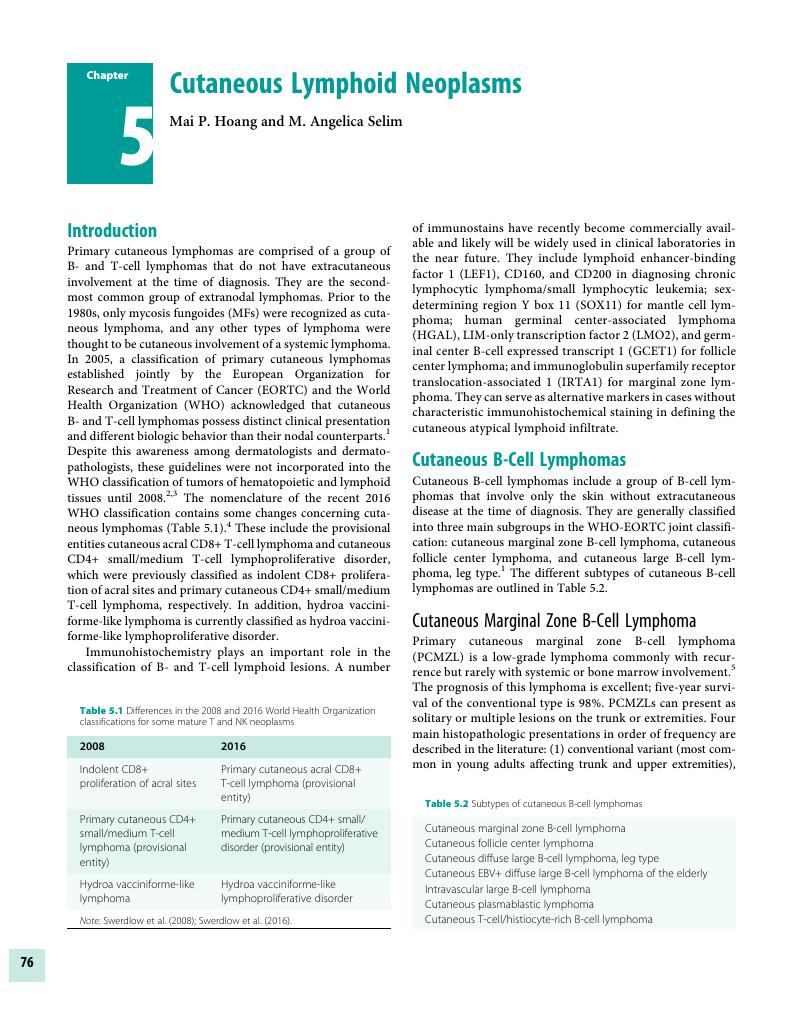
Chapter 5 - Cutaneous Lymphoid Neoplasms

Published online by Cambridge University Press:  04 November 2017

Mai P. Hoang
Affiliation:
Harvard Medical School, Boston
Get access

Summary

Image of the first page of this content. For PDF version, please use the ‘Save PDF’ preceeding this image.'
Type
Chapter
Information
Publisher: Cambridge University Press
Print publication year: 2017

Access options

Get access to the full version of this content by using one of the access options below. (Log in options will check for institutional or personal access. Content may require purchase if you do not have access.)

References

Willemze, R, Jaffe, ES, Burg, G, et al. WHO-EORTC classification for cutaneous lymphomas. Blood 2005; 105(10):3768–85.CrossRefGoogle ScholarPubMed
Senff, NJ, Hoefnagel, JJ, Jansen, PM, et al. Reclassification of 300 primary cutaneous B-Cell lymphomas according to the new WHO-EORTC classification for cutaneous lymphomas: Comparison with previous classifications and identification of prognostic markers. J Clin Oncol 2007; 25(12):1581–87.CrossRefGoogle Scholar
Swerdlow, SH, Campo, E, Harris, NL, et al. WHO classification of tumors haematopoietic and lymphoid tissues. Lyon: IARC, 2008.Google Scholar
Swerdlow, SH, Campo, E, Pileri, SA, et al. The 2016 revision of the World Health Organization classification of lymphoid neoplasms. Blood 2016; 127(20):2375–90.CrossRefGoogle ScholarPubMed
Cerroni, L, Signoretti, S, Hofler, G, et al. Primary cutaneous marginal B-cell lymphoma: A recently described entity of low-grade malignant cutaneous B-cell lymphoma. Am J Surg Pathol 1997; 21(11):1307–15.Google Scholar
Servitje, O, Gallardo, F, Estrach, T, et al. Primary cutaneous marginal zone B-cell lymphoma: A clinical, histopathological, immunophenotypic and molecular genetic study of 22 cases. Br J Dermatol 2002; 147(6):1147–58.CrossRefGoogle Scholar
de Leval, L, Harris, NL, Longtine, J, Ferry, JA, Duncan, LM. Cutaneous B-cell lymphomas of follicular and marginal zone types: Use of Bcl-6, CD10, Bcl-2, and CD21 in differential diagnosis and classification. Am J Surg Pathol 2001; 25(6):732–41.CrossRefGoogle ScholarPubMed
Cerroni, L, Arzberger, E, Putz, B, et al. Primary cutaneous follicular center cell lymphoma with follicular growth pattern. Blood 2000; 95(12):3922–28.CrossRefGoogle ScholarPubMed
Hoefnagel, JJ, Vermeer, MH, Janssen, PM, et al. Bcl-2, Bcl-6 and CD10 expression in cutaneous B-cell lymphoma: Further support for a follicle centre cell origin and differential diagnostic significance. Br J Dermatol 2003; 149(6):1183–91.CrossRefGoogle ScholarPubMed
Brenner, I, Roth, S, Puppe, B, et al. Primary cutaneous marginal zone lymphomas with plasmacytic differentiation show frequent IgG4 expression. Mod Pathol 2013; 26(12):1568–76.CrossRefGoogle ScholarPubMed
Falini, B, Agostinelli, C, Bigerna, B, et al. IRTA1 is selectively expressed in nodal and extranodal marginal zone lymphomas. Histopathology 2012; 61(5):930–41.CrossRefGoogle ScholarPubMed
Charli-Joseph, Y, Cerroni, L, LeBoit, PE. Cutaneous spindle-cell B-cell lymphomas: Most are neoplasms of follicular center cell origin. Am J Surg Pathol 2015; 39(6):737–43.CrossRefGoogle ScholarPubMed
Lawnicki, LC, Weisenburger, DD, Aoun, P, et al. The t(14;18) and bcl-2 expression are present in a subset of primary cutaneous follicular lymphoma: Association with lower grade. Am J Clin Pathol 2002; 118(5):765–72.Google Scholar
Szablewski, V, Ingen-Housz-Oro, S, Baia, M, et al. Primary cutaneous follicle center lymphomas expression Bcl2 protein frequently harbor Bcl2 gene break and may present 1p36 deletion: A study of 20 cases. Am J Surg Pathol 2016; 40(1):127–36.Google Scholar
Koens, L, Vermeer, MH, Willemze, R, Jansen, PM. IgM expression on paraffin sections distinguishes primary cutaneous large B-cell lymphoma, leg type from primary cutaneous follicle center lymphoma. Am J Surg Pathol 2010; 34(7):1043–48.Google Scholar
Robson, A, Shukur, Z, Ally, M, et al. Immunocytochemical p63 expression discriminates between primary cutaneous follicle center cell and diffuse large B-cell lymphoma-leg type, and is of the TAp63 isoform. Histopathology 2016; 69(1):1119.CrossRefGoogle ScholarPubMed
Fink-Puches, R, Wolf, IH, Zalaudek, I, Kerl, H, Cerroni, L. Treatment of primary cutaneous B-cell lymphoma with rituximab. J Am Acad Dermatol 2005; 52(5):847–53.CrossRefGoogle ScholarPubMed
Kempf, W, Kazakov, DV, Rutten, A, et al. Primary cutaneous follicle center lymphoma with diffuse CD30 expression: A report of 4 cases of a rare variant. J Am Acad Dermatol 2014; 71(3):548–54.Google Scholar
Grange, F, Beylot-Barry, M, Courville, P, et al. Primary cutaneous diffuse large B-cell lymphoma, leg type: Clinicopathologic features and prognostic analysis in 60 cases. Arch Dermatol 2007; 143(9):1144–50.Google Scholar
Hoefnagel, JJ, Dijkman, R, Basso, K, et al. Distinct types of primary cutaneous large B-cell lymphoma identified by gene expression profiling. Blood 2005; 105(9):3671–78.Google Scholar
Oyama, T, Ichimura, K, Suzuki, R, et al. Senile EBV+ B-cell lymphoproliferative disorders. A clinicopathologic study of 22 patients. Am J Surg Pathol 2003; 27(1):1626.Google Scholar
Gibson, SE, Hsi, ED. Epstein-Barr virus-positive B-cell lymphoma of the elderly at a United States tertiary medical center: An uncommon aggressive lymphoma with a nongerminal center B-cell phenotype. Hum Pathol 2009; 40(5):653–61.Google Scholar
Ponzoni, M, Ferreri, AJM, Campo, E, et al. Definition, diagnosis, and management of intravascular large B-cell lymphoma: Proposals and perspectives from an international consensus meeting. J Clin Oncol 2007; 25(21):3168–73.CrossRefGoogle ScholarPubMed
Glass, J, Hochberg, FH, Miller, DC. Intravascular lymphomatosis: A systemic disease with neurologic manifestations. Cancer 1993; 71(10):3156–64.3.0.CO;2-O>CrossRefGoogle ScholarPubMed
Murase, T, Yamaguchi, M, Suzuki, R, et al. Intravascular large B-cell lymphoma (IVLBCL): A clinicopahologic study of 96 cases with special reference to the immunophenotypic heterogeneity of CD5. Blood 2007; 109(2):895902.CrossRefGoogle Scholar
Morscio, J, Dierickx, D, Nijs, J, et al. Clinicopathologic comparison of plasmablastic lymphoma in HIV-positive, immunocompetent, and posttransplant patients: Single-center series of 25 cases and meta-analysis of 277 reported cases. Am J Surg Pathol 2014; 38(7):875–86.Google Scholar
Gilaberte, M, Gallardo, F, Bellosillo, B, et al. Recurrent and self-healing cutaneous monoclonal plasmablastic infiltrates in a patient with AIDS and Kaposi sarcoma. Br J Dermatol 2005; 153(4):828–32.CrossRefGoogle Scholar
Delecluse, HJ, Anagnostopoulos, I, Dallenbach, F, et al. Plasmablastic lymphomas of the oral cavity: A new entity associated with the human immunodeficiency virus infection. Blood 1997; 89(4):1413–20.CrossRefGoogle ScholarPubMed
Liu, W, Lacouture, ME, Jiang, J, et al. KSHV/HHV8-associated primary cutaneous plasmablastic lymphoma in a patient with Castleman’s disease and Kaposi’s sarcoma. J Cutan Pathol 2006; 33(Suppl 2):4651.CrossRefGoogle Scholar
Sander, CA, Kaudewitz, P, Kutzner, H, et al. T-cell-rich B-cell lymphoma presenting in skin: A clinicopathologic analysis of six cases. J Cutan Pathol 1996; 23(2):101–8.Google Scholar
Dunphy, CH, Nahass, GT. Primary cutaneous T-cell-rich B-cell lymphomas with flow cytometric immunophenotypic findings: Report of 3 cases and review of the literature. Arch Pathol Lab Med 1999; 123(12):1236–40.Google Scholar
Rodriguez-Pinilla, SM, Ortiz-Romero, PL, et al. TCR-gamma expression in primary cutaneous T-cell lymphomas. Am J Surg Pathol 2016; 37(3):375–84.Google Scholar
Falini, B, Flenghi, L, Pileri, S, et al. Distribution of T-cells bearing different forms of the T-cell receptor gamma/delta in normal and pathological human tissues. J Immunol 1989; 143(8):2480–88.Google Scholar
Zackheim, HS, Amin, S, Kashani-Sabet, M, McMillan, A. Prognosis in cutaneous T-cell lymphoma by skin stage: Long-term survival in 489 patients. J Am Acad Dermatol 1999; 40(3):418–25.Google Scholar
Quaglino, P, Pimpinelli, N, Berti, E, et al; Gruppo Italiano Linfomi Cutanei. Time course, clinical pathways, and long-term hazards risk trends of disease progression in patients with classic mycosis fungoides: A multicenter, retrospective follow-up study from the Italian Group of Cutaneous Lymphomas. Cancer 2012; 118(23):5830–39.Google Scholar
Cerroni, L, Fink-Puches, R, Back, B, Kerl, H. Follicular mucinosis: A critical reappraisal of clinicopathologic features and association with mycosis fungoides and Sezary syndrome. Arch Dermatol 2002; 138(2):182–89.CrossRefGoogle ScholarPubMed
Pileri, A, Facchetti, F, Rutten, A, et al. Syringotropic mycosis fungoides: A rare variant of the disease with peculiar clinicopathologic features. Am J Surg Pathol 2011; 35(1):100–9.Google Scholar
Haghighi, B, Smoller, BR, LeBoit, PE, Warnke, RA, Sander, CA, Kohler, S. Pagetoid reticulosis (Woringer-Kolopp disease): An immunophenotypic, molecular, and clinicopathologic study. Mod Pathol 2000; 13(5):502–10.Google Scholar
El Shabrawi-Caelen, L, Cerroni, L, Medeiros, LJ, McCalmont, TH. Hypopigmented mycosis fungoides: Frequent expression of a CD8 T-cell phenotype. Am J Surg Pathol 2002; 26(4):450–57.CrossRefGoogle ScholarPubMed
Benner, MA, Jansen, PM, Vermeer, MH, et al. Prognostic factors in transformed mycosis fungoides: A retrospective analysis of 100 cases. Blood 2012; 119(7):1643–49.CrossRefGoogle ScholarPubMed
Cerroni, L, Rieger, E, Hodl, S, Kerl, H. Clinicopathologic and immunologic features associated with transformation of mycosis fungoides to large cell lymphoma. Am J Surg Pathol 1992; 16(6):543–52.Google Scholar
Vergier, B, De Muret, A, Beylot-Barry, M, et al. Transformation of mycosis fungoides: Clinicopathologic and prognostic features of 45 cases. Blood 2009; 95(7):2212–18.Google Scholar
Jullié, ML, Carlotti, M, Vivot, A Jr, et al. CD20 antigen may be expressed by reactive or lymphomatous cells of transformed mycosis fungoides: Diagnostic and prognostic impact. Am J Surg Pathol 2013; 37(12):1845–54.Google Scholar
Vonderheid, EC, Bernengo, MG, Burg, G, et al. Update on erythrodermic cutaneous T-cell lymphoma: Report of the International Society for Cutaneous Lymphomas. J Am Acad Dermatol 2002; 46(1):95106.CrossRefGoogle ScholarPubMed
Klemke, CD, Booken, N, Weiss, C, et al. Histopathological and immunophenotypical criteria for the diagnosis of Sezary syndrome in differentiation from other erythrodermic skin diseases: A European Organisation for Research and Treatment of Cancer (EORTC) Cutaneous Lymphoma Task Force Study of 97 cases. Br J Dermatol 2015; 173(1):93105.Google Scholar
Cetinozman, F, Jansen, PM, Vermeer, MH, et al. Differential expression of programmed death-1 (PD-1) in Sezary syndrome and mycosis fungoides. Arch Dermatol 2012; 148(12):1379–85.CrossRefGoogle ScholarPubMed
Capriotti, E, Vonderheid, EC, Thoburn, CJ, et al. Expression of T-plastin, FoxP3 and other tumor-associated markers by leukemic T-cells of cutaneous T-cell lymphoma. Leuk Lymphoma 2008; 49(6):1190–201.Google Scholar
El Shabrawi-Caelen, L, Kerl, H, Cerroni, L. Lymphomatoid papulosis: Reappraisal of clinicopathologic presentation and classification into subtypes A, B, and C. Arch Dermatol 2004; 140(4):441–47.Google Scholar
Saggini, A, Gulia, A, Argenyi, Z, et al. A variant of lymphomatoid papulosis simulating primary cutaneous aggressive epidermotropic CD8+ cytotoxic T-cell lymphoma. Description of 9 cases. Am J Surg Pathol 2010; 34(8):1168–75.Google Scholar
Sharaf, MA, Romanelli, P, Kirsner, R, Miteva, M. Angioinvasive lymphomatoid papulosis: Another case of a newly described variant. Am J Dermatopathol 2014; 36(3):e7577.CrossRefGoogle ScholarPubMed
Karai, LJ, Kadin, ME, Hsi, ED, et al. Chromosomal rearrangments of 6p25.3 define a new subtype of lymphomatoid papulosis. Am J Surg Pathol 2013; 37(8):1173–81.Google Scholar
Smoller, BR, Longacre, TA, Warnke, RA. Ki-1 (CD30) expression in differentiation of lymphomatoid papulosis from arthropod bite reactions. Mod Pathol 1992; 5(5):492–96.Google ScholarPubMed
Werner, B, Massone, C, Kerl, H, Cerroni, L. Large CD30-positive cells in benign, atypical lymphoid infiltrates of the skin. J Cutan Pathol 2008; 35(12):1100–07.CrossRefGoogle ScholarPubMed
Cepeda, LT, Pieretti, M, Chapman, SF, Horenstein, MG. CD30-positive atypical lymphoid cells in common non-neoplastic cutaneous infiltrates rich in neutrophils and eosinophils. Am J Surg Pathol 2003; 27(7):912–18.Google Scholar
Kempf, W, Kazakov, DV, Palmedo, G, et al. Pityriasis lichenoides et varioliformis acuta with numerous CD30(+) cells: A variant mimicking lymphomatoid papulosis and other cutaneous lymphomas. A clinicopathologic, immunohistochemical, and molecular biological study of 13 cases. Am J Surg Pathol 2012; 36(7):1021–29.CrossRefGoogle Scholar
Kempf, W. CD30+ lymphoproliferative disorders: Histopathology, differential diagnosis, new variants, and simulators. J Cutan Pathol 2006; 33(Suppl 1):5870.Google Scholar
Kempf, W, Kutzner, H, Cozzio, A, et al. MUM1 expression in cutaneous CD30+ lymphoproliferative disorders: A valuable tool for the distinction between lymphomatoid papulosis and primary cutaneous anaplastic large-cell lymphoma. Br J Dermatol 2008; 158(6):1280–87.Google Scholar
Kempf, W, Pfaltz, K, Vermeer, MH, et al. EORTC, ISCL, and USCLC consensus recommendations for the treatment of primary cutaneous CD30-positive lymphoproliferative disorders: Lymphomatoid papulosis and primary cutaneous anaplastic large-cell lymphoma. Blood 2011; 118(15):4024–35.Google Scholar
Massone, C, Cerroni, L. Phenotypic variability in primary cutaneous anaplastic large T-cell lymphoma: A study on 35 patients. Am J Dermatopathol 2014; 36(2):153–57.Google Scholar
ten Berge, RL, Oudejans, JJ, Ossenkoppele, GJ, et al. ALK expression in extranodal anaplastic large cell lymphoma favours systemic disease with (primary) nodal involvement and a good prognosis and occurs before dissemination. J Clin Pathol 2000; 53(6):445–50.Google Scholar
Oschlies, I, Lisfeld, J, Lamant, L, et al. ALK-positive anaplastic large cell lymphoma limited to the skin: Clinical, histopathological and molecular analysis of 6 pediatric cases: A report from the ALCL99 study. Haematologica 2013; 98(1):5056.Google Scholar
de Bruin, PC, Beljaards, RC van Heerde, P, et al. Differences in clinical behaviour and immunophenotype between primary cutaneous and primary nodal anaplastic large cell lymphoma of T-cell or null cell phenotype. Histopathology 1993; 23(2):127–35.Google Scholar
Ferrara, G, Ena, L, Cota, C, Cerroni, L. Intralymphatic spread is a common finding in cutaneous CD30+ lymphoproliferative disorders. Am J Surg Pathol 2015; 39(11):1511–17.CrossRefGoogle ScholarPubMed
Samols, MA, Su, A, Ra, S, Cappel, MA, et al. Intralymphatic cutaneous anaplastic large cell lymphoma/lymphomatoid papulosis: Expanding the spectrum of CD30-positive lymphoproliferative disorders. Am J Surg Pathol 2014; 38(9):1203–11.Google Scholar
Hagiwara, M, Takata, K, Shimoyama, Y, et al. Primary cutaneous T-cell lymphoma of unspecified type with cytotoxic phenotype: Clinicopathological analysis of 27 patients. Cancer Sci 2009; 100(1):3341.Google Scholar
Savage, KJ, Ferreri, AJ, Zinzani, PL, Pilari, SA. Peripheral T-cell lymphoma– Not otherwise specified. Crit Rev Oncol Hematol 2011; 79(3):321–29.Google Scholar
Marchetti, MA, Pulitzer, MP, Myskowski, PL, et al. Cutaneous manifestations of human T-cell lymphotrophic virus type-1-associated adult T-cell leukemia/lymphoma: A single-center, retrospective study. J Am Acad Dermatol 2015; 72(2):293301.Google Scholar
Bittencourt, AL, Barbosa, HS, Vieira, MD, Farre, L. Adult T-cell leukemia/lymphoma (ATL) presenting in the skin: Clinical, histological and immunohistochemical features of 52 cases. Acta Oncol 2009; 48(4):598604.Google Scholar
Tokura, Y, Sawada, Y, Shimauchi, T. Skin manifestations of adult T-cell leukemia/lymphoma: Clinical, cytological and immunological features. J Dermatol 2014; 41(1):1925.CrossRefGoogle ScholarPubMed
Willemze, R, Jansen, PM, Cerroni, L, et al. Subcutaneous panniculitis-like T-cell lymphoma: Definition, classification, and prognostic factors: An EORTC Cutaneous Lymphoma Group Study of 83 cases. Blood 2008; 111(2):838–45.CrossRefGoogle Scholar
Salhany, KE, Macon, WR, Choi, JK, et al. Subcutaneous panniculitis-like T cell lymphoma: Clinicopathologic, immunophenotypic, and genotypic analysis of alpha/beta and gamma/delta subtypes. Am J Surg Pathol 1998; 22(7):881–93.Google Scholar
Go, RS, Wester, SM. Immunophenotypic and molecular features, clinical outcomes, treatments, and prognostic factors associated with subcutaneous panniculitis-like T-cell lymphoma. Cancer 2004; 101(6):1404–13.Google Scholar
Kong, YY, Dai, B, Kong, JC, et al. Subcutaneous panniculitis-like T-cell lymphoma: A clinicopathologic, immunophenotypic, and molecular study of 22 Asian cases according to WHO-EORTC classification. Am J Surg Pathol 2008; 32(10):1495–502.Google Scholar
Bosisio, F, Boi, S, Caputo, V, et al. Lobular panniculitic infiltrates with overlapping histopathologic features of lupus panniculitis (lupus profundus) and subcutaneous T-cell lymphoma: A conceptual and practical dilemma. Am J Surg Pathol 2015; 39(2):206–11.Google Scholar
Magro, CM, Schaefer, JT, Morrison, C, Porcu, P. Atypical lymphocytic lobular panniculitis: A clonal subcutaneous T-cell dyscrasia. J Cutan Pathol 2008; 35(10):947–54.Google Scholar
LeBlanc, RE, Tavallaee, M, Kim, YH, Kim, J. Useful parameters for distinguishing subcutaneous panniculitis-like T-Cell lymphoma from lupus erythematosus panniculitis. Am J Surg Pathol 2016; 40(6):745–54.Google Scholar
Guitart, J, Weisenburger, DD, Subtil, A, et al. Cutaneous gamma/delta T-cell lymphomas. A spectrum of presentations with overlap with other cytotoxic lymphomas. Am J Surg Pathol 2012; 36(11):1656–65.Google Scholar
Berti, E, Tomasini, D, Vermeer, MH, et al. Primary cutaneous CD8-positive epidermotropic cytotoxic T-cell lymphomas: A distinct clinicopathological entity with an aggressive clinical behavior. Am J Pathol 1999; 155(2):483–92.Google Scholar
Robson, A, Assaf, C, Bagot, M, et al. Aggressive epidermotropic cutaneous CD8+ lymphoma: A cutaneous lymphoma with distinct clinical and pathological features. Report of an EORTC Cutaneous Lymphoma Task Force Workshop. Histopathology 2015; 67(4):425–41.Google Scholar
Saggini, A, Gulia, A, Argenyi, Z, et al. A variant of lymphomatoid papulosis simulating primary cutaneous aggressive epidermotropic CD8+ cytotoxic T-cell lymphoma. Description of 9 cases. Am J Surg Pathol 2010; 34(8):1168–75.Google Scholar
Takata, K, Hong, ME, Sitthinamsuwan, P, et al. Primary cutaneous NK/T-cell lymphoma, nasal type and CD56-positive peripheral T-cell lymphoma: A cellular lineage and clinicopathologic study of 60 patients from Asia. Am J Surg Pathol 2015; 39(1):112.Google Scholar
Lee, WJ, Jung, JM, Won, CH, et al. Cutaneous extranodal natural killer/T-cell lymphoma: A comparative clinicohistopathologic and survival outcome analysis of 45 cases according to the primary tumor site. J Am Acad Dermatol 2014; 70(6):1002–9.Google Scholar
Gualco, G, Domeny-Duarte, P, Chioato, L, et al. Clinicopathologic and molecular features of 122 Brazilian cases of nodal and extranodal NK/T-cell lymphoma, nasal type, with EBV subtyping analysis. Am J Surg Pathol 2011; 35(8):1195–203.Google Scholar
Schwartz, EJ, Molina-Kirsch, H, Zhao, S, et al. Immunohistochemical characterization of nasal-type extranodal NK/T-cell lymphoma using a tissue microarray: An analysis of 84 cases. Am J Clin Pathol 2008; 130(3):343–51.Google Scholar
Ohno, T, Yamaguchi, M, Oka, K, et al. Frequent expression of CD3 epsilon in CD3 (Leu 4)-negative nasal T-cell lymphoma. Leukemia 1995; 9(1):4452.Google Scholar
Petrella, T, Maubec, E, Cornillet-Lefebvre, P, et al. Indolent CD8-positive lymphoid proliferation of the ear: A distinct primary cutaneous T-cell lymphoma? Am J Surg Pathol 2007; 31(12):1887–92.CrossRefGoogle Scholar
Greenblatt, D, Ally, M, Child, F, et al. Indolent CD8+ lymphoid proliferation of acral sites: A clinicopathologic study of six patients with some atypical features. J Cutan Pathol 2013; 40(2):248–58.Google Scholar
Wobser, M, Petrella, T, Kneitz, H, et al. Extrafacial indolent CD8-positive cutaneous lymphoid proliferation with unusual symmetrical presentation involving both feet. J Cutan Pathol 2013; 40(11):955–61.CrossRefGoogle ScholarPubMed
Hagen, JW, Magro, CM. Indolent CD8+ lymphoid proliferation of the face with eyelid involvement. Am J Dermatopathol 2014; 36(2):137–41.CrossRefGoogle ScholarPubMed
Beltraminelli, H, Mullegger, R, Cerroni, L. Indolent, CD8+ lymphoid proliferation of the ear: A phenotypic variant of the small-medium pleomorphic cutaneous T-cell lymphoma? J Cutan Pathol 2010; 37(1):8184.Google Scholar
Berti, E, Tomasini, D, Vermeer, MH, et al. Primary cutaneous CD8-positive epidermotropic cytotoxic T cell lymphomas. A distinct clinicopathological entity with an aggressive clinical behavior. Am J Pathol 1999; 155(2):483–92.Google Scholar
Li, JY, Guitart, J, Pulitzer, MP, et al. Multicenter case series of indolent small/medium-sized CD8+ lymphoid proliferations with predilection for the ear and face. Am J Dermatopathol 2014; 36(5):402–8.Google Scholar
Wobser, M, Roth, S, Reinartz, T, et al. CD68 expression is a discriminative feature of indolent cutaneous CD8-positive lymphoid proliferation and distinguishes this lymphoma subtype from other CD8-positive cutaneous lymphomas. Br J Dermatol 2015; 172(6):1573–80.Google Scholar
Beltraminelli, H, Leinweber, B, Kerl, H, Cerroni, L. Primary cutaneous CD4+ small/medium-sized pleomorphic T-cell lymphoma: A cutaneous nodular proliferation of pleomorphic T lymphocytes of undetermined significance? A study of 136 cases. Am J Dermatopathol 2009; 31(4):317–22.Google Scholar
William, VL, Torres-Cabala, CA, Duvic, M. Primary cutaneous small- to medium-sized CD4+ pleomorphic T-cell lymphoma: A retrospective case series and review of the provisional cutaneous lymphoma category. Am J Clin Dermatol 2011; 12(6):389401.Google Scholar
Rodriguez-Pinilla, SM, Roncador, G, Rodriguez-Peralto, IJ, et al. Primary cutaneous CD4+ small/medium-sized pleomorphic T-cell lymphoma expresses follicular T-cell markers. Am J Surg Pathol 2009; 33(1):8190.Google Scholar
Magana, M, Massone, C, Magana, P, Cerroni, L. Clinicopathologic features of hydroa vacciniforme-like lymphoma: A series of 9 patients. Am J Dermatopathol 2016; 38(1):2025.Google Scholar
Sangueza, M, Plaza, JA. Hydroa vacciniforme-like cutaneous T-cell lymphoma: Clinicopathologic and immunohistochemical study of 12 cases. J Am Acad Dermatol 2013; 69(1):112–19.Google Scholar
Quintanilla-Martinez, L, Ridaura, C, Nagl, F, et al. Hydroa vacciniforme-like lymphoma: A chronic EBV+ lymphoproliferative disorder with risk to develop a systemic lymphoma. Blood 2013; 122(18):3101–10.Google Scholar
Cota, C, Vale, E, Viana, I, et al. Cutaneous manifestations of blastic plasmacytoid dendritic cells neoplasm – morphologic and phenotypic variability in a series of 33 patients. Am J Surg Pathol 2010; 34(1):7587.Google Scholar
Julia, F, Petrella, T, Beylot-Barry, M, et al. Blastic plasmacytoid dendritic cell neoplasm: Clinical features in 90 patients. Br J Dermatol 2013; 169(3):579–86.Google Scholar
Julia, F, Dalle, S, Duru, G, et al. Blastic plasmacytoid dendritic cell neoplasms. Clinico-immunohistochemical correlations in a series of 91 patients. Am J Surg Pathol 2014; 38(5):673–80.Google Scholar
Leroux, D, Mugneret, F, Callanan, M, et al. CD4(+) CD56(+) DC2 acute leukemia is characterized by recurrent clonal chromosomal changes affecting 6 major targets: A study of 21 cases by the Groupe Francais de Ctyogenetique Hematologique. Blood 2002; 99(11):4154–59.CrossRefGoogle ScholarPubMed
Sangle, NA, Schmidt, RL, Patel, JL, et al. Optimized immunohistochemical panel to differentiate myeloid sarcoma from blastic plasmacytoid dendritic cell neoplasm. Mod Pathol 2014; 27(8):1137–43.Google Scholar
Sen, F, Medeiros, LJ, Lu, D, et al. Mantle cell lymphoma involving skin: Cutaneous lesions may be the first manifestation of disease and tumors often have blastoid cytologic features. Am J Surg Pathol 2002; 26(10):1312–18.Google Scholar
Soldini, D, Valera, A, Sole, C, et al. Assessment of Sox11 expression in routine lymphoma tissue sections: Characterization of new monoclonal antibodies for diagnosis of mantle cell lymphoma. Am J Surg Pathol 2014; 38(1):8693.Google Scholar
Jares, P, Colomer, D, Campo, E. Molecular pathogenesis of mantle cell lymphoma. J Clin Invest 2012; 122(10):3416–23.CrossRefGoogle ScholarPubMed
Scarisbrick, JJ, Prince, HM, Vermeer, MH, et al. Cutaneous Lymphoma International Consortium study of outcome in advanced stages of mycosis fungoides and Sezary syndrome: Effect of specific prognostic markers on survival and development of a prognostic model. J Clin Oncol 2015; 33(32):3766–73.Google Scholar
Salhany, KE, Sousar, JB, Greer, JP, et al. Transformation of cutaneous T-cell lymphoma to large cell lymphoma. A clinicopathologic and immunologic study. Am J Pathol 1988; 132(2):265–77.Google Scholar
Hussong, JW, Perkins, SL, Schnitzer, B, Hargreaves, H, Frizzera, G. Extramedullary plasmacytoma. A form of marginal zone cell lymphoma? Am J Clin Pathol 1999; 111(1):111–16.Google Scholar
Jurczyszyn, A, Olszewska-Szopa, M, Hundria, V, et al. Cutaneous involvement in multiple myeloma: A multi-institutional retrospective study of 53 patients. Leuk Lymphoma 2016; 57(9):2071–76.Google Scholar
Deshpande, V, Zen, Y, Chan, JK, et al. Consensus statement on the pathology of IgG4-related disease. Mod Pathol 2012; 25(9):1181–92.CrossRefGoogle ScholarPubMed
Stenzinger, A, Endris, V, Pfarr, N, et al. Targeted ultra-deep sequencing reveals recurrent and mutually exclusive mutations of cancer genes in blastic plasmacytoid dendritic cell neoplasm. Oncotarget 2014; 5(15):6404–13.Google Scholar
Botros, N, Cerroni, L, Shawwa, A, et al. Cutaneous manifestations of angioimmunoblastic T-cell lymphoma: Clinical and pathological characteristics. Am J Dermatopathol 2015; 37(4):274–83.Google Scholar
Yu, H, Shahsafaei, A, Dorfman, DM. Germinal-center T-helper-cell markers PD-1 and CXCL13 are both expressed by neoplastic cells in angioimmunoblastic T-cell lymphoma. Am J Clin Pathol 2009; 131(1):3341.Google Scholar
Brown, HA, Macon, WR, Kurtin, PJ, Gibson, LE. Cutaneous involvement by angioimmunoblastic T-cell lymphoma with remarkable heterogeneous Epstein-Barr virus expression. J Cutan Pathol 2001; 28(8):432–38.Google Scholar

Save book to Kindle

To save this book to your Kindle, first ensure coreplatform@cambridge.org is added to your Approved Personal Document E-mail List under your Personal Document Settings on the Manage Your Content and Devices page of your Amazon account. Then enter the ‘name’ part of your Kindle email address below. Find out more about saving to your Kindle.

Note you can select to save to either the @free.kindle.com or @kindle.com variations. ‘@free.kindle.com’ emails are free but can only be saved to your device when it is connected to wi-fi. ‘@kindle.com’ emails can be delivered even when you are not connected to wi-fi, but note that service fees apply.

Find out more about the Kindle Personal Document Service.

Available formats
×

Save book to Dropbox

To save content items to your account, please confirm that you agree to abide by our usage policies. If this is the first time you use this feature, you will be asked to authorise Cambridge Core to connect with your account. Find out more about saving content to Dropbox.

Available formats
×

Save book to Google Drive

To save content items to your account, please confirm that you agree to abide by our usage policies. If this is the first time you use this feature, you will be asked to authorise Cambridge Core to connect with your account. Find out more about saving content to Google Drive.

Available formats
×