
Book contents
- Immunohistochemistry in Diagnostic Dermatopathology
- Immunohistochemistry in Diagnostic Dermatopathology
- Copyright page
- Dedication
- Contents
- Contributors
- Preface and Acknowledgments
- Chapter 1 Introduction to Immunohistochemistry
- Chapter 2 Epithelial or Squamous Neoplasms
- Chapter 3 Neoplasms of Cutaneous Appendages
- Chapter 4 Inflammatory Dermatoses Mimicking Lymphomas
- Chapter 5 Cutaneous Lymphoid Neoplasms
- Chapter 6 Melanocytic neoplasms
- Chapter 7 Soft Tissue Neoplasms
- Chapter 8 Miscellaneous Tumors
- Chapter 9 Detection of Genetic Syndromes
- Chapter 10 Immunobullous Disorders
- Chapter 11 Cutaneous Infections
- Chapter 12 Therapeutic and Prognostic Applications
- Index
- References
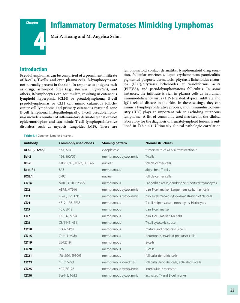
Chapter 4 - Inflammatory Dermatoses Mimicking Lymphomas
Published online by Cambridge University Press: 04 November 2017
Edited by

Book contents
- Immunohistochemistry in Diagnostic Dermatopathology
- Immunohistochemistry in Diagnostic Dermatopathology
- Copyright page
- Dedication
- Contents
- Contributors
- Preface and Acknowledgments
- Chapter 1 Introduction to Immunohistochemistry
- Chapter 2 Epithelial or Squamous Neoplasms
- Chapter 3 Neoplasms of Cutaneous Appendages
- Chapter 4 Inflammatory Dermatoses Mimicking Lymphomas
- Chapter 5 Cutaneous Lymphoid Neoplasms
- Chapter 6 Melanocytic neoplasms
- Chapter 7 Soft Tissue Neoplasms
- Chapter 8 Miscellaneous Tumors
- Chapter 9 Detection of Genetic Syndromes
- Chapter 10 Immunobullous Disorders
- Chapter 11 Cutaneous Infections
- Chapter 12 Therapeutic and Prognostic Applications
- Index
- References
Summary
A summary is not available for this content so a preview has been provided. Please use the Get access link above for information on how to access this content.
- Type
- Chapter
- Information
- Immunohistochemistry in Diagnostic Dermatopathology , pp. 55 - 75Publisher: Cambridge University PressPrint publication year: 2017
References
Cerroni, L, Arzberger, E, Putz, B, et al. Primary cutaneous follicular center cell lymphoma with follicular growth pattern. Blood 2000;95(12):3922–28.Google Scholar
Bergman, R, Khamaysi, K, Khamayi, Z, Ben Arie, Y. A study of histologic and immunophenotypical staining patterns in cutaneous lymphoid hyperplasia. J Am Acad Dermatol 2011;65(1):112–24.Google Scholar
Colli, C, Leinweber, B, Mullegger, R, et al. Borrelia burgdorferi-associated lymphocytoma cutis: Clinicopathologic, immunophenotypic, and molecular study of 106 cases. J Cutan Pathol 2004;31(3):232–40.Google Scholar
Arai, E, Shimizu, M, Hirose, T. A review of 55 cases of cutaneous lymphoid hyperplasia: Reassessment of the histopathologic findings leading to reclassification of 4 lesions as cutaneous marginal zone lymphoma and 19 as pseudolymphomatous folliculitis. Hum Pathol 2005;36(5):505–11.Google Scholar
Servitje, O, Gallardo, F, Estrach, T, et al. Primary cutaneous marginal zone B-cell lymphoma: A clinical, histopathological, immunophenotypic and molecular genetic study of 22 cases. Br J Dermatol 2002;147(6):1147–58.Google Scholar
de Leval, L, Harris, NL, Longtine, J, Ferry, JA, Duncan, LM. Cutaneous B-cell lymphomas of follicular and marginal zone types: Use of Bcl-6, CD10, Bcl-2, and CD21 in differential diagnosis and classification. Am J Surg Pathol 2001;25(6):732–41.Google Scholar
LeBoit, PE, McNutt, NS, Reed, JA, Jacobson, M, Weiss, LM. Primary cutaneous immunocytoma. A B-cell lymphoma that can easily be mistaken for cutaneous lymphoid hyperplasia. Am J Surg Pathol 1994;18(10):969–78.Google Scholar
Goodlad, JR, Krajewski, AS, Batstone, PJ, et al. Primary cutaneous follicular lymphoma: A clinicopathologic and molecular study of 16 cases in support of a distinct entity. Am J Surg Pathol 2002;26(6):733–41.Google Scholar
Orbaneja, JG, Diez, LI, Lozano, JL, Salazar, LC. Lymphomatoid contact dermatitis: A syndrome produced by epicutaneous hypersensitivity with clinical features and a histopathologic picture similar to that of mycosis fungoides. Contact Dermatitis 1976;2(3):139–43.Google Scholar
Smolle, J, Torne, R, Soyer, HP, Kerl, H. Immunohistochemical classification of cutaneous pseudolymphomas: Delineation of distinct patterns. J Cutan Pathol 1990;17(3):149–59.Google Scholar
Haynes, BF, Hensley, LL, Jegasothy, BV. Phenotypic characterization of skin-infiltrating T cells in cutaneous T-cell lymphoma: Comparison with benign cutaneous T-cell infiltrates. Blood 1982;60(2):463–73.Google Scholar
Souteyrand, P, d’Incan, M. Drug induced mycosis fungoides like lesions. Curr Probl Dermatol 1990;19:176–82.Google Scholar
Ploysangam, T, Breneman, DL, Mutasim, DF. Cutaneous pseudolymphomas. J Am Acad Dermatol 1998;38(6 Pt 1):877–95.Google Scholar
Magro, CM, Crowson, AN. Drug-induced immune dysregulation as a cause of atypical cutaneous lymphoid infiltrates: A hypothesis. Hum Pathol 1996;27(2):125–32.Google Scholar
Murphy, M, Fullen, D, Carlson, JA. Low CD7 expression in benign and malignant cutaneous lymphocytic infiltrates: Experience with an antibody reactive with paraffin-embedded tissue. Am J Dermatopathol 2002;24(1):6–16.Google Scholar
Florell, SR, Cessna, M, Lundell, RB, et al. Usefulness (or lack thereof) of immunophenotyping in atypical cutaneous T-cell infiltrates. Am J Clin Pathol 2006;125(5):727–36.Google Scholar
Michie, SA, Abel, EA, Hoppe, RT, Warnke, RA, Wood, GS. Expression of T-cell receptor antigens in mycosis fungoides and inflammatory skin lesions. J Invest Dermatol 1989;93(1):116–20.Google Scholar
Ormsby, A, Bergfeld, WF, Tubbs, RR, Hsi, ED. Evaluation of a new paraffin-reactive CD7 T-cell deletion marker and a polymerase chain reaction-based T-cell receptor gene rearrangement assay: Implications for diagnosis of mycosis fungoides in community clinical practice. J Am Acad Dermatol 2001;45(3):405–13.Google Scholar
Ortonne, N, Buyukbabani, N, Delfau-Larue, MH, Bago, M, Wechsler, J. Value of the CD8-CD3 ratio for the diagnosis of mycosis fungoides. Mod Pathol 2003;16(9):857–62.Google Scholar
Pulitzer, MP, Nolan, KA, Oshman, RG, Phelps, RG. CD30+ lymphomatoid drug reactions. Am J Dermatopathol 2013;35(3):343–50.Google Scholar
Cerroni, L, Fink-Puches, R, Back, B, Kerl, H. Follicular mucinosis: A critical reappraisal of clinicopathologic features and association with mycosis fungoides and Sezary syndrome. Arch Dermatol 2002;138(2):182–89.Google Scholar
Brown, HA, Gibson, LE, Pujol, RM, Lust, JA, Pittelkow, MR. Primary follicular mucinosis: Long-term follow-up of patients younger than 40 years with and without clonal T-cell receptor gene rearrangement. J Am Acad Dermatol 2002;47(6):856–62.Google Scholar
Gerami, P, Rosen, S, Kuzel, T, Boone, SL, Guitart, J. Folliculotropic mycosis fungoides: An aggressive variant of cutaneous T-cell lymphoma. Arch Dermatol 2008;144(6):738–46.Google Scholar
Burg, G, Kempf, W, Cozzio, A, et al. WHO/EORTC classification of cutaneous lymphomas 2005: Histological and molecular aspects. J Cutan Pathol 2005;32(10):647–74.Google Scholar
Rongioletti, F, De Lucchi, S, Meyes, D, et al. Follicular mucinosis: A clinicopathologic, histochemical, immunohistochemical and molecular study comparing the primary benign form and the mycosis fungoides-associated follicular mucinosis. J Cutan Pathol 2010;37(1):15–19.Google Scholar
Demirkesen, C, Esirgen, G, Engin, B, Songur, A, Oquz, O. The clinical features and histopathologic patterns of folliculotropic mycosis fungoides in a series of 38 cases. J Cutan Pathol 2015;42(1):22–31.Google Scholar
van Doorn, R, Scheffer, E, Willemze, R. Follicular mycosis fungoides, a distinct disease entity with or without associated follicular mucinosis: A clinicopathologic and follow-up study of 51 patients. Arch Dermatol 2002;138(2):191–98.Google Scholar
Smoller, BR, Kamel, OW. Pigmented purpuric eruptions: Immunopathologic studies supportive of a common immunophenotype. J Cutan Pathol 1991;18(6):423–27.Google Scholar
Lipsker, D. The pigmented and purpuric dermatitis and the many faces of mycosis fungoides. Dermatology 2003;207(3):246–47.Google Scholar
Geller, L, Antonov, NK, Lauren, CT, Morel, KD, Garzon, MC. Pityriasis lichenoides in childhood: Review of clinical presentation and treatment options. Pediatr Dermatol 2015;32(5):579–92.Google Scholar
Magro, C, Crowson, AN, Kovatich, A, Burns, F. Pityriasis lichenoides: A clonal T cell lymphoproliferative disorder. Hum Pathol 2002;33(8):788–95.Google Scholar
Kempf, W, Kazakov, DV, Palmedo, G, et al. Pityriasis lichenoides et varioliformis acuta with numerous CD30(+) cells: A variant mimicking lymphomatoid papulosis and other cutaneous lymphomas. A clinicopathologic, immunohistochemical, and molecular biological study of 13 cases. Am J Surg Pathol 2012;36(7):1021–29.Google Scholar
Martinez-Escala, ME, Sidiropoulos, M, Deonizio, J, et al. Gamma delta T-cell rich variants of lichenoides and lymphomatoid papulosis: Benign cutaneous disorders to be distinguished from aggressive cutaneous alpha beta T-cell lymphomas. Br J Dermatol 2015;172(2):372–79.Google Scholar
Magro, CM, Crowson, AN, Kovatich, AJ, Burns, F. Lupus profundus, indeterminate lymphocytic lobular panniculitis and subcutaneous T-cell lymphoma: A spectrum of subcuticular T-cell lymphoid dyscrasia. J Cutan Pathol 2001;28(5):235–47.Google Scholar
Aguilera, P, Mascaro, JM, Martinez, A, et al. Cutaneous gamma/delta T-cell lymphoma: A histopathologic mimicker of lupus erythematosus profundus (lupus panniculitis). J Am Acad Dermatol 2007;56(4):643–47.Google Scholar
Massone, C, Kodama, K, Salmhofer, W, et al. Lupus erythematosus panniculitis (lupus profundus): Clinical, histopathological, and molecular analysis of nine cases. J Cutan Pathol 2005;32(6):396–404.Google Scholar
Park, HS, Choi, JW, Kim, BK, Cho, KH. Lupus erythematosus panniculitis: Clinicopathological, immunophenotypic, and molecular studies. Am J Dermatopathol 2010;32(1):24–30.Google Scholar
LeBlanc, RE, Tavllaee, M, Kim, YH, Kim, J. Useful parameters for distinguishing subcutaneous panniculitis-like T-cell lymphoma from lupus erythematosus panniculitis. Am J Surg Pathol 2016;40(6):745–54.Google Scholar
Kumar, S, Krenacs, L, Medeiros, J, et al. Subcutaneous panniculitic T-cell lymphoma is a tumor of cytotoxic T lymphocytes. Hum Pathol 1998;29(4):397–403.Google Scholar
Hoque, SR, Child, FJ, Whittaker, SJ, et al. Subcutaneous panniculitis-like T-cell lymphoma: A clinicopathological, immunophenotypic and molecular analysis of six patients. Br J Dermatol 2003;148(3):516–25.Google Scholar
Massone, C, Chott, A, Metze, D, et al. Subcutaneous, blastic natural killer (NK), NK/T-cell, and other cytotoxic lymphomas of the skin: A morphologic, immunophenotypic, and molecular study of 50 patients. Am J Surg Pathol 2004;28(6):719–35.Google Scholar
Willemze, R, Jansen, PM, Cerroni, L, et al. Subcutaneous panniculitis-like T-cell lymphoma: Definition, classification, and prognostic factors: An EORTC Cutaneous Lymphoma Group Study of 83 cases. Blood 2008;111(2):838–45.Google Scholar
Go, RS, Wester, SM. Immunophenotypic and molecular features, clinical outcomes, treatments, and prognostic factors associated with subcutaneous panniculitis-like T-cell lymphoma. Cancer 2004;101(6):1404–13.Google Scholar
Willemze, R, Jaffe, ES, Burg, G, et al. WHO-EORTC classification for cutaneous lymphomas. Blood 2005;105(10):3768–85.Google Scholar
Bosisio, F, Boi, S, Caputo, V, et al. Lobular panniculitic infiltrates with overlapping histopathologic features of lupus panniculitis (lupus profundus) and subcutaneous T-cell lymphoma: A conceptual and practical dilemma. Am J Surg Pathol 2015;39(2):206–11.Google Scholar
Magro, CM, Schaefer, JT, Morrison, C, Porcu, P. Atypical lymphocytic lobular panniculitis: A clonal subcutaneous T-cell dyscrasia. J Cutan Pathol 2008;35(10):947–54.Google Scholar
Garcia-Herrera, A, Song, JY, Chuang, SS, et al. Nonhepatosplenic gamma delta T-cell lymphomas represent a spectrum of aggressive cytotoxic T-cell lymphomas with a mainly extranodal presentation. Am J Surg Pathol 2011;35(8): 1214–25.Google Scholar
Kong, YY, Dai, B, Kong, JC, et al. Subcutaneous panniculitis-like T-cell lymphoma: A clinicopathologic, immunophenotypic, and molecular study of 22 Asian cases according to WHO-EORTC classification. Am J Surg Pathol 2008;32(10):1495–502.Google Scholar
Arai, E, Okubo, H, Tetsuya, T, Kitamura, K, Katayama, I. Pseudolymphomatous folliculitis: A clinicopathologic study of 15 cases of cutaneous pseudolymphoma with follicular invasion. Am J Surg Pathol 1999;23(11):1313–19.Google Scholar
Kwon, EJ, Kristjansson, AK, Meyerson, HJ, et al. A case of recurrent pseudolymphomatous folliculitis: A mimic of cutaneous lymphoma. J Am Acad Dermatol 2009;60(6):994–1000.Google Scholar
Kazakov, DV, Belousova, IE, Kacerovska, D, et al. Hyperplasia of hair follicles and other adnexal structures in cutaneous lymphoproliferative disorders: A study of 53 cases, including so-called pseudolymphomatous folliculitis and overt lymphomas. Am J Surg Pathol 2008;32(10):1468–78.Google Scholar
Goyal, A, Moore, JB, Gimbel, D, et al. PD-1, S-100 and CD1a expression in pseudolymphomatous folliculitis, primary cutaneous marginal zone B-cell lymphoma (MALT lymphoma) and cutaneous lymphoid hyperplasia. J Cutan Pathol 2015;42(1):6–15.Google Scholar
Kempf, W, Kazakov, DV, Baumgartner, HP, Kutzner, H. Follicular lymphomatoid papulosis revisited: A study of 11 cases, with new histopathological findings. J Am Acad Dermatol 2013;68(5):809–16.Google Scholar
Guitart, J, Variakojis, D, Kuzel, T, Rosen, S. Cutaneous CD8+ T-cell infiltrates in advanced HIV infection. J Am Acad Dermatol 1999;41(5 Pt 1):722–77.Google Scholar
Zhang, P, Chiriboga, L, Jacobson, M, et al. Mycosis fungoides like T-cell cutaneous lymphoid infiltrates in patients with HIV infection. Am J Dermatopathol 1995;17(1):29–35.Google Scholar
Friedler, S, Parisi, MT, Waldo, E, et al. Atypical cutaneous lymphoproliferative disorder in patients with HIV infection. Int J Dermatol 1999;38(2):111–18.Google Scholar
Lin, W, Lu, S, Chen, H, et al. Clinical characteristics of immunoglobulin G4-related disease: A prospective study of 118 Chinese patients. Rheumatology 2015;54(11):1982–90.Google Scholar
Deshpande, V, Zen, Y, Chan, JK, et al. Consensus statement on the pathology of IgG4-related disease. Mod Pathol 2012;25(9):1181–92.Google Scholar
Deng, C, Li, W, Chen, S, et al. Histolopathologic diagnostic value of the IgG4+/IgG+ ratio of plasmacytic infiltration for IgG4-related diseases: A PRISMA-compliant systematic review and meta-analysis. Medicine 2015;94(9):e579. doi: 10.1097/MD.00000000000000579.Google Scholar
Sato, Y, Takeuchi, M, Takata, K, et al. Clinicopathologic analysis of IgG4-related skin disease. Mod Pathol 2013;26(4):523–32.Google Scholar
Cheuk, W, Lee, K-C, Chong, L-Y, Yuen, S-T, Chan, JKC. IgG4-related sclerosing disease: A potential new etiology of cutaneous pseudolymphoma. Am J Surg Pathol 2009;33(11):1713–19.Google Scholar
Hattori, T, Miyanaga, T, Tago, O, et al. Isolated cutaneous manifestation of IgG4-related disease. J Clin Pathol 2012;65(9):815–18.Google Scholar
Lehman, JS, Smyrk, TC, Pittelkow, MR. Increased immunoglobulin (Ig) G4-positive plasma cell density and IgG4/IgG ratio are not specific for IgG4-related disease in the skin. Am J Clin Pathol 2014;141(2):234–38.Google Scholar
Aggarwal, N, Parwani, AV, Ho, J, Cook, JR, Swerdlow, SH. Plasma cell (Zoon) balanitis. Another inflammatory disorder that can be rich in IgG4+ plasma cells. Am J Surg Pathol 2014;38(10):1437–43.Google Scholar
Pincus, LB, LeBoit, PE, McCalmont, TH, et al. Subcutaneous panniculitis-like T-cell lymphoma with overlapping clinicopathologic features of lupus erythematosus: Coexistence of 2 entities? Am J Dermatopathol 2009;31(6):520–26.Google Scholar
Shiau, CJ, Abi Daoud, MS, Wong, SM, Crawford, RI. Lymphocytic panniculitis: An algorithmic approach to lymphocytes in subcutaneous tissue. J Clin Pathol 2015;68(12):954–62.Google Scholar
Lozzi, GP, Massone, C, Citarella, L, Kerl, H, Cerroni, L. Rimming of adipocytes by neoplastic lymphocytes: A histopathologic feature not restricted to subcutaneous T-cell lymphoma. Am J Dermatopathol 2006;28(1):9–12.Google Scholar
Requena, L, Kutzner, H, Palmedo, G, et al. Histiocytoid Sweet syndrome: A dermal infiltration of immature neutrophilic granulocytes. Arch Dermatol 2005;141(7):834–42.Google Scholar
Ghoufi, L, Ortonne, N, Ingen-Housz-Ora, S, et al. Histiocytoid Sweet syndrome is more frequently associated with myelodysplastic syndromes than the classical neutrophilic variant: A comparative series of 62 patients. Medicine 2016;95(15):e3033.Google Scholar