Skip to main content Accessibility help
×
Hostname: page-component-788cddb947-xdx58 Total loading time: 0 Render date: 2024-10-09T19:45:31.943Z Has data issue: false hasContentIssue false

Surgical Correction of Neural Tube Anomalies

from Section 2: - Fetal Disease: Pathogenesis and Treatment

Published online by Cambridge University Press:  21 October 2019

Mark D. Kilby
Affiliation:
University of Birmingham
Anthony Johnson
Affiliation:
University of Texas Medical School at Houston
Dick Oepkes
Affiliation:
Leids Universitair Medisch Centrum
Get access

Summary

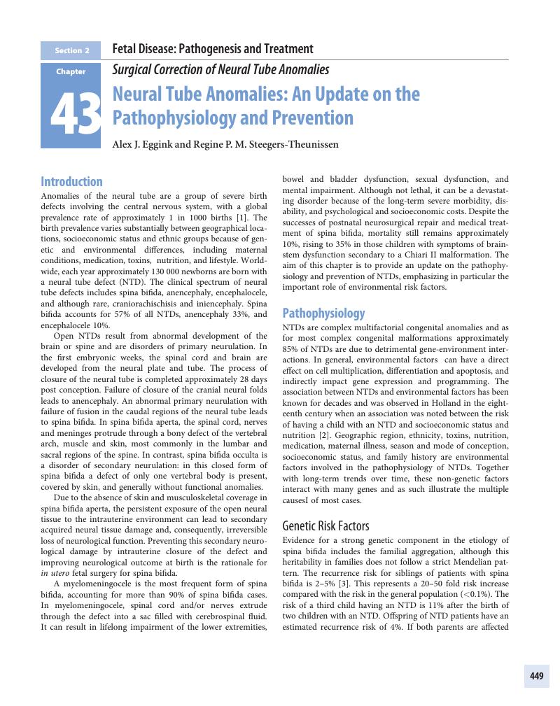
Image of the first page of this content. For PDF version, please use the ‘Save PDF’ preceeding this image.'
Type
Chapter
Information
Fetal Therapy
Scientific Basis and Critical Appraisal of Clinical Benefits
, pp. 449 - 479
Publisher: Cambridge University Press
Print publication year: 2020

Access options

Get access to the full version of this content by using one of the access options below. (Log in options will check for institutional or personal access. Content may require purchase if you do not have access.)

References

References

Botto, LD, Moore, CA, Khoury, MJ, et al. Neural-tube defects. N Engl J Med. 1999; 341: 1509–19.CrossRefGoogle ScholarPubMed
Michie, CA. Neural tube defects in 18th century. Lancet. 1991; 337: 504.Google Scholar
Padmanabhan, R. Etiology, pathogenesis and prevention of neural tube defects. Congenit Anom (Kyoto). 2006; 46: 5567.CrossRefGoogle ScholarPubMed
Deak, KL, Siegel, DG, George, TM, et al. Further evidence for a maternal genetic effect and a sex-influenced effect contributing to risk for human neural tube defects. Birth Defects Res A Clin Mol Teratol 2008; 82: 662–9.Google Scholar
Wilde, JJ, Petersen, JR, Niswander, L. Genetic, epigenetic, and environmental contributions to neural tube closure. Annu Rev Genet. 2014; 48: 583611.CrossRefGoogle ScholarPubMed
Au, KS, Ashley-Koch, A, Northrup, H. Epidemiologic and genetic aspects of spina bifida and other neural tube defects. Dev Disabil Res Rev. 2010; 16: 615.CrossRefGoogle ScholarPubMed
Copp, AJ, Adzick, NS, Chitty, LS, et al. Spina bifida. Nat Rev Dis Primers. 2015; 1: 15007.CrossRefGoogle ScholarPubMed
Viswanathan, M, Treiman, KA, Doto, JK, et al. Folic Acid Supplementation: An Evidence Review for the U.S. Preventive Services Task Force: U.S. Preventive Services Task Force Evidence Syntheses. Rockville: Agency for Healthcare Research and Quality, 2017.Google Scholar
van der Put, NM, Steegers-Theunissen, RP, Frosst, P, et al. Mutated methylenetetrahydrofolate reductase as a risk factor for spina bifida. Lancet. 1995; 346: 1070–1.Google Scholar
Besser, LM, Williams, LJ, Cragan, JD. Interpreting changes in the epidemiology of anencephaly and spina bifida following folic acid fortification of the U.S. grain supply in the setting of long-term trends, Atlanta, Georgia, 1968–2003. Birth Defects Res A Clin Mol Teratol. 2007; 79: 730–6.CrossRefGoogle ScholarPubMed
Becerra, JE, Khoury, MJ, Cordero, JF, et al. Diabetes mellitus during pregnancy and the risks for specific birth defects: a population-based case-control study. Pediatrics. 1990; 85: 19.Google Scholar
Mitchell, LE, Adzick, NS, Melchionne, J, et al. Spina bifida. Lancet. 2004; 364: 1885–95.CrossRefGoogle ScholarPubMed
Groenen, PM, Peer, PG, Wevers, RA, et al. Maternal myo-inositol, glucose, and zinc status is associated with the risk of offspring with spina bifida. Am J Obstet Gynecol. 2003; 189: 1713–19.CrossRefGoogle ScholarPubMed
Carmichael, SL, Rasmussen, SA, Shaw, GM. Prepregnancy obesity: a complex risk factor for selected birth defects. Birth Defects Res A Clin Mol Teratol. 2010; 88: 804–10.Google Scholar
Parker, SE, Yazdy, MM, Tinker, SC, et al. The impact of folic acid intake on the association among diabetes mellitus, obesity, and spina bifida. Am J Obstet Gynecol. 2013; 209: 239. e1–8.Google Scholar
Desrosiers, TA, Siega-Riz, AM, Mosley, BS, et al. Low carbohydrate diets may increase risk of neural tube defects. Birth Defects Res. 2018; 110: 901–9.Google Scholar
Vujkovic, M, Steegers, EA, Looman, CW, et al. The maternal Mediterranean dietary pattern is associated with a reduced risk of spina bifida in the offspring. BJOG. 2009; 116: 408–15.Google Scholar
Suarez, L, Cardarelli, K, Hendricks, K. Maternal stress, social support, and risk of neural tube defects among Mexican Americans. Epidemiology. 2003; 14: 612–16.Google Scholar
Moretti, ME, Bar-Oz, B, Fried, S, et al. Maternal hyperthermia and the risk for neural tube defects in offspring: systematic review and meta-analysis. Epidemiology. 2005; 16: 216–19.CrossRefGoogle ScholarPubMed
Molloy, AM, Pangilinan, F, Brody, LC. Genetic risk factors for folate-responsive neural tube defects. Annu Rev Nutr. 2017; 37: 269–91.Google Scholar
Smithells, RW, Sheppard, S, Schorah, CJ, et al. Apparent prevention of neural tube defects by periconceptional vitamin supplementation. Arch Dis Child. 1981; 56: 911–18.CrossRefGoogle ScholarPubMed
MRC Vitamin Study Research Group. Prevention of neural tube defects: results of the Medical Research Council Vitamin Study. MRC Vitamin Study Research Group. Lancet. 1991; 338: 131–7.Google Scholar
Czeizel, AE, Dudas, I. Prevention of the first occurrence of neural-tube defects by periconceptional vitamin supplementation. N Engl J Med. 1992; 327: 1832–5.Google Scholar
Giardini, V, Russo, FM, Ornaghi, S, et al. Seasonal impact in the frequency of isolated spina bifida. Prenat Diagn. 2013; 33: 1007–9.Google Scholar
Ray, JG, Wyatt, PR, Thompson, MD, et al. Vitamin B12 and the risk of neural tube defects in a folic-acid-fortified population. Epidemiology. 2007; 18: 362–6.Google Scholar
Steegers-Theunissen, RP, Boers, GH, Trijbels, FJ, et al. Maternal hyperhomocysteinemia: a risk factor for neural-tube defects? Metabolism. 1994; 43: 1475–80.Google Scholar
Steegers-Theunissen, RP, Boers, GH, Trijbels, FJ, et al. Neural-tube defects and derangement of homocysteine metabolism. N Engl J Med. 1991; 324: 199200.Google Scholar
Yang, M, Li, W, Wan, Z, et al. Elevated homocysteine levels in mothers with neural tube defects: a systematic review and meta-analysis. J Matern Fetal Neonatal Med. 2017; 30: 2051–7.CrossRefGoogle ScholarPubMed
Shaw, GM, Finnell, RH, Blom, HJ, et al. Choline and risk of neural tube defects in a folate-fortified population. Epidemiology. 2009; 20: 714–19.Google Scholar
Greene, ND, Leung, KY, Copp, AJ. Inositol, neural tube closure and the prevention of neural tube defects. Birth Defects Res. 2017; 109: 6880.Google Scholar
Grewal, J, Carmichael, SL, Ma, C, et al. Maternal periconceptional smoking and alcohol consumption and risk for select congenital anomalies. Birth Defects Res A Clin Mol Teratol. 2008; 82: 519–26.CrossRefGoogle ScholarPubMed
Schmidt, RJ, Romitti, PA, Burns, TL, et al. Maternal caffeine consumption and risk of neural tube defects. Birth Defects Res A Clin Mol Teratol. 2009; 85: 879–89.Google Scholar
Vajda, FJ, Eadie, MJ. Maternal valproate dosage and foetal malformations. Acta Neurol Scand. 2005; 112: 137–43.Google Scholar
Padula, AM, Tager, IB, Carmichael, SL, et al. The association of ambient air pollution and traffic exposures with selected congenital anomalies in the San Joaquin Valley of California. Am J Epidemiol. 2013; 177: 1074–85.Google Scholar
Lupo, PJ, Symanski, E, Waller, DK, et al. Maternal exposure to ambient levels of benzene and neural tube defects among offspring: Texas, 1999-2004. Environ Health Perspect. 2011; 119: 397402.Google Scholar
Li, Z, Zhang, L, Ye, R, et al. Indoor air pollution from coal combustion and the risk of neural tube defects in a rural population in Shanxi Province, China. Am J Epidemiol. 2011; 174: 451–8.Google Scholar
Righi, E, Bechtold, P, Tortorici, D, et al. Trihalomethanes, chlorite, chlorate in drinking water and risk of congenital anomalies: a population-based case-control study in Northern Italy. Environ Res. 2012; 116: 6673.Google Scholar
Brender, JD, Felkner, M, Suarez, L, et al. Maternal pesticide exposure and neural tube defects in Mexican Americans. Ann Epidemiol. 2010; 20: 1622.Google Scholar
Honein, MA, Paulozzi, LJ, Mathews, TJ, et al. Impact of folic acid fortification of the US food supply on the occurrence of neural tube defects. JAMA. 2001; 285: 2981–6.Google Scholar
Williams, J, Mai, CT, Mulinare, J, et al. Updated estimates of neural tube defects prevented by mandatory folic acid fortification – United States, 1995–2011. MMWR Morb Mortal Wkly Rep. 2015; 64: 15.Google Scholar
De Wals, P, Tairou, F, Van Allen, MI, et al. Reduction in neural-tube defects after folic acid fortification in Canada. N Engl J Med. 2007; 357: 135–42.Google Scholar
Bentley, TG, Weinstein, MC, Willett, WC, et al. A cost-effectiveness analysis of folic acid fortification policy in the United States. Public Health Nutr. 2009; 12: 455–67.Google Scholar
Atta, CA, Fiest, KM, Frolkis, AD, et al. Global birth prevalence of spina bifida by folic acid fortification status: a systematic review and meta-analysis. Am J Public Health. 2016; 106: e24–34.Google Scholar
Mills, JL, Von Kohorn, I, Conley, MR, et al. Low vitamin B-12 concentrations in patients without anemia: the effect of folic acid fortification of grain. Am J Clin Nutr. 2003; 77: 1474–7.Google Scholar
Vollset, SE, Clarke, R, Lewington, S, et al. Effects of folic acid supplementation on overall and site-specific cancer incidence during the randomised trials: meta-analyses of data on 50,000 individuals. Lancet. 2013; 381: 1029–36.CrossRefGoogle Scholar
Tinker, SC, Hamner, HC, Qi, YP, et al. U.S. women of childbearing age who are at possible increased risk of a neural tube defect-affected pregnancy due to suboptimal red blood cell folate concentrations, National Health and Nutrition Examination Survey 2007 to 2012. Birth Defects Res A Clin Mol Teratol. 2015; 103: 517–26.Google Scholar
Centers for Disease Control and Prevention. Racial/ethnic differences in the birth prevalence of spina bifida – United States, 1995–2005. MMWR Morb Mortal Wkly Rep. 2009; 57: 1409–13.Google Scholar
Greene, ND, Leung, KY, Gay, V, et al. Inositol for prevention of neural tube defects: a pilot randomised controlled trial – CORRIGENDUM. Br J Nutr. 2016; 115: 1697.Google Scholar
Khoshnood, B, Loane, M, de Walle, H, et al. Long term trends in prevalence of neural tube defects in Europe: population based study. BMJ. 2015; 351: h5949.Google Scholar
Busby, A, Abramsky, L, Dolk, H, et al. Preventing neural tube defects in Europe: population based study. BMJ. 2005; 330: 574–5.CrossRefGoogle ScholarPubMed

References

Bowman, RM, McLone, DG, Grant, JA, et al. Spina bifida outcome: a 25-year prospective. Pediatr Neurosurg. 2001; 34: 114–20.Google Scholar
Shin, M, Kucik, JE, Siffel, C, et al. Improved survival among children with spina bifida in the United States. J Pediatrics. 2012; 161: 1132–7. e3.Google Scholar
Rintoul, NE, Sutton, LN, Hubbard, AM, et al. A new look at myelomeningoceles: Functional level, vertebral level, shunting and the implications for fetal intervention. Pediatrics. 2002; 109: 409–13.Google Scholar
Caldarelli, M, Di Rocco, C, La Marca, F. Shunt complications in the first postoperative year in children with meningomyelocele. Childs Nerv Syst. 1996; 12: 748–54.Google Scholar
McLone, DG, Dias, MS. The Chiari II malformation: cause and impact. Childs Nerv Sys. 2003; 19: 540–50.Google Scholar
Just, M, Schwarz, M, Ludwig, B, et al. Cerebral and spinal MR-findings in patients with postrepair myelomeningocele. Pediatr Radiol. 1990; 20: 262–6.Google Scholar
Mitchell, LE, Adzick, NS, Melchionne, J, et al. Spina bifida. Lancet. 2004; 364: 1885–95.Google Scholar
Cochrane, DD, Wilson, RD, Steinbok, P, et al. Prenatal spinal evaluation and functional outcome of patients born with myelomeningocele: information for improved prenatal counselling and outcome prediction. Fetal Diagn Ther. 1996; 11: 159–68.Google Scholar
Cass, AS, Luxenberg, M, Johnson, CF, et al. Incidence of urinary tract complications with myelomeningocele. Urology. 1985; 25: 374–8.Google Scholar
Drennan, JC. Foot deformities in myelomeningocele. In Thilos, HS, ed., Instruction Course Lectures. Park Ridge, IL: American Academy of Orthopedic Surgeons, 1991.Google Scholar
Oakeshott, P, Hunt, GM, Poulton, A, et al. Expectation of life and unexpected death in open spina bifida: a 40-year complete, nonselective, longitudinal cohort study. Dev Med Child Neurol. 2010; 52: 749–53.Google Scholar
Oakeshott, P, Hunt, GM. Long-term outcome in spina bifida. Br J Gen Pract. 2003; 53: 632–6.Google Scholar
Parker, SE, Mai, CT, Canfield, MA, et al. Updated national birth prevalence estimates for selected birth defects in the United States, 2004–2006. Birth Defects Res A Clin Mol Teratol. 2010; 88: 1008–16.CrossRefGoogle ScholarPubMed
Christianson, A, Modell, B, Howson, C. March of Dimes Global Report on Birth Defects: The Hidden Toll of Dying and Disabled Children. White Plains, NY: March of Dimes, 2006.Google Scholar
Centers for Disease Control and Prevention. Hospital stays, hospital charges, and in-hospital deaths among infants with selected birth defects – United States, 2003. MMWR Morb Mortal Wkly Rep. 2007; 56: 25–9.Google Scholar
Radcliff, E, Cassell, CHTanner, JP, et al. Hospital use, associated costs, and payer status for infants born with spina bifida. Birth Defects Res A Clin Mol Teratol. 2012; 94: 1044–53.Google Scholar
Ouyang, L, Grosse, SD, Armour, BS, et al. Health care expenditures of children and adults with spina bifida in a privately insured U.S. population. Birth Defects Res A Clin Mol Teratol. 2007; 79: 552–8.Google Scholar
Werner, EF, Han, CS, Burd, I, et al. Evaluating the cost-effectiveness of prenatal surgery for myelomeningocele: a decision analysis. Ultrasound Obstet Gynecol. 2012; 40: 158–64.Google Scholar
Michejda, M. Intrauterine treatment of spina bifida: primate model. Z Kinderchir. 1984; 39: 259–61.Google ScholarPubMed
Heffez, DS, Aryanpur, J, Rotellini, NA, et al. Intrauterine repair of experimental surgically created dysraphism. Neurosurgery. 1993; 32: 1005–10.Google Scholar
Heffez, DS, Aryanpur, J, Hutchins, GM, et al. The paralysis associated with myelomeningocele: clinical and experimental data implicating a preventable spinal cord injury. Neurosurgery 1990; 26: 987–92.Google Scholar
Meuli, M, Meuli-Simmen, C, Yingling, CD, et al. Creation of myelomeningocele in utero: a model of functional damage from spinal cord exposure in fetal sheep. J Pediatr Surg. 1995; 30: 1028–32.Google Scholar
Meuli, M, Meuli-Simmen, C, Hutchins, GM, et al. In utero surgery rescues neurological function at birth in sheep with spina bifida. Nat Med. 1995; 1: 342–7.Google Scholar
Paek, BW, Farmer, DL, Wilkinson, CC, et al. Hindbrain herniation develops in surgically created myelomeningocele but is absent after repair in fetal lambs. Am J Obstet Gynecol. 2000; 183: 1119–23.Google Scholar
Bouchard, S, Davey, MG, Rintoul, NE, et al. Correction of hindbrain herniation and anatomy of the vermis after in utero repair of myelomeningocele in sheep. J Pediatr Surg. 2003; 38: 451–8.CrossRefGoogle ScholarPubMed
Bruner, JP, Tulipan, NB, Richards, WO. Endoscopic coverage of fetal open myelomeningocele in utero. Am J Obstet Gynecol. 1997; 176: 256–7.Google Scholar
Adzick, NS, Sutton, LN, Crombleholme, TM, et al. Successful fetal surgery for spina bifida. Lancet. 1998; 352: 1675–6.Google Scholar
Tulipan, N, Hernanz-Schulman, M, Bruner, JP. Reduced hindbrain herniation after intrauterine myelomeningocele repair: a report of four cases. Pediatr Neurosurg. 1998; 29: 274–8.Google Scholar
Sutton, LN, Adzick, NS, Bilaniuk, LT, et al. Improvement in hindbrain herniation demonstrated by serial fetal magnetic resonance imaging following fetal surgery for myelomeningocele. JAMA. 1999; 282: 1826–31.CrossRefGoogle ScholarPubMed
Adzick, NS, Thom, EA, Spong, CY, et al. A randomized trial of prenatal versus postnatal repair of myelomeningocele. N Engl J Med. 2011; 364: 9931004. (Full study protocol available in the Supplementary Appendix at NEJM.org)Google Scholar
Tulipan, N, Wellons, JC III, Thom, EA, et al. Prenatal surgery for myelomeningocele and the need for cerebrospinal fluid shunt placement. J Neurosurg Pediatr. 2015; 16: 613–20.Google Scholar
Farmer, DL, Thom, EA, Brock, JW, et al. The Management of the Myelomeningocele Study: Full cohort 30 month pediatric outcomes. Am J Obstet Gyn. 2018; 218: 256. e1–13.Google Scholar
Brock, JW III, Carr, MC, Adzick, NS, et al. Bladder function after fetal surgery for myelomeningocele. Pediatrics. 2015; 136: e906–13.Google Scholar
Johnson, MP, Bennett, KA, Rand, L, et al. The Management of Myelomeningocele Study: obstetrical outcomes and risk factors for obstetrical complications following prenatal surgery. Am J Obstet Gyn. 2016; 215: 778–9.Google Scholar
Antiel, RM, Adzick, NS, Thom, EA, et al. Impact on family and parental stress of prenatal versus postnatal repair of myelomeningocele. Am J Obstet Gyn. 2016; 215: 522. e1–6.Google Scholar
Moldenhauer, JS, Soni, S, Rintoul, NE, et al. Fetal myelomeningocele repair: the post-MOMS experience at the Children’s Hospital of Philadelphia. Fetal Diagn Ther. 2015; 37: 235–40.Google Scholar
Soni, S, Moldenhauer, JS, Spinner, SS, et al. Chorioamniotic membrane separation and preterm premature rupture of membranes complicating in utero myelomeningocele repair. Am J Obstet Gynecol. 2016; 214: 647. e1–7.Google Scholar
Wilson, RD, Johnson, MP, Crombleholme, TM, et al. Chorioamniotic membrane separation following open fetal surgery: pregnancy outcome. Fetal Diagn Ther. 2003; 18: 314–20.CrossRefGoogle ScholarPubMed
Heuer, GG, Adzick, NS, Sutton, LN. Fetal myelomeningocele closure: technical considerations. Fetal Diagn Ther. 2015; 37: 166–71.Google Scholar
Flanders, TM, Heuer, GG, Madsen, PJ, et al. Modified myofascial technique for open fetal myelomeningocele results in improved outcomes. Presented at the 88th meeting of the American Association of Neurological Surgery, New Orleans, LA, May 1, 2018.Google Scholar
American College of Obstetricians and Gynecologists. ACOG Committee Opinion No. 550: Maternal-fetal surgery for myelomeningocele. Obstet Gynecol. 2013; 121: 218–19.Google Scholar
Simpson, JL, Greene, MF. Fetal surgery for myelomeningocele? N Engl J Med. 2011; 364: 1076–7.Google Scholar
Cohen, AR, Couto, J, Cummings, JJ, et al. Position statement on fetal myelomeningocele repair. Am J Obstet Gynecol. 2014; 210: 107–11.Google Scholar
Bennett, KA, Carroll, MA, Shannon, CN, et al. Reducing perinatal complications and preterm delivery for patients undergoing in utero closure of fetal myelomeningocele: further modifications to the multidisciplinary surgical technique. J Neurosurg Pediatr. 2014; 14: 108–14.Google Scholar
Moise, KJ Jr, Moldenhauer, JS, Bennett, KA, et al. Current selection criteria and perioperative therapy used for fetal myelomeningocele surgery. Obstet Gynecol. 2016; 127: 593–7.Google Scholar
Thom, EA. Maternal reproductive outcomes after in-utero repair of myelomeningocele. Am J Obstet Gynecol. 2016; 214: S36.Google Scholar
Wilson, RD, Lemerand, K, Johnson, MP, et al. Reproductive outcomes in subsequent pregnancies after a pregnancy complicated by open maternal-fetal surgery (1996–2007). Am J Obstet Gynecol. 2010; 203: 209. e1–6.Google Scholar
Kohl, T. Percutaneous minimally invasive fetoscopic surgery for spina bifida aperta. Part I: surgical technique and perioperative outcome. Ultrasound Obstet Gynecol. 2014; 44: 515–24.Google Scholar
Degenhardt, J, Schürg, R, Winarno, A, et al. Percutaneous minimal-access fetoscopic surgery for spina bifida aperta. Part II: maternal management and outcome. Ultrasound Obstet Gynecol. 2014; 44: 525–31.CrossRefGoogle ScholarPubMed
Graf, K, Kohl, T, Neubauer, BA, et al. Percutaneous minimally invasive fetoscopic surgery for spina bifida aperta – Part III: postnatal neurosurgical interventions in the first year of life. Ultrasound Obstet Gynecol. 2016; 47: 158–61.Google Scholar
Flake, AW. Percutaneous minimal-access fetoscopic surgery for myelomeningocele – not so minimal! Ultrasound Obstet Gynecol. 2014; 44: 499500.Google Scholar
Pedreira, DA, Zanon, N, Nishikuni, K, et al. Endoscopic surgery for the antenatal treatment of myelomeningocele: the CECAM trial. Am J Obstet Gynecol. 2016; 214: 111.e1111.e11.Google Scholar
Belfort, MA, Whitehead, WE, Shamshirsaz, AA, et al. Fetoscopic open neural tube defect repair: Development and refinement of a two-port, carbon dioxide insufflation technique. Obstet Gynecol. 2017; 129: 734–43.Google Scholar
Luks, FI, Deprest, J, Marcus, M, et al. Carbon dioxide pneumoamnios causes acidosis in fetal lamb. Fetal Diagn Ther. 1994; 9: 105–9.Google Scholar
Skinner, S, DeKoninck, P, Crossley, K, et al. Partial amniotic carbon dioxide insufflation for fetal surgery. Prenat Diagn. 2018; 38: 983–93.Google ScholarPubMed
Lawrence, KM, Rossidis, AC, Baumgarten, HD et al. Safety of prolonged intra-amniotic carbon dioxide insufflation in a fetal sheep model. Presented at the 49th meeting of the American Pediatric Surgical Association, Palm Desert, CA, May 4, 2018.Google Scholar
Joyeux, L, Engels, AC, Russo, FM, et al. Fetoscopic versus open repair for spina bifida aperta: a systematic review of outcomes. Fetal Diagn Ther. 2016; 39: 161–71.Google Scholar
Araujo Junior, E, Eggink, AJ, van den Dobbelsteen, J, et al. Procedure-related complications of open vs. endoscopic fetal surgery for treatment of spina bifida in an era of intrauterine myelomeningocele repair: systematic review and meta-analysis. Ultrasound Obstet Gynecol. 2016; 48: 151–60.Google Scholar
Kabagambe, SK, Jensen, GW, Chen, YJ, et al. Fetal surgery for myelomeningocele: a systematic review and meta-analysis of outcomes in fetoscopic versus open repair. Fetal Diagn Ther. 2018; 43: 161–74.Google Scholar
Watanabe, M, Kim, AG, Flake, AW. Tissue engineering strategies for fetal myelomeningocele repair in animal models. Fetal Diagn Ther. 2015; 37: 197205.Google Scholar
Brown, EG, Saadai, P, Pivetti, CD, et al. In utero repair of myelomeningocele with autologous amniotic membrane in the fetal lamb model. J Pediatr Surg. 2014; 49: 133–8.Google Scholar

References

Williams, J, Mai, CT, Mulinare, J, et al. Updated estimates of neural tube defects prevented by mandatory folic acid fortification – United States, 1995–2011. MMWR Morb Mortal Wkly Rep. 2015; 64: 15.Google ScholarPubMed
Bowman, RM, Boshnjaku, V, McLone, DG. The changing incidence of myelomeningocele and its impact on pediatric neurosurgery: a review from the Children’s Memorial Hospital. Childs Nerv Syst. 2009; 25: 801–6.Google Scholar
Meuli, M, Meuli-Simmen, C, Yingling, CD, et al. In utero repair of experimental myelomeningocele saves neurological function at birth. J Pediatr Surg. 1996; 31: 397402.Google Scholar
Paek, BW, Farmer, DL, Wilkinson, CC, et al. Hindbrain herniation develops in surgically created myelomeningocele but is absent after repair in fetal lambs. Am J Obstet Gynecol. 2000; 183: 1119–23.Google Scholar
Tulipan, N, Bruner, JP. Myelomeningocele repair in utero: a report of three cases. Pediatr Neurosurg. 1998;28: 177–80.Google Scholar
Adzick, NS, Thom, EA, Spong, CY, et al. A randomized trial of prenatal versus postnatal repair of myelomeningocele. N Engl J Med. 2011; 364: 9931004.CrossRefGoogle ScholarPubMed
Farmer, DL, Thom, EA, Brock, JW 3rd, Burrows, PK, Johnson, MP, Howell, LJ, et al. The Management of Myelomeningocele Study: full cohort 30-month pediatric outcomes. Am J Obstet Gynecol. 2018; 218: 256. e1–256. e13.Google Scholar
Brock, JW 3rd, Carr, MC, Adzick, NS, Burrows, PK, Thomas, JC, Thom, EA, et al. Bladder function after fetal surgery for myelomeningocele. Pediatrics. 2015; 136: e906–13.CrossRefGoogle ScholarPubMed
Bruner, JP, Tulipan, NE, Richards, WO. Endoscopic coverage of fetal open myelomeningocele in utero. Am J Obstet Gynecol. 1997; 176: 256–7.Google Scholar
Bruner, JP, Richards, WO, Tulipan, NB, Arney, TL. Endoscopic coverage of fetal myelomeningocele in utero. Am J Obstet Gynecol. 1999; 180: 153–8.Google Scholar
Farmer, DL, von Koch, CS, Peacock, WJ, et al. In utero repair of myelomeningocele: experimental pathophysiology, initial clinical experience, and outcomes. Arch Surg. 2003; 138: 872–8.Google Scholar
Bruner, JP, Tulipan, NB, Richards, WO, Walsh, WF, Boehm, FH, Vrabcak, EK. In utero repair of myelomeningocele: a comparison of endoscopy and hysterotomy. Fetal Diagn Ther. 2000; 15: 83–8.Google Scholar
Kohl, T, Hering, R, Heep, A, et al. Percutaneous fetoscopic patch coverage of spina bifida aperta in the human – early clinical experience and potential. Fetal Diagn Ther. 2006; 21: 185–93.Google Scholar
Kohl, T. Percutaneous minimally invasive fetoscopic surgery for spina bifida aperta. Part I: Surgical technique and perioperative outcome. Ultrasound Obstet Gynecol. 2014; 44: 515–24.Google Scholar
Degenhardt, J, Schurg, R, Winarno, A, et al. Percutaneous minimal-access fetoscopic surgery for spina bifida aperta. Part II: maternal management and outcome. Ultrasound Obstet Gynecol. 2014; 44: 525–31.Google Scholar
Graf, K, Kohl, T, Neubauer, BA, et al. Percutaneous minimally invasive fetoscopic surgery for spina bifida aperta. Part III: neurosurgical intervention in the first postnatal year. Ultrasound Obstet Gynecol. 2016; 47: 158–61.Google Scholar
Ziemann, M, Fimmers, R, Khaleeva, A, Schürg, R, Weigand, MA, Kohl, T. Partial amniotic carbon dioxide insufflation (PACI) during minimally invasive fetoscopic interventions on fetuses with spina bifida aperta. Surg Endosc. 2018; 32: 3138–48.CrossRefGoogle ScholarPubMed
Pedreira, DA, Zanon, N, de Sa, RA, et al. Fetoscopic single-layer repair of open spina bifida using a cellulose patch: preliminary clinical experience. J Mat Fetal Neonat Med. 2014; 27: 1613–19.Google Scholar
Pedreira, DA, Zanon, N,Nishikuni, K, et al. Endoscopic surgery for the antenatal treatment of myelomeningocele: the CECAM trial. Am J Obstet Gynecol. 2016; 214: 111. e1–111. e11.Google Scholar
Lapa Pedreira, DA, Acacio, GL, Gonçalves, RT, , RAM, Brandt, RA, Chmait, R, et al. Percutaneous fetoscopic closure of large open spina bifida using a bilaminar skin substitute. Ultrasound Obstet Gynecol. 2018; 52: 458–66.Google Scholar
Belfort, M, Deprest, J, Hecher, K. Current controversies in prenatal diagnosis 1: in utero therapy for spina bifida is ready for endoscopic repair. Prenat Diagn. 2016; 36: 1161–6.Google Scholar
Peiro, JL, Fontecha, CG, Ruano, R, Esteves, M, Fonseca, C, Marotta, M, et al. Single-access fetal endoscopy (SAFE) for myelomeningocele in sheep model I: amniotic carbon dioxide gas approach. Surg Endosc. 2013; 27: 3835–40.Google Scholar
Belfort, MA, Whitehead, WE, Bednov, A, Shamshirsaz, AA. Low-fidelity simulator for the standardized training of fetoscopic meningomyelocele repair. Obstet Gynecol. 2018; 131: 125–9.Google Scholar
Belfort, MA, Whitehead, WE, Shamshirsaz, AA, et al. Fetoscopic repair of meningomyelocele. Obstet Gynecol. 2015; 126: 881–4.Google Scholar
Pham, P. (2018). Sophisticated tools lead to breakthroughs in prenatal surgery. Wired. www.wired.com/story/nicu-tools-fetal-surgeryGoogle Scholar
Belfort, MA, Whitehead, WE, Shamshirsaz, AA, et al. Fetoscopic open neural tube defect repair: development and refinement of a two-port, carbon dioxide insufflation technique. Obstet Gynecol. 2017; 129: 734–43.Google Scholar
Kohn, JR, Rao, V, Sellner, AA, Sharhan, D, Espinoza, J, Shamshirsaz, AA, et al. Management of labor and delivery after fetoscopic repair of an open neural tube defect. Obstet Gynecol. 2018; 131: 1062–8.Google Scholar
Sanz Cortes, M, Torres, O, Sharhan, D, Yepez, M, Espinoza, J, Shamshirsaz, AA, et al. Neurodevelopmental assessment in patients who underwent prenatal fetoscopic and open fetal neural tube defect repair. Am J Obstet Gynecol. 2018; 1 (Suppl.): S294–5.Google Scholar
Kohl, T, Reckers, J, Strümper, D, Große Hartlage, M, Gogarten, W, Gembruch, U, et al. Amniotic air insufflation during minimally invasive fetoscopic fetal cardiac interventions is safe for the fetal brain in sheep. J Thorac Cardiovasc Surg. 2004; 128: 467–71.Google Scholar
Moise, KJ Jr., Tsao, K, Papanna, RM, Bebbington, MW. Fetoscopic repair of meningomyelocele. Obstet Gynecol. 2015; 126: 674.Google Scholar
Moise, KJ Jr., Flake, A. Fetoscopic open neural tube defect repair: development and refinement of a two-port, carbon dioxide insufflation technique. Obstet Gynecol. 2017; 130: 648.Google Scholar
Luks, FI, Deprest, J, Marcus, M, Vandenberghe, K, Vertommen, JD, Lerut, T, et al. Carbon dioxide pneumoamnios causes acidosis in fetal lamb. Fetal Diagn Ther. 1994; 9: 105–9.Google Scholar
Gratacós, E, Wu, J, Devlieger, R, Van de Velde, M, Deprest, JA. Effects of amniodistension with carbon dioxide on fetal acid–base status during fetoscopic surgery in the sheep model. Surg Endosc. 2001; 15: 368–72.Google Scholar
Gardner, DS, Fletcher, AJ, Bloomfield, MR, Fowden, AL, Giussani, DA. Effects of prevailing hypoxaemia, acidaemia or hypoglycaemia upon the cardiovascular, endocrine and metabolic responses to acute hypoxaemia in the ovine fetus. J Physiol. 2002; 540: 351–66.Google Scholar
Sabik, JF, Assad, RS, Hanley, FL. Halothane as an anesthetic for fetal surgery. J Pediatr Surg. 1993; 28: 542–6.Google Scholar
Saiki, Y, Litwin, DE, Bigras, JL, Waddell, J, Konig, A, Baik, S, et al. Reducing the deleterious effects of intrauterine CO2 during fetoscopic surgery. J Surg Res. 1997; 69: 51–4.Google Scholar
Akbar, SA, Brown, PR. Human erythrocyte CAI and CAII isoenzymes in hypoxemic and anemic fetuses. Clin Biochem. 1996; 29: 5762.Google Scholar
Lönnerholm, G, Wistrand, PJ. Carbonic anhydrase in the human fetal kidney. Pediatr Res. 1983; 17: 390–7.Google Scholar
Supuran, CT. Structure and function of carbonic anhydrases. Biochem J. 2016; 473: 2023–32.Google Scholar
Sly, WS, Hu, PY. Human carbonic anhydrases and carbonic anhydrase deficiencies. Annu Rev Biochem. 1995; 64: 375401.Google Scholar
Schipain, E, Mangiavini, L, Merceron, C. HIF-1a and growth plate development: what we really know. Bonekey Rep. 2015; 4: 730.Google Scholar
Thakor, AS, Giussani, DA. Effects of acute acidemia on the fetal cardiovascular defense to acute hypoxemia. Am J Physiol Regul Integr Comp Physiol. 2009; 296: R90–9.Google Scholar
Kohl, T. Impact of partial amniotic carbon dioxide insufflation (PACI) on middle cerebral artery blood flow in mid-gestation human fetuses undergoing fetoscopic surgery for spina bifida aperta. Ultrasound Obstet Gynecol. 2016; 47: 521–2.Google Scholar
Kassir, E, Belfort, MA, Shamshirsaz, AA, et al. Doppler changes in umbilical artery and ductus venosus during fetoscopic prenatal surgical repair of myelomeningocele. Ultrasound Obstet Gynecol. 2019; 53: 335–9.Google Scholar
Mann, DG, Nassr, AA, Whitehead, WE, Espinoza, J, Belfort, MA, Shamshirsaz, AA. Fetal bradycardia associated with maternal hypothermia after fetoscopic repair of neural tube defect. Ultrasound Obstet Gynecol. 2018; 51: 411–12.Google Scholar
Dean, M, Ramsay, R, Heriot, A, Mackay, J, Hiscock, R, Lynch, AC. Warmed, humidified CO2 insufflation benefits intraoperative core temperature during laparoscopic surgery: a meta-analysis. Asian J Endosc Surg. 2017; 10: 128–36.Google Scholar
Baschat, AA, Ahn, ES, Murphy, J, Miller, JL. Fetal blood gas values during fetoscopic myelomeningocele repair performed under carbon dioxide insufflation. Ultrasound Obstet Gynecol. 2018; 52: 400–2.Google Scholar
Partridge, EA, Davey, MG, Hornick, MA, McGovern, PE, Mejaddam, AY, Vrecenak, JD, et al. An extra-uterine system to physiologically support the extreme premature lamb. Nat Commun. 2017; 8: 15112.CrossRefGoogle ScholarPubMed
Davey, AK, Hayward, J, Marshall, JK, Woods, AE. The effects of insufflation conditions on rat mesothelium. Int J Inflam. 2013; 2013: 816283.Google Scholar
Peng, Y, Zheng, M, Ye, Q, Chen, X, Yu, B, Liu, B. Heated and humidified CO2 prevents hypothermia, peritoneal injury, and intra-abdominal adhesions during prolonged laparoscopic insufflations. J Surg Res. 2009; 151: 40–7.CrossRefGoogle ScholarPubMed
Sanz Cortes, M, Castro, E, Sharhan, D, Torres, P, Yepez, M, Espinoza, J, Shamshirsaz, AA, Nassr, AA, Popek, E, Whitehead, W, Belfort, MA. Amniotic membrane and placental histopathological findings after open and fetoscopic prenatal neural tube defect repair. Prenat Diagn. 2019 Mar; 39(4): 269279.Google Scholar
Erikoglu, M, Yol, S, Avunduk, MC, Erdemli, E, Can, A. Electron-microscopic alterations of the peritoneum after both cold and heated carbon dioxide pneumoperitoneum. J Surg Res. 2005; 125: 73–7.Google Scholar
Papanna, R, Mann, LK, Moise, KJ Jr., Kyriakides, T, Johnson, A, Garcia, E, et al. Histologic changes of the fetal membranes after fetoscopic laser surgery for twin-twin transfusion syndrome. Pediatr Res. 2015; 78: 247–55.CrossRefGoogle ScholarPubMed

Save book to Kindle

To save this book to your Kindle, first ensure coreplatform@cambridge.org is added to your Approved Personal Document E-mail List under your Personal Document Settings on the Manage Your Content and Devices page of your Amazon account. Then enter the ‘name’ part of your Kindle email address below. Find out more about saving to your Kindle.

Note you can select to save to either the @free.kindle.com or @kindle.com variations. ‘@free.kindle.com’ emails are free but can only be saved to your device when it is connected to wi-fi. ‘@kindle.com’ emails can be delivered even when you are not connected to wi-fi, but note that service fees apply.

Find out more about the Kindle Personal Document Service.

Available formats
×

Save book to Dropbox

To save content items to your account, please confirm that you agree to abide by our usage policies. If this is the first time you use this feature, you will be asked to authorise Cambridge Core to connect with your account. Find out more about saving content to Dropbox.

Available formats
×

Save book to Google Drive

To save content items to your account, please confirm that you agree to abide by our usage policies. If this is the first time you use this feature, you will be asked to authorise Cambridge Core to connect with your acco