
Book contents
- Pediatric Head and Neck Pathology
- Pediatric Head and Neck Pathology
- Copyright page
- Contents
- Contributors
- Preface
- Acknowledgments
- Chapter 1 Diagnostic Methods and Specimen Handling Techniques in Pediatric Surgical Pathology
- Chapter 2 Soft Tissue Tumors and Reactive and Inflammatory Lesions of the Oral Cavity and Head and Neck
- Chapter 3 Cutaneous Tumors and Pseudotumors of the Head and Neck
- Chapter 4 Mucocutaneous Pigmented Lesions, Nevi and Melanoma
- Chapter 5 Vesiculo-Erosive and Ulcerative Lesions of Oral Cavity and Skin
- Chapter 6 Oral Epithelial Neoplasms
- Chapter 7 Odontogenic and Non-Odontogenic Cysts
- Chapter 8 Odontogenic Tumors
- Chapter 9 Non-Neoplastic Diseases of Salivary Glands
- Chapter 10 Benign Neoplasms of Salivary Glands
- Chapter 11 Malignant Neoplasm of Salivary Glands
- Chapter 12 Non-Neoplastic Disorders of the Nasal Cavity, Paranasal Sinuses and Nasopharynx
- Chapter 13 Benign Neoplasms of the Nasal Cavity, Paranasal Sinuses and Nasopharynx
- Chapter 14 Malignant Neoplasms of the Nasal Cavity, Paranasal Sinuses and Nasopharynx
- Chapter 15 Disorders of the Larynx and Hypopharynx
- Chapter 16 Neoplasms of Bone and Fibro-Osseous Lesions of the Craniofacial Skeleton
- Chapter 17 Disorders of the Ear
- Chapter 18 Disorders of the Thyroid Gland and Parathyroid Glands
- Chapter 19 Reactive and Malignant Diseases of Hematopoietic and Lymphoid Tissues
- Chapter 20 Developmental and Syndromic Disturbances of the Craniofacial Region
- Index
- References
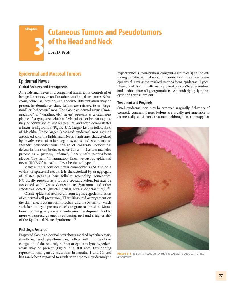
Chapter 3 - Cutaneous Tumors and Pseudotumors of the Head and Neck
Published online by Cambridge University Press: 26 June 2017

Book contents
- Pediatric Head and Neck Pathology
- Pediatric Head and Neck Pathology
- Copyright page
- Contents
- Contributors
- Preface
- Acknowledgments
- Chapter 1 Diagnostic Methods and Specimen Handling Techniques in Pediatric Surgical Pathology
- Chapter 2 Soft Tissue Tumors and Reactive and Inflammatory Lesions of the Oral Cavity and Head and Neck
- Chapter 3 Cutaneous Tumors and Pseudotumors of the Head and Neck
- Chapter 4 Mucocutaneous Pigmented Lesions, Nevi and Melanoma
- Chapter 5 Vesiculo-Erosive and Ulcerative Lesions of Oral Cavity and Skin
- Chapter 6 Oral Epithelial Neoplasms
- Chapter 7 Odontogenic and Non-Odontogenic Cysts
- Chapter 8 Odontogenic Tumors
- Chapter 9 Non-Neoplastic Diseases of Salivary Glands
- Chapter 10 Benign Neoplasms of Salivary Glands
- Chapter 11 Malignant Neoplasm of Salivary Glands
- Chapter 12 Non-Neoplastic Disorders of the Nasal Cavity, Paranasal Sinuses and Nasopharynx
- Chapter 13 Benign Neoplasms of the Nasal Cavity, Paranasal Sinuses and Nasopharynx
- Chapter 14 Malignant Neoplasms of the Nasal Cavity, Paranasal Sinuses and Nasopharynx
- Chapter 15 Disorders of the Larynx and Hypopharynx
- Chapter 16 Neoplasms of Bone and Fibro-Osseous Lesions of the Craniofacial Skeleton
- Chapter 17 Disorders of the Ear
- Chapter 18 Disorders of the Thyroid Gland and Parathyroid Glands
- Chapter 19 Reactive and Malignant Diseases of Hematopoietic and Lymphoid Tissues
- Chapter 20 Developmental and Syndromic Disturbances of the Craniofacial Region
- Index
- References
Summary
A summary is not available for this content so a preview has been provided. Please use the Get access link above for information on how to access this content.
- Type
- Chapter
- Information
- Pediatric Head and Neck Pathology , pp. 77 - 99Publisher: Cambridge University PressPrint publication year: 2016
References
Primary Sources
Brandling-Bennett, HA, Morel, KD. Epidermal nevi. Pediatr Clin North Am 2010; 57(5): 1177–98.Google Scholar
Khachemoune, A, Janjua, SA, Guldbakke, KK. Inflammatory linear verrucous epidermal nevus: a case report and short review of the literature. Cutis 2006; 78(4): 261–7.Google Scholar
Reddy, BS. Generalized epidermolytic hyperkeratosis in a child born to a parent with systematized epidermolytic linear epidermal nevus. Int J Dermatol 1997; 36(3): 198–200.Google Scholar
Idriss, MH, Elston, DM. Secondary neoplasms associated with nevus sebaceus of Jadassohn: a study of 707 cases. J Am Acad Dermatol 2014; 70(2): 332–7.CrossRefGoogle ScholarPubMed
Sellhyer, K, et al. Basaloid tumors in nevus sebaceus revisited: the follicular stem cell marker PHLDA1 (TDAG51) indicates that most are basal cell carcinomas and not trichoblastomas. J Cutan Pathol 2013; 40(5): 455–62.Google Scholar
Rankin, JS, Schwarts, RA. Accessory tragus: a possible sign of Goldenhar syndrome. Cutis 2011; 88(2): 62–4.Google Scholar
Songu, M, et al. White sponge nevus: clinical suspicion and diagnosis. Pediatr Dermatol 2012; 29(4): 495–7.Google Scholar
Castori, M, et al. Genetic skin diseases predisposing to basal cell carcinoma. Eur J Dermatol 2012; 22(3): 299–309.Google Scholar
Griffin, JR, et al. Basal cell carcinoma in childhood: case report and literature review. J Am Acad Dermatol 2007; 57(5): S97–102.Google Scholar
Newman, JC, Leffell, DJ. Correlation of embryonic fusion planes with the anatomical distribution of basal cell carcinoma. Dermatol Surg 2007; 33(8): 957–64.Google Scholar
Lam, C, Ou, JC, Billingsley, EM. “PTCH”-ing it together: a basal cell nevus syndrome review. Dermatol Surg 2013; 39(11): 1557–72.Google Scholar
Sellheyer, K, et al. The immunohistochemical differential diagnosis of microcystic adnexal carcinoma, desmoplastic trichoepithelioma and morpheaform basal cell carcinoma using BerEP4 and stem cell markers. J Cutan Pathol 2013; 40(4): 363–70.Google Scholar
Ali, FR, Lear, JT. Systemic treatments for basal cell carcinoma (BCC): the advent of dermato-oncology for BCC. Br J Dermatol 2013; 169(1): 53–7.Google Scholar
Secondary Sources
Brandling-Bennett, HA, Morel, KD. Epidermal nevi. Pediatr Clin North Am 2010; 57(5): 1177–98.Google Scholar
Khachemoune, A, Janjua, SA, Guldbakke, KK. Inflammatory linear verrucous epidermal nevus: a case report and short review of the literature. Cutis 2006; 78(4): 261–7.Google Scholar
Reddy, BS. Generalized epidermolytic hyperkeratosis in a child born to a parent with systematized epidermolytic linear epidermal nevus. Int J Dermatol 1997; 36(3): 198–200.Google Scholar
Idriss, MH, Elston, DM. Secondary neoplasms associated with nevus sebaceus of Jadassohn: a study of 707 cases. J Am Acad Dermatol 2014; 70(2): 332–7.CrossRefGoogle ScholarPubMed
Sellhyer, K, et al. Basaloid tumors in nevus sebaceus revisited: the follicular stem cell marker PHLDA1 (TDAG51) indicates that most are basal cell carcinomas and not trichoblastomas. J Cutan Pathol 2013; 40(5): 455–62.Google Scholar
Rankin, JS, Schwarts, RA. Accessory tragus: a possible sign of Goldenhar syndrome. Cutis 2011; 88(2): 62–4.Google Scholar
Songu, M, et al. White sponge nevus: clinical suspicion and diagnosis. Pediatr Dermatol 2012; 29(4): 495–7.Google Scholar
Castori, M, et al. Genetic skin diseases predisposing to basal cell carcinoma. Eur J Dermatol 2012; 22(3): 299–309.Google Scholar
Griffin, JR, et al. Basal cell carcinoma in childhood: case report and literature review. J Am Acad Dermatol 2007; 57(5): S97–102.Google Scholar
Newman, JC, Leffell, DJ. Correlation of embryonic fusion planes with the anatomical distribution of basal cell carcinoma. Dermatol Surg 2007; 33(8): 957–64.Google Scholar
Lam, C, Ou, JC, Billingsley, EM. “PTCH”-ing it together: a basal cell nevus syndrome review. Dermatol Surg 2013; 39(11): 1557–72.Google Scholar
Sellheyer, K, et al. The immunohistochemical differential diagnosis of microcystic adnexal carcinoma, desmoplastic trichoepithelioma and morpheaform basal cell carcinoma using BerEP4 and stem cell markers. J Cutan Pathol 2013; 40(4): 363–70.Google Scholar
Ali, FR, Lear, JT. Systemic treatments for basal cell carcinoma (BCC): the advent of dermato-oncology for BCC. Br J Dermatol 2013; 169(1): 53–7.Google Scholar
Brooke, JD, Fitzpatrick, JE, Golitz, LE. Papillary mesenchymal bodies: a histologic finding useful in differentiating trichoepitheliomas from basal cell carcinomas. J Am Acad Dermatol 1989; 21: 523–8.Google Scholar
Arits, AH, et al. Differentiation between basal cell carcinoma and trichoepithelioma by immunohistochemical staining of the androgen receptor: an overview. Eur J Dermatol 2011; 21(6): 870–3.Google Scholar
Afshar, M, Lee, RA, Jiang, SI. Desmoplastic trichilemmoma – a report of successful treatment with Mohs micrographic surgery and a review and update of the literature. Dermatol Surg 2012; 38(11): 1867–71.CrossRefGoogle Scholar
Misago, N, et al. A reevaluation of trichofolliculoma: the histopathological and immunohistochemical features. Am J Dermatopathol 2010; 32(1): 35–43.Google Scholar
Hassan, SF, et al. Characterizing pilomatricomas in children: a single institution experience. J Pediatr Surg 2013; 48(7): 1551–6.Google Scholar
Kazakov, DV, et al. Mutations in exon 3 of the CTNNB1 gene (beta-catenin gene) in cutaneous adnexal tumors. Am J Dermatopathol 2009; 31(3): 248–55.Google Scholar
Ghanadan, A, Khosravi, M. Cutaneous syringoma: a clinicopathologic study of 34 new cases and review of the literature. Indian J Dermatol 2013; 58(4): 326.CrossRefGoogle ScholarPubMed
Daneshpazhoo, M, et al. Mucocutaneous findings in 100 children with Down syndrome. Pediatr Dermatol 2007; 24(3): 317–20.Google Scholar
Chen, M, et al. Brooke-Spiegler syndrome associated with cylindroma, trichoepithelioma and eccrine spiradenoma. Int J Dermatol 2013; 52(12): 1602–4.Google Scholar
Liang, S, Liu, YH, Fang, K. Juvenile xanthogranuloma with ocular involvement. Pediatr Dermatol 2009; 26(2): 232–4.CrossRefGoogle ScholarPubMed
Ngendahayo, P, de Saint Aubain, N. Mitotically active xanthogranuloma: a case report with review of the literature. Am J Dermatopathol 2012; 34(3): 27–30.Google Scholar
Black, J, Coffin, CM, Dehner, LP. Fibrohistiocytic tumors and related neoplasms in children and adolescents. Pediatr Dev Pathol 2012; 15(1 Suppl): 181–210.CrossRefGoogle ScholarPubMed
West, KL, et al. Immunohistochemical markers in fibrohistiocytic lesions: factor XIIIa, CD34, S-100 and p75. Am J Dermatopathol 2014; 36(5): 414–9.Google Scholar
Doyle, LA, Fletcher, D. Metastasizing “benign” cutaneous fibrous histiocytoma: a clinicopathologic analysis of 16 cases. Am J Surg Pathol 2013; 37(4): 484–95.Google Scholar
Shmookler, BM, Enzinger, FM, Weiss, SW. Giant cell fibroblastoma. A juvenile form of dermatofibrosarcoma protuberans. Cancer 1989; 64(10): 2154–61.Google Scholar
Diwan, AH, et al. Dermatofibrosarcoma protuberans and giant cell fibroblastoma exhibit CD99 positivity. J Cutan Pathol 2008; 35(7): 647–50.Google Scholar
Nirvikalpa, N, Narayanan, V. Intraosseous infantile myofibroma of the mandible. Ann Maxillofac Surg 2011; 1(1): 87–90.Google Scholar
Mynatt, CJ, Feldman, KA, Thompson, LD. Orbital infantile myofibroma: a case report and clinicopathologic review of 24 cases from the literature. Head Neck Pathol 2011; 5(3): 205–15.Google Scholar
Garcia-Perla, A, et al. Upper airway distress due to an oropharyngeal infantile myofibroma. J Craniomaxillofac Surg 2012; 40(4): 112–4.Google Scholar
Friedman, BJ, et al. Congenital myofibroma masquerading as an ulcerated infantile hemangioma in a neonate. Pediatr Dermatol 2013; 30(6):248–9.Google Scholar
Bansal, C, Stewart, D, Li, A, Cockerell, CJ. Histologic variants of fibrous papule. J Cutan Pathol 2005; 32(6): 424–8.Google Scholar
Foster, RS, Bint, LJ, Halbert, AR. Topical 0.1% rapamycin for angiofibromas in paediatric patients with tuberous sclerosis: a pilot study of four patients. Australas J Dermatol 2012; 53(1): 52–6.Google Scholar
Seguier-Lipszyc, E, et al. Fibrous hamartoma of infancy. J Pediatr Surg 2011; 46(4): 753–5.Google Scholar
Marque, M, et al. Nevus anemicus in neurofibromatosis type 1: a potential new diagnostic criterion. J Am Acad Dermatol 2013; 69(5): 768–75.CrossRefGoogle ScholarPubMed
Szudek, J, Evans, DG, Friedman, JM. Patterns of associations of clinical features in neurofibromatosis 1. Hum Genet 2003; 112: 289–97.CrossRefGoogle ScholarPubMed
Sharma, DK, et al. Schwannomas of head and neck and review of literature. Indian J Otolaryngol Head Neck Surg 2012; 64(2): 177–80.Google Scholar
Fanburg-Smith, JC, Majidi, M, Miettinen, M. Keratin expression in schwannoma; a study of 115 retroperitoneal and 22 peripheral schwannomas. Mod Pathol Jan 2006; 19(1): 115–21.Google Scholar
Moore, RL, White, CR multiple palisaded encapsulated neuromas in a child without other associated abnormalities. J Am Acad Dermatol 2010; 62(2): 358–9.Google Scholar
Jokinen, CH, Ragsdale, BD, Argenyi, ZB. Expanding the clinicopathologic spectrum of palisaded encapsulated neuroma. J Cutan Pathol 2010; 37(1): 43–8.Google Scholar
Moak, Set al. Alar rim neurothekeoma in a child. J Craniofac Surg 2012; 23(6): 1914–7.Google Scholar
Zelger, BG, Steiner, H, Kutzner, H, Maier, H, Zelger, B. Cellular ‘neurothekeoma’: an epithelioid variant of dermatofibroma?. Histopathology 1998; 32(5): 414–22.Google Scholar
Fetsch, JF, Laskin, WB, Hallman, JR, Lupton, GP, Miettinen, M. Neurothekeoma: an analysis of 178 tumors with detailed immunohistochemical data and long-term patient follow-up information. Am J Surg Pathol Jul 2007; 31(7): 1103–14.Google Scholar
Suarez, A, High, WA. Immunohistochemical analysis of KBA.62 in 18 neurothekeomas: a potential marker for differentiating neurothekeoma, but a marker that may lead to confusion with melanocytic tumors. J Cutan Pathol 2014; 41(1): 36–41.Google Scholar
Bang, KO, Bodhade, AS, Dive, AM. Congenital granular cell epulis of a newborn. Dent Res J 2012; 9(Suppl 1): S136–8.Google Scholar
Henry, M, Perry, A. Multiple cutaneous granular cell tumors: case report of a 19-year-old African American female. J Cutan Med Surg 2011; 15(6): 344–6.Google Scholar
Rejas, RA, Campos, MS, Cortes, AR, Pinto, DD, de Sousa, SC. The neural histogenetic origin of the oral granular cell tumor: an immunohistochemical evidence. Med Oral Patol Oral Cir Bucal 2011; 16(1): 6–10.Google Scholar
Lazar, AJ, Fletcher, CD. Primitive nonneural granular cell tumors of skin: clinicopathologic analysis of 13 cases. Am J Surg Pathol 2005; 29(7): 927–34.Google Scholar
Rahbar, R, et al. Nasal glioma and encephalocele: diagnosis and management. Laryngoscope 2003; 113(12): 2069–77.Google Scholar
Kurban, Y, et al. Heterotopic brain tissue on the face and neck in a neonate: a rare case report and literature review. J Matern Fetal Neonatal Med 2013; 26(6): 619–21.Google Scholar
Comi, AM. Pathophysiology of Sturge-Weber syndrome. J Child Neurol 2003; 18(8): 509–16.Google Scholar
Sudarsanam, A, Ardern-Holmes, SL. Sturge-Weber syndrome: from the past to the present. Eur J Paediatr Neurol 2014; 18(3): 257–66.Google Scholar
Van Drooge, AM, et al. Port-wine stain progression: is prevention by pulsed dye laser therapy possible? Eur J Dermatol 2013; 23(2): 282–3.Google Scholar
Finn, SM, et al. The significance of cutaneous spider naevi in children. Arch Dis Child 2006; 91(7): 604–5.Google ScholarPubMed
Baglin, AC. Vascular tumors and pseudotumors. Pyogenic granuloma (lobular capillary hemangioma). Ann Pathol 2011; 31(4): 266–70.Google Scholar
Hoeger, PH, Colmenero, I. Vascular tumours in infants. Part I: benign vascular tumours other than infantile haemangioma. Br J Dermatol 2014; 171(3): 466–73.Google Scholar
Tritton, SM, et al. Pyogenic granuloma in ten children treated with topical imiquimod. Pediatr Dermatol 2009; 26(3): 269–72.Google Scholar
Huoh, KC, Rosbe, KW. Infantile hemangiomas of the head and neck. Pediatr Clin North Am 2013; 60(4): 937–49.Google Scholar
Kwon, EK, Seefeldt, M, Drolet, BA. Infantile hemangiomas: an update. Am J Clin Dermatol 2013; 14(2): 111–23.CrossRefGoogle ScholarPubMed
Orozco-Covarrubias, L, et al. Dermoid cysts: a report of 75 pediatric patients. Pediatr Dermatol 2013; 30(6): 706–11.CrossRefGoogle ScholarPubMed
Kurokawa, I, et al. Cutaneous dermoid cyst: cytokeratin and filaggrin expression suggesting differentiation towards follicular infundibulum and mature sebaceous gland. Oncol Rep 2006; 16(2): 295–9.Google Scholar
Wood, J, Couture, D, David, LR. Midline dermoid cyst resulting in frontal bone erosion. J Craniofac Surg 2012; 23(1): 131–4.Google Scholar
Park, JS, Ko, DK. A histopathologic study of epidermoid cysts in Korea: comparison between ruptured and unruptured epidermal cyst. Int J Clin Exp Pathol 2013; 6(2): 242–8.Google Scholar
Al-Khateeb, TH, Al-Masri, NM, Al-Zoubi, F. Cutaneous cysts of the head and neck. J Oral Maxillofac Surg 2009; 67(1): 52–7.Google Scholar
Thornsberry, LA, English, JC 3rd. Etiology, diagnosis, and therapeutic management of granuloma annulare: an update. Am J Clin Dermatol 2013; 14(4): 279–90.Google Scholar
Rakheja, D, et al. A subset of cranial fasciitis is associated with dysregulation of the Wnt/beta-catenin pathway. Mod Pathol 2008; 21(11): 1330–6.Google Scholar
Sarangarajan, R, Dehner, LP. Cranial and extracranial fasciitis of childhood: a clinicopathologic and immunohistochemical study. Hum Pathol 1999; 30(1): 87–92.Google Scholar
Alaiti, S, Nelson, FP, Ryoo, JW. Solitary cutaneous myxoma. J Am Acad Dermatol 2000; 43(2 Pt 2): 377–9.CrossRefGoogle ScholarPubMed
Martí, N. Multiple cutaneous angiomyxomas in a child. Pediatr Dermatol 2011; 28(4): 462–4.CrossRefGoogle ScholarPubMed
Bran, GM, et al. Keloids: current concepts of pathogenesis (review). Int J Mol Med 2009; 24(3): 283–93.Google Scholar
Abdou, AG, et al. Immunohistochemical evaluation of COX-1 and COX-2 expression in keloid and hypertrophic scar. Am J Dermatopathol 2014; 36(4): 311–17.Google Scholar
Brandling-Bennett, HA, Morel, KD. Epidermal nevi. Pediatr Clin North Am 2010; 57(5): 1177–98.Google Scholar
Khachemoune, A, Janjua, SA, Guldbakke, KK. Inflammatory linear verrucous epidermal nevus: a case report and short review of the literature. Cutis 2006; 78(4): 261–7.Google Scholar
Reddy, BS. Generalized epidermolytic hyperkeratosis in a child born to a parent with systematized epidermolytic linear epidermal nevus. Int J Dermatol 1997; 36(3): 198–200.Google Scholar
Idriss, MH, Elston, DM. Secondary neoplasms associated with nevus sebaceus of Jadassohn: a study of 707 cases. J Am Acad Dermatol 2014; 70(2): 332–7.CrossRefGoogle ScholarPubMed
Sellhyer, K, et al. Basaloid tumors in nevus sebaceus revisited: the follicular stem cell marker PHLDA1 (TDAG51) indicates that most are basal cell carcinomas and not trichoblastomas. J Cutan Pathol 2013; 40(5): 455–62.Google Scholar
Rankin, JS, Schwarts, RA. Accessory tragus: a possible sign of Goldenhar syndrome. Cutis 2011; 88(2): 62–4.Google Scholar
Songu, M, et al. White sponge nevus: clinical suspicion and diagnosis. Pediatr Dermatol 2012; 29(4): 495–7.Google Scholar
Castori, M, et al. Genetic skin diseases predisposing to basal cell carcinoma. Eur J Dermatol 2012; 22(3): 299–309.Google Scholar
Griffin, JR, et al. Basal cell carcinoma in childhood: case report and literature review. J Am Acad Dermatol 2007; 57(5): S97–102.Google Scholar
Newman, JC, Leffell, DJ. Correlation of embryonic fusion planes with the anatomical distribution of basal cell carcinoma. Dermatol Surg 2007; 33(8): 957–64.Google Scholar
Lam, C, Ou, JC, Billingsley, EM. “PTCH”-ing it together: a basal cell nevus syndrome review. Dermatol Surg 2013; 39(11): 1557–72.Google Scholar
Sellheyer, K, et al. The immunohistochemical differential diagnosis of microcystic adnexal carcinoma, desmoplastic trichoepithelioma and morpheaform basal cell carcinoma using BerEP4 and stem cell markers. J Cutan Pathol 2013; 40(4): 363–70.Google Scholar
Ali, FR, Lear, JT. Systemic treatments for basal cell carcinoma (BCC): the advent of dermato-oncology for BCC. Br J Dermatol 2013; 169(1): 53–7.Google Scholar
Brooke, JD, Fitzpatrick, JE, Golitz, LE. Papillary mesenchymal bodies: a histologic finding useful in differentiating trichoepitheliomas from basal cell carcinomas. J Am Acad Dermatol 1989; 21: 523–8.Google Scholar
Arits, AH, et al. Differentiation between basal cell carcinoma and trichoepithelioma by immunohistochemical staining of the androgen receptor: an overview. Eur J Dermatol 2011; 21(6): 870–3.Google Scholar
Afshar, M, Lee, RA, Jiang, SI. Desmoplastic trichilemmoma – a report of successful treatment with Mohs micrographic surgery and a review and update of the literature. Dermatol Surg 2012; 38(11): 1867–71.CrossRefGoogle Scholar
Misago, N, et al. A reevaluation of trichofolliculoma: the histopathological and immunohistochemical features. Am J Dermatopathol 2010; 32(1): 35–43.Google Scholar
Hassan, SF, et al. Characterizing pilomatricomas in children: a single institution experience. J Pediatr Surg 2013; 48(7): 1551–6.Google Scholar
Kazakov, DV, et al. Mutations in exon 3 of the CTNNB1 gene (beta-catenin gene) in cutaneous adnexal tumors. Am J Dermatopathol 2009; 31(3): 248–55.Google Scholar
Ghanadan, A, Khosravi, M. Cutaneous syringoma: a clinicopathologic study of 34 new cases and review of the literature. Indian J Dermatol 2013; 58(4): 326.CrossRefGoogle ScholarPubMed
Daneshpazhoo, M, et al. Mucocutaneous findings in 100 children with Down syndrome. Pediatr Dermatol 2007; 24(3): 317–20.Google Scholar
Chen, M, et al. Brooke-Spiegler syndrome associated with cylindroma, trichoepithelioma and eccrine spiradenoma. Int J Dermatol 2013; 52(12): 1602–4.Google Scholar
Liang, S, Liu, YH, Fang, K. Juvenile xanthogranuloma with ocular involvement. Pediatr Dermatol 2009; 26(2): 232–4.CrossRefGoogle ScholarPubMed
Ngendahayo, P, de Saint Aubain, N. Mitotically active xanthogranuloma: a case report with review of the literature. Am J Dermatopathol 2012; 34(3): 27–30.Google Scholar
Black, J, Coffin, CM, Dehner, LP. Fibrohistiocytic tumors and related neoplasms in children and adolescents. Pediatr Dev Pathol 2012; 15(1 Suppl): 181–210.CrossRefGoogle ScholarPubMed
West, KL, et al. Immunohistochemical markers in fibrohistiocytic lesions: factor XIIIa, CD34, S-100 and p75. Am J Dermatopathol 2014; 36(5): 414–9.Google Scholar
Doyle, LA, Fletcher, D. Metastasizing “benign” cutaneous fibrous histiocytoma: a clinicopathologic analysis of 16 cases. Am J Surg Pathol 2013; 37(4): 484–95.Google Scholar
Shmookler, BM, Enzinger, FM, Weiss, SW. Giant cell fibroblastoma. A juvenile form of dermatofibrosarcoma protuberans. Cancer 1989; 64(10): 2154–61.Google Scholar
Diwan, AH, et al. Dermatofibrosarcoma protuberans and giant cell fibroblastoma exhibit CD99 positivity. J Cutan Pathol 2008; 35(7): 647–50.Google Scholar
Nirvikalpa, N, Narayanan, V. Intraosseous infantile myofibroma of the mandible. Ann Maxillofac Surg 2011; 1(1): 87–90.Google Scholar
Mynatt, CJ, Feldman, KA, Thompson, LD. Orbital infantile myofibroma: a case report and clinicopathologic review of 24 cases from the literature. Head Neck Pathol 2011; 5(3): 205–15.Google Scholar
Garcia-Perla, A, et al. Upper airway distress due to an oropharyngeal infantile myofibroma. J Craniomaxillofac Surg 2012; 40(4): 112–4.Google Scholar
Friedman, BJ, et al. Congenital myofibroma masquerading as an ulcerated infantile hemangioma in a neonate. Pediatr Dermatol 2013; 30(6):248–9.Google Scholar
Bansal, C, Stewart, D, Li, A, Cockerell, CJ. Histologic variants of fibrous papule. J Cutan Pathol 2005; 32(6): 424–8.Google Scholar
Foster, RS, Bint, LJ, Halbert, AR. Topical 0.1% rapamycin for angiofibromas in paediatric patients with tuberous sclerosis: a pilot study of four patients. Australas J Dermatol 2012; 53(1): 52–6.Google Scholar
Seguier-Lipszyc, E, et al. Fibrous hamartoma of infancy. J Pediatr Surg 2011; 46(4): 753–5.Google Scholar
Marque, M, et al. Nevus anemicus in neurofibromatosis type 1: a potential new diagnostic criterion. J Am Acad Dermatol 2013; 69(5): 768–75.CrossRefGoogle ScholarPubMed
Szudek, J, Evans, DG, Friedman, JM. Patterns of associations of clinical features in neurofibromatosis 1. Hum Genet 2003; 112: 289–97.CrossRefGoogle ScholarPubMed
Sharma, DK, et al. Schwannomas of head and neck and review of literature. Indian J Otolaryngol Head Neck Surg 2012; 64(2): 177–80.Google Scholar
Fanburg-Smith, JC, Majidi, M, Miettinen, M. Keratin expression in schwannoma; a study of 115 retroperitoneal and 22 peripheral schwannomas. Mod Pathol Jan 2006; 19(1): 115–21.Google Scholar
Moore, RL, White, CR multiple palisaded encapsulated neuromas in a child without other associated abnormalities. J Am Acad Dermatol 2010; 62(2): 358–9.Google Scholar
Jokinen, CH, Ragsdale, BD, Argenyi, ZB. Expanding the clinicopathologic spectrum of palisaded encapsulated neuroma. J Cutan Pathol 2010; 37(1): 43–8.Google Scholar
Moak, Set al. Alar rim neurothekeoma in a child. J Craniofac Surg 2012; 23(6): 1914–7.Google Scholar
Zelger, BG, Steiner, H, Kutzner, H, Maier, H, Zelger, B. Cellular ‘neurothekeoma’: an epithelioid variant of dermatofibroma?. Histopathology 1998; 32(5): 414–22.Google Scholar
Fetsch, JF, Laskin, WB, Hallman, JR, Lupton, GP, Miettinen, M. Neurothekeoma: an analysis of 178 tumors with detailed immunohistochemical data and long-term patient follow-up information. Am J Surg Pathol Jul 2007; 31(7): 1103–14.Google Scholar
Suarez, A, High, WA. Immunohistochemical analysis of KBA.62 in 18 neurothekeomas: a potential marker for differentiating neurothekeoma, but a marker that may lead to confusion with melanocytic tumors. J Cutan Pathol 2014; 41(1): 36–41.Google Scholar
Bang, KO, Bodhade, AS, Dive, AM. Congenital granular cell epulis of a newborn. Dent Res J 2012; 9(Suppl 1): S136–8.Google Scholar
Henry, M, Perry, A. Multiple cutaneous granular cell tumors: case report of a 19-year-old African American female. J Cutan Med Surg 2011; 15(6): 344–6.Google Scholar
Rejas, RA, Campos, MS, Cortes, AR, Pinto, DD, de Sousa, SC. The neural histogenetic origin of the oral granular cell tumor: an immunohistochemical evidence. Med Oral Patol Oral Cir Bucal 2011; 16(1): 6–10.Google Scholar
Lazar, AJ, Fletcher, CD. Primitive nonneural granular cell tumors of skin: clinicopathologic analysis of 13 cases. Am J Surg Pathol 2005; 29(7): 927–34.Google Scholar
Rahbar, R, et al. Nasal glioma and encephalocele: diagnosis and management. Laryngoscope 2003; 113(12): 2069–77.Google Scholar
Kurban, Y, et al. Heterotopic brain tissue on the face and neck in a neonate: a rare case report and literature review. J Matern Fetal Neonatal Med 2013; 26(6): 619–21.Google Scholar
Comi, AM. Pathophysiology of Sturge-Weber syndrome. J Child Neurol 2003; 18(8): 509–16.Google Scholar
Sudarsanam, A, Ardern-Holmes, SL. Sturge-Weber syndrome: from the past to the present. Eur J Paediatr Neurol 2014; 18(3): 257–66.Google Scholar
Van Drooge, AM, et al. Port-wine stain progression: is prevention by pulsed dye laser therapy possible? Eur J Dermatol 2013; 23(2): 282–3.Google Scholar
Finn, SM, et al. The significance of cutaneous spider naevi in children. Arch Dis Child 2006; 91(7): 604–5.Google ScholarPubMed
Baglin, AC. Vascular tumors and pseudotumors. Pyogenic granuloma (lobular capillary hemangioma). Ann Pathol 2011; 31(4): 266–70.Google Scholar
Hoeger, PH, Colmenero, I. Vascular tumours in infants. Part I: benign vascular tumours other than infantile haemangioma. Br J Dermatol 2014; 171(3): 466–73.Google Scholar
Tritton, SM, et al. Pyogenic granuloma in ten children treated with topical imiquimod. Pediatr Dermatol 2009; 26(3): 269–72.Google Scholar
Huoh, KC, Rosbe, KW. Infantile hemangiomas of the head and neck. Pediatr Clin North Am 2013; 60(4): 937–49.Google Scholar
Kwon, EK, Seefeldt, M, Drolet, BA. Infantile hemangiomas: an update. Am J Clin Dermatol 2013; 14(2): 111–23.CrossRefGoogle ScholarPubMed
Orozco-Covarrubias, L, et al. Dermoid cysts: a report of 75 pediatric patients. Pediatr Dermatol 2013; 30(6): 706–11.CrossRefGoogle ScholarPubMed
Kurokawa, I, et al. Cutaneous dermoid cyst: cytokeratin and filaggrin expression suggesting differentiation towards follicular infundibulum and mature sebaceous gland. Oncol Rep 2006; 16(2): 295–9.Google Scholar
Wood, J, Couture, D, David, LR. Midline dermoid cyst resulting in frontal bone erosion. J Craniofac Surg 2012; 23(1): 131–4.Google Scholar
Park, JS, Ko, DK. A histopathologic study of epidermoid cysts in Korea: comparison between ruptured and unruptured epidermal cyst. Int J Clin Exp Pathol 2013; 6(2): 242–8.Google Scholar
Al-Khateeb, TH, Al-Masri, NM, Al-Zoubi, F. Cutaneous cysts of the head and neck. J Oral Maxillofac Surg 2009; 67(1): 52–7.Google Scholar
Thornsberry, LA, English, JC 3rd. Etiology, diagnosis, and therapeutic management of granuloma annulare: an update. Am J Clin Dermatol 2013; 14(4): 279–90.Google Scholar
Rakheja, D, et al. A subset of cranial fasciitis is associated with dysregulation of the Wnt/beta-catenin pathway. Mod Pathol 2008; 21(11): 1330–6.Google Scholar
Sarangarajan, R, Dehner, LP. Cranial and extracranial fasciitis of childhood: a clinicopathologic and immunohistochemical study. Hum Pathol 1999; 30(1): 87–92.Google Scholar
Alaiti, S, Nelson, FP, Ryoo, JW. Solitary cutaneous myxoma. J Am Acad Dermatol 2000; 43(2 Pt 2): 377–9.CrossRefGoogle ScholarPubMed
Martí, N. Multiple cutaneous angiomyxomas in a child. Pediatr Dermatol 2011; 28(4): 462–4.CrossRefGoogle ScholarPubMed
Bran, GM, et al. Keloids: current concepts of pathogenesis (review). Int J Mol Med 2009; 24(3): 283–93.Google Scholar
Abdou, AG, et al. Immunohistochemical evaluation of COX-1 and COX-2 expression in keloid and hypertrophic scar. Am J Dermatopathol 2014; 36(4): 311–17.Google Scholar
Brandling-Bennett, HA, Morel, KD. Epidermal nevi. Pediatr Clin North Am 2010; 57(5): 1177–98.Google Scholar
Khachemoune, A, Janjua, SA, Guldbakke, KK. Inflammatory linear verrucous epidermal nevus: a case report and short review of the literature. Cutis 2006; 78(4): 261–7.Google Scholar
Reddy, BS. Generalized epidermolytic hyperkeratosis in a child born to a parent with systematized epidermolytic linear epidermal nevus. Int J Dermatol 1997; 36(3): 198–200.Google Scholar
Idriss, MH, Elston, DM. Secondary neoplasms associated with nevus sebaceus of Jadassohn: a study of 707 cases. J Am Acad Dermatol 2014; 70(2): 332–7.CrossRefGoogle ScholarPubMed
Sellhyer, K, et al. Basaloid tumors in nevus sebaceus revisited: the follicular stem cell marker PHLDA1 (TDAG51) indicates that most are basal cell carcinomas and not trichoblastomas. J Cutan Pathol 2013; 40(5): 455–62.Google Scholar
Rankin, JS, Schwarts, RA. Accessory tragus: a possible sign of Goldenhar syndrome. Cutis 2011; 88(2): 62–4.Google Scholar
Songu, M, et al. White sponge nevus: clinical suspicion and diagnosis. Pediatr Dermatol 2012; 29(4): 495–7.Google Scholar
Castori, M, et al. Genetic skin diseases predisposing to basal cell carcinoma. Eur J Dermatol 2012; 22(3): 299–309.Google Scholar
Griffin, JR, et al. Basal cell carcinoma in childhood: case report and literature review. J Am Acad Dermatol 2007; 57(5): S97–102.Google Scholar
Newman, JC, Leffell, DJ. Correlation of embryonic fusion planes with the anatomical distribution of basal cell carcinoma. Dermatol Surg 2007; 33(8): 957–64.Google Scholar
Lam, C, Ou, JC, Billingsley, EM. “PTCH”-ing it together: a basal cell nevus syndrome review. Dermatol Surg 2013; 39(11): 1557–72.Google Scholar
Sellheyer, K, et al. The immunohistochemical differential diagnosis of microcystic adnexal carcinoma, desmoplastic trichoepithelioma and morpheaform basal cell carcinoma using BerEP4 and stem cell markers. J Cutan Pathol 2013; 40(4): 363–70.Google Scholar
Ali, FR, Lear, JT. Systemic treatments for basal cell carcinoma (BCC): the advent of dermato-oncology for BCC. Br J Dermatol 2013; 169(1): 53–7.Google Scholar
Brooke, JD, Fitzpatrick, JE, Golitz, LE. Papillary mesenchymal bodies: a histologic finding useful in differentiating trichoepitheliomas from basal cell carcinomas. J Am Acad Dermatol 1989; 21: 523–8.Google Scholar
Arits, AH, et al. Differentiation between basal cell carcinoma and trichoepithelioma by immunohistochemical staining of the androgen receptor: an overview. Eur J Dermatol 2011; 21(6): 870–3.Google Scholar
Afshar, M, Lee, RA, Jiang, SI. Desmoplastic trichilemmoma – a report of successful treatment with Mohs micrographic surgery and a review and update of the literature. Dermatol Surg 2012; 38(11): 1867–71.CrossRefGoogle Scholar
Misago, N, et al. A reevaluation of trichofolliculoma: the histopathological and immunohistochemical features. Am J Dermatopathol 2010; 32(1): 35–43.Google Scholar
Hassan, SF, et al. Characterizing pilomatricomas in children: a single institution experience. J Pediatr Surg 2013; 48(7): 1551–6.Google Scholar
Kazakov, DV, et al. Mutations in exon 3 of the CTNNB1 gene (beta-catenin gene) in cutaneous adnexal tumors. Am J Dermatopathol 2009; 31(3): 248–55.Google Scholar
Ghanadan, A, Khosravi, M. Cutaneous syringoma: a clinicopathologic study of 34 new cases and review of the literature. Indian J Dermatol 2013; 58(4): 326.CrossRefGoogle ScholarPubMed
Daneshpazhoo, M, et al. Mucocutaneous findings in 100 children with Down syndrome. Pediatr Dermatol 2007; 24(3): 317–20.Google Scholar
Chen, M, et al. Brooke-Spiegler syndrome associated with cylindroma, trichoepithelioma and eccrine spiradenoma. Int J Dermatol 2013; 52(12): 1602–4.Google Scholar
Liang, S, Liu, YH, Fang, K. Juvenile xanthogranuloma with ocular involvement. Pediatr Dermatol 2009; 26(2): 232–4.CrossRefGoogle ScholarPubMed
Ngendahayo, P, de Saint Aubain, N. Mitotically active xanthogranuloma: a case report with review of the literature. Am J Dermatopathol 2012; 34(3): 27–30.Google Scholar
Black, J, Coffin, CM, Dehner, LP. Fibrohistiocytic tumors and related neoplasms in children and adolescents. Pediatr Dev Pathol 2012; 15(1 Suppl): 181–210.CrossRefGoogle ScholarPubMed
West, KL, et al. Immunohistochemical markers in fibrohistiocytic lesions: factor XIIIa, CD34, S-100 and p75. Am J Dermatopathol 2014; 36(5): 414–9.Google Scholar
Doyle, LA, Fletcher, D. Metastasizing “benign” cutaneous fibrous histiocytoma: a clinicopathologic analysis of 16 cases. Am J Surg Pathol 2013; 37(4): 484–95.Google Scholar
Shmookler, BM, Enzinger, FM, Weiss, SW. Giant cell fibroblastoma. A juvenile form of dermatofibrosarcoma protuberans. Cancer 1989; 64(10): 2154–61.Google Scholar
Diwan, AH, et al. Dermatofibrosarcoma protuberans and giant cell fibroblastoma exhibit CD99 positivity. J Cutan Pathol 2008; 35(7): 647–50.Google Scholar
Nirvikalpa, N, Narayanan, V. Intraosseous infantile myofibroma of the mandible. Ann Maxillofac Surg 2011; 1(1): 87–90.Google Scholar
Mynatt, CJ, Feldman, KA, Thompson, LD. Orbital infantile myofibroma: a case report and clinicopathologic review of 24 cases from the literature. Head Neck Pathol 2011; 5(3): 205–15.Google Scholar
Garcia-Perla, A, et al. Upper airway distress due to an oropharyngeal infantile myofibroma. J Craniomaxillofac Surg 2012; 40(4): 112–4.Google Scholar
Friedman, BJ, et al. Congenital myofibroma masquerading as an ulcerated infantile hemangioma in a neonate. Pediatr Dermatol 2013; 30(6):248–9.Google Scholar
Bansal, C, Stewart, D, Li, A, Cockerell, CJ. Histologic variants of fibrous papule. J Cutan Pathol 2005; 32(6): 424–8.Google Scholar
Foster, RS, Bint, LJ, Halbert, AR. Topical 0.1% rapamycin for angiofibromas in paediatric patients with tuberous sclerosis: a pilot study of four patients. Australas J Dermatol 2012; 53(1): 52–6.Google Scholar
Seguier-Lipszyc, E, et al. Fibrous hamartoma of infancy. J Pediatr Surg 2011; 46(4): 753–5.Google Scholar
Marque, M, et al. Nevus anemicus in neurofibromatosis type 1: a potential new diagnostic criterion. J Am Acad Dermatol 2013; 69(5): 768–75.CrossRefGoogle ScholarPubMed
Szudek, J, Evans, DG, Friedman, JM. Patterns of associations of clinical features in neurofibromatosis 1. Hum Genet 2003; 112: 289–97.CrossRefGoogle ScholarPubMed
Sharma, DK, et al. Schwannomas of head and neck and review of literature. Indian J Otolaryngol Head Neck Surg 2012; 64(2): 177–80.Google Scholar
Fanburg-Smith, JC, Majidi, M, Miettinen, M. Keratin expression in schwannoma; a study of 115 retroperitoneal and 22 peripheral schwannomas. Mod Pathol Jan 2006; 19(1): 115–21.Google Scholar
Moore, RL, White, CR multiple palisaded encapsulated neuromas in a child without other associated abnormalities. J Am Acad Dermatol 2010; 62(2): 358–9.Google Scholar
Jokinen, CH, Ragsdale, BD, Argenyi, ZB. Expanding the clinicopathologic spectrum of palisaded encapsulated neuroma. J Cutan Pathol 2010; 37(1): 43–8.Google Scholar
Moak, Set al. Alar rim neurothekeoma in a child. J Craniofac Surg 2012; 23(6): 1914–7.Google Scholar
Zelger, BG, Steiner, H, Kutzner, H, Maier, H, Zelger, B. Cellular ‘neurothekeoma’: an epithelioid variant of dermatofibroma?. Histopathology 1998; 32(5): 414–22.Google Scholar
Fetsch, JF, Laskin, WB, Hallman, JR, Lupton, GP, Miettinen, M. Neurothekeoma: an analysis of 178 tumors with detailed immunohistochemical data and long-term patient follow-up information. Am J Surg Pathol Jul 2007; 31(7): 1103–14.Google Scholar
Suarez, A, High, WA. Immunohistochemical analysis of KBA.62 in 18 neurothekeomas: a potential marker for differentiating neurothekeoma, but a marker that may lead to confusion with melanocytic tumors. J Cutan Pathol 2014; 41(1): 36–41.Google Scholar
Bang, KO, Bodhade, AS, Dive, AM. Congenital granular cell epulis of a newborn. Dent Res J 2012; 9(Suppl 1): S136–8.Google Scholar
Henry, M, Perry, A. Multiple cutaneous granular cell tumors: case report of a 19-year-old African American female. J Cutan Med Surg 2011; 15(6): 344–6.Google Scholar
Rejas, RA, Campos, MS, Cortes, AR, Pinto, DD, de Sousa, SC. The neural histogenetic origin of the oral granular cell tumor: an immunohistochemical evidence. Med Oral Patol Oral Cir Bucal 2011; 16(1): 6–10.Google Scholar
Lazar, AJ, Fletcher, CD. Primitive nonneural granular cell tumors of skin: clinicopathologic analysis of 13 cases. Am J Surg Pathol 2005; 29(7): 927–34.Google Scholar
Rahbar, R, et al. Nasal glioma and encephalocele: diagnosis and management. Laryngoscope 2003; 113(12): 2069–77.Google Scholar
Kurban, Y, et al. Heterotopic brain tissue on the face and neck in a neonate: a rare case report and literature review. J Matern Fetal Neonatal Med 2013; 26(6): 619–21.Google Scholar
Comi, AM. Pathophysiology of Sturge-Weber syndrome. J Child Neurol 2003; 18(8): 509–16.Google Scholar
Sudarsanam, A, Ardern-Holmes, SL. Sturge-Weber syndrome: from the past to the present. Eur J Paediatr Neurol 2014; 18(3): 257–66.Google Scholar
Van Drooge, AM, et al. Port-wine stain progression: is prevention by pulsed dye laser therapy possible? Eur J Dermatol 2013; 23(2): 282–3.Google Scholar
Finn, SM, et al. The significance of cutaneous spider naevi in children. Arch Dis Child 2006; 91(7): 604–5.Google ScholarPubMed
Baglin, AC. Vascular tumors and pseudotumors. Pyogenic granuloma (lobular capillary hemangioma). Ann Pathol 2011; 31(4): 266–70.Google Scholar
Hoeger, PH, Colmenero, I. Vascular tumours in infants. Part I: benign vascular tumours other than infantile haemangioma. Br J Dermatol 2014; 171(3): 466–73.Google Scholar
Tritton, SM, et al. Pyogenic granuloma in ten children treated with topical imiquimod. Pediatr Dermatol 2009; 26(3): 269–72.Google Scholar
Huoh, KC, Rosbe, KW. Infantile hemangiomas of the head and neck. Pediatr Clin North Am 2013; 60(4): 937–49.Google Scholar
Kwon, EK, Seefeldt, M, Drolet, BA. Infantile hemangiomas: an update. Am J Clin Dermatol 2013; 14(2): 111–23.CrossRefGoogle ScholarPubMed
Orozco-Covarrubias, L, et al. Dermoid cysts: a report of 75 pediatric patients. Pediatr Dermatol 2013; 30(6): 706–11.CrossRefGoogle ScholarPubMed
Kurokawa, I, et al. Cutaneous dermoid cyst: cytokeratin and filaggrin expression suggesting differentiation towards follicular infundibulum and mature sebaceous gland. Oncol Rep 2006; 16(2): 295–9.Google Scholar
Wood, J, Couture, D, David, LR. Midline dermoid cyst resulting in frontal bone erosion. J Craniofac Surg 2012; 23(1): 131–4.Google Scholar
Park, JS, Ko, DK. A histopathologic study of epidermoid cysts in Korea: comparison between ruptured and unruptured epidermal cyst. Int J Clin Exp Pathol 2013; 6(2): 242–8.Google Scholar
Al-Khateeb, TH, Al-Masri, NM, Al-Zoubi, F. Cutaneous cysts of the head and neck. J Oral Maxillofac Surg 2009; 67(1): 52–7.Google Scholar
Thornsberry, LA, English, JC 3rd. Etiology, diagnosis, and therapeutic management of granuloma annulare: an update. Am J Clin Dermatol 2013; 14(4): 279–90.Google Scholar
Rakheja, D, et al. A subset of cranial fasciitis is associated with dysregulation of the Wnt/beta-catenin pathway. Mod Pathol 2008; 21(11): 1330–6.Google Scholar
Sarangarajan, R, Dehner, LP. Cranial and extracranial fasciitis of childhood: a clinicopathologic and immunohistochemical study. Hum Pathol 1999; 30(1): 87–92.Google Scholar
Alaiti, S, Nelson, FP, Ryoo, JW. Solitary cutaneous myxoma. J Am Acad Dermatol 2000; 43(2 Pt 2): 377–9.CrossRefGoogle ScholarPubMed
Martí, N. Multiple cutaneous angiomyxomas in a child. Pediatr Dermatol 2011; 28(4): 462–4.CrossRefGoogle ScholarPubMed
Bran, GM, et al. Keloids: current concepts of pathogenesis (review). Int J Mol Med 2009; 24(3): 283–93.Google Scholar
Abdou, AG, et al. Immunohistochemical evaluation of COX-1 and COX-2 expression in keloid and hypertrophic scar. Am J Dermatopathol 2014; 36(4): 311–17.Google Scholar
Brandling-Bennett, HA, Morel, KD. Epidermal nevi. Pediatr Clin North Am 2010; 57(5): 1177–98.Google Scholar
Khachemoune, A, Janjua, SA, Guldbakke, KK. Inflammatory linear verrucous epidermal nevus: a case report and short review of the literature. Cutis 2006; 78(4): 261–7.Google Scholar
Reddy, BS. Generalized epidermolytic hyperkeratosis in a child born to a parent with systematized epidermolytic linear epidermal nevus. Int J Dermatol 1997; 36(3): 198–200.Google Scholar
Idriss, MH, Elston, DM. Secondary neoplasms associated with nevus sebaceus of Jadassohn: a study of 707 cases. J Am Acad Dermatol 2014; 70(2): 332–7.CrossRefGoogle ScholarPubMed
Sellhyer, K, et al. Basaloid tumors in nevus sebaceus revisited: the follicular stem cell marker PHLDA1 (TDAG51) indicates that most are basal cell carcinomas and not trichoblastomas. J Cutan Pathol 2013; 40(5): 455–62.Google Scholar
Rankin, JS, Schwarts, RA. Accessory tragus: a possible sign of Goldenhar syndrome. Cutis 2011; 88(2): 62–4.Google Scholar
Songu, M, et al. White sponge nevus: clinical suspicion and diagnosis. Pediatr Dermatol 2012; 29(4): 495–7.Google Scholar
Castori, M, et al. Genetic skin diseases predisposing to basal cell carcinoma. Eur J Dermatol 2012; 22(3): 299–309.Google Scholar
Griffin, JR, et al. Basal cell carcinoma in childhood: case report and literature review. J Am Acad Dermatol 2007; 57(5): S97–102.Google Scholar
Newman, JC, Leffell, DJ. Correlation of embryonic fusion planes with the anatomical distribution of basal cell carcinoma. Dermatol Surg 2007; 33(8): 957–64.Google Scholar
Lam, C, Ou, JC, Billingsley, EM. “PTCH”-ing it together: a basal cell nevus syndrome review. Dermatol Surg 2013; 39(11): 1557–72.Google Scholar
Sellheyer, K, et al. The immunohistochemical differential diagnosis of microcystic adnexal carcinoma, desmoplastic trichoepithelioma and morpheaform basal cell carcinoma using BerEP4 and stem cell markers. J Cutan Pathol 2013; 40(4): 363–70.Google Scholar
Ali, FR, Lear, JT. Systemic treatments for basal cell carcinoma (BCC): the advent of dermato-oncology for BCC. Br J Dermatol 2013; 169(1): 53–7.Google Scholar
Brooke, JD, Fitzpatrick, JE, Golitz, LE. Papillary mesenchymal bodies: a histologic finding useful in differentiating trichoepitheliomas from basal cell carcinomas. J Am Acad Dermatol 1989; 21: 523–8.Google Scholar
Arits, AH, et al. Differentiation between basal cell carcinoma and trichoepithelioma by immunohistochemical staining of the androgen receptor: an overview. Eur J Dermatol 2011; 21(6): 870–3.Google Scholar
Afshar, M, Lee, RA, Jiang, SI. Desmoplastic trichilemmoma – a report of successful treatment with Mohs micrographic surgery and a review and update of the literature. Dermatol Surg 2012; 38(11): 1867–71.CrossRefGoogle Scholar
Misago, N, et al. A reevaluation of trichofolliculoma: the histopathological and immunohistochemical features. Am J Dermatopathol 2010; 32(1): 35–43.Google Scholar
Hassan, SF, et al. Characterizing pilomatricomas in children: a single institution experience. J Pediatr Surg 2013; 48(7): 1551–6.Google Scholar
Kazakov, DV, et al. Mutations in exon 3 of the CTNNB1 gene (beta-catenin gene) in cutaneous adnexal tumors. Am J Dermatopathol 2009; 31(3): 248–55.Google Scholar
Ghanadan, A, Khosravi, M. Cutaneous syringoma: a clinicopathologic study of 34 new cases and review of the literature. Indian J Dermatol 2013; 58(4): 326.CrossRefGoogle ScholarPubMed
Daneshpazhoo, M, et al. Mucocutaneous findings in 100 children with Down syndrome. Pediatr Dermatol 2007; 24(3): 317–20.Google Scholar
Chen, M, et al. Brooke-Spiegler syndrome associated with cylindroma, trichoepithelioma and eccrine spiradenoma. Int J Dermatol 2013; 52(12): 1602–4.Google Scholar
Liang, S, Liu, YH, Fang, K. Juvenile xanthogranuloma with ocular involvement. Pediatr Dermatol 2009; 26(2): 232–4.CrossRefGoogle ScholarPubMed
Ngendahayo, P, de Saint Aubain, N. Mitotically active xanthogranuloma: a case report with review of the literature. Am J Dermatopathol 2012; 34(3): 27–30.Google Scholar
Black, J, Coffin, CM, Dehner, LP. Fibrohistiocytic tumors and related neoplasms in children and adolescents. Pediatr Dev Pathol 2012; 15(1 Suppl): 181–210.CrossRefGoogle ScholarPubMed
West, KL, et al. Immunohistochemical markers in fibrohistiocytic lesions: factor XIIIa, CD34, S-100 and p75. Am J Dermatopathol 2014; 36(5): 414–9.Google Scholar
Doyle, LA, Fletcher, D. Metastasizing “benign” cutaneous fibrous histiocytoma: a clinicopathologic analysis of 16 cases. Am J Surg Pathol 2013; 37(4): 484–95.Google Scholar
Shmookler, BM, Enzinger, FM, Weiss, SW. Giant cell fibroblastoma. A juvenile form of dermatofibrosarcoma protuberans. Cancer 1989; 64(10): 2154–61.Google Scholar
Diwan, AH, et al. Dermatofibrosarcoma protuberans and giant cell fibroblastoma exhibit CD99 positivity. J Cutan Pathol 2008; 35(7): 647–50.Google Scholar
Nirvikalpa, N, Narayanan, V. Intraosseous infantile myofibroma of the mandible. Ann Maxillofac Surg 2011; 1(1): 87–90.Google Scholar
Mynatt, CJ, Feldman, KA, Thompson, LD. Orbital infantile myofibroma: a case report and clinicopathologic review of 24 cases from the literature. Head Neck Pathol 2011; 5(3): 205–15.Google Scholar
Garcia-Perla, A, et al. Upper airway distress due to an oropharyngeal infantile myofibroma. J Craniomaxillofac Surg 2012; 40(4): 112–4.Google Scholar
Friedman, BJ, et al. Congenital myofibroma masquerading as an ulcerated infantile hemangioma in a neonate. Pediatr Dermatol 2013; 30(6):248–9.Google Scholar
Bansal, C, Stewart, D, Li, A, Cockerell, CJ. Histologic variants of fibrous papule. J Cutan Pathol 2005; 32(6): 424–8.Google Scholar
Foster, RS, Bint, LJ, Halbert, AR. Topical 0.1% rapamycin for angiofibromas in paediatric patients with tuberous sclerosis: a pilot study of four patients. Australas J Dermatol 2012; 53(1): 52–6.Google Scholar
Seguier-Lipszyc, E, et al. Fibrous hamartoma of infancy. J Pediatr Surg 2011; 46(4): 753–5.Google Scholar
Marque, M, et al. Nevus anemicus in neurofibromatosis type 1: a potential new diagnostic criterion. J Am Acad Dermatol 2013; 69(5): 768–75.CrossRefGoogle ScholarPubMed
Szudek, J, Evans, DG, Friedman, JM. Patterns of associations of clinical features in neurofibromatosis 1. Hum Genet 2003; 112: 289–97.CrossRefGoogle ScholarPubMed
Sharma, DK, et al. Schwannomas of head and neck and review of literature. Indian J Otolaryngol Head Neck Surg 2012; 64(2): 177–80.Google Scholar
Fanburg-Smith, JC, Majidi, M, Miettinen, M. Keratin expression in schwannoma; a study of 115 retroperitoneal and 22 peripheral schwannomas. Mod Pathol Jan 2006; 19(1): 115–21.Google Scholar
Moore, RL, White, CR multiple palisaded encapsulated neuromas in a child without other associated abnormalities. J Am Acad Dermatol 2010; 62(2): 358–9.Google Scholar
Jokinen, CH, Ragsdale, BD, Argenyi, ZB. Expanding the clinicopathologic spectrum of palisaded encapsulated neuroma. J Cutan Pathol 2010; 37(1): 43–8.Google Scholar
Moak, Set al. Alar rim neurothekeoma in a child. J Craniofac Surg 2012; 23(6): 1914–7.Google Scholar
Zelger, BG, Steiner, H, Kutzner, H, Maier, H, Zelger, B. Cellular ‘neurothekeoma’: an epithelioid variant of dermatofibroma?. Histopathology 1998; 32(5): 414–22.Google Scholar
Fetsch, JF, Laskin, WB, Hallman, JR, Lupton, GP, Miettinen, M. Neurothekeoma: an analysis of 178 tumors with detailed immunohistochemical data and long-term patient follow-up information. Am J Surg Pathol Jul 2007; 31(7): 1103–14.Google Scholar
Suarez, A, High, WA. Immunohistochemical analysis of KBA.62 in 18 neurothekeomas: a potential marker for differentiating neurothekeoma, but a marker that may lead to confusion with melanocytic tumors. J Cutan Pathol 2014; 41(1): 36–41.Google Scholar
Bang, KO, Bodhade, AS, Dive, AM. Congenital granular cell epulis of a newborn. Dent Res J 2012; 9(Suppl 1): S136–8.Google Scholar
Henry, M, Perry, A. Multiple cutaneous granular cell tumors: case report of a 19-year-old African American female. J Cutan Med Surg 2011; 15(6): 344–6.Google Scholar
Rejas, RA, Campos, MS, Cortes, AR, Pinto, DD, de Sousa, SC. The neural histogenetic origin of the oral granular cell tumor: an immunohistochemical evidence. Med Oral Patol Oral Cir Bucal 2011; 16(1): 6–10.Google Scholar
Lazar, AJ, Fletcher, CD. Primitive nonneural granular cell tumors of skin: clinicopathologic analysis of 13 cases. Am J Surg Pathol 2005; 29(7): 927–34.Google Scholar
Rahbar, R, et al. Nasal glioma and encephalocele: diagnosis and management. Laryngoscope 2003; 113(12): 2069–77.Google Scholar
Kurban, Y, et al. Heterotopic brain tissue on the face and neck in a neonate: a rare case report and literature review. J Matern Fetal Neonatal Med 2013; 26(6): 619–21.Google Scholar
Comi, AM. Pathophysiology of Sturge-Weber syndrome. J Child Neurol 2003; 18(8): 509–16.Google Scholar
Sudarsanam, A, Ardern-Holmes, SL. Sturge-Weber syndrome: from the past to the present. Eur J Paediatr Neurol 2014; 18(3): 257–66.Google Scholar
Van Drooge, AM, et al. Port-wine stain progression: is prevention by pulsed dye laser therapy possible? Eur J Dermatol 2013; 23(2): 282–3.Google Scholar
Finn, SM, et al. The significance of cutaneous spider naevi in children. Arch Dis Child 2006; 91(7): 604–5.Google ScholarPubMed
Baglin, AC. Vascular tumors and pseudotumors. Pyogenic granuloma (lobular capillary hemangioma). Ann Pathol 2011; 31(4): 266–70.Google Scholar
Hoeger, PH, Colmenero, I. Vascular tumours in infants. Part I: benign vascular tumours other than infantile haemangioma. Br J Dermatol 2014; 171(3): 466–73.Google Scholar
Tritton, SM, et al. Pyogenic granuloma in ten children treated with topical imiquimod. Pediatr Dermatol 2009; 26(3): 269–72.Google Scholar
Huoh, KC, Rosbe, KW. Infantile hemangiomas of the head and neck. Pediatr Clin North Am 2013; 60(4): 937–49.Google Scholar
Kwon, EK, Seefeldt, M, Drolet, BA. Infantile hemangiomas: an update. Am J Clin Dermatol 2013; 14(2): 111–23.CrossRefGoogle ScholarPubMed
Orozco-Covarrubias, L, et al. Dermoid cysts: a report of 75 pediatric patients. Pediatr Dermatol 2013; 30(6): 706–11.CrossRefGoogle ScholarPubMed
Kurokawa, I, et al. Cutaneous dermoid cyst: cytokeratin and filaggrin expression suggesting differentiation towards follicular infundibulum and mature sebaceous gland. Oncol Rep 2006; 16(2): 295–9.Google Scholar
Wood, J, Couture, D, David, LR. Midline dermoid cyst resulting in frontal bone erosion. J Craniofac Surg 2012; 23(1): 131–4.Google Scholar
Park, JS, Ko, DK. A histopathologic study of epidermoid cysts in Korea: comparison between ruptured and unruptured epidermal cyst. Int J Clin Exp Pathol 2013; 6(2): 242–8.Google Scholar
Al-Khateeb, TH, Al-Masri, NM, Al-Zoubi, F. Cutaneous cysts of the head and neck. J Oral Maxillofac Surg 2009; 67(1): 52–7.Google Scholar
Thornsberry, LA, English, JC 3rd. Etiology, diagnosis, and therapeutic management of granuloma annulare: an update. Am J Clin Dermatol 2013; 14(4): 279–90.Google Scholar
Rakheja, D, et al. A subset of cranial fasciitis is associated with dysregulation of the Wnt/beta-catenin pathway. Mod Pathol 2008; 21(11): 1330–6.Google Scholar
Sarangarajan, R, Dehner, LP. Cranial and extracranial fasciitis of childhood: a clinicopathologic and immunohistochemical study. Hum Pathol 1999; 30(1): 87–92.Google Scholar
Alaiti, S, Nelson, FP, Ryoo, JW. Solitary cutaneous myxoma. J Am Acad Dermatol 2000; 43(2 Pt 2): 377–9.CrossRefGoogle ScholarPubMed
Martí, N. Multiple cutaneous angiomyxomas in a child. Pediatr Dermatol 2011; 28(4): 462–4.CrossRefGoogle ScholarPubMed
Bran, GM, et al. Keloids: current concepts of pathogenesis (review). Int J Mol Med 2009; 24(3): 283–93.Google Scholar
Abdou, AG, et al. Immunohistochemical evaluation of COX-1 and COX-2 expression in keloid and hypertrophic scar. Am J Dermatopathol 2014; 36(4): 311–17.Google Scholar