

Book contents
- Frontmatter
- Contents
- List of contributors
- Preface
- Acknowledgments
- 1 Cytology of the uterine cervix and corpus
- 2 Cervix: squamous cell carcinoma and precursors
- 3 Cervix: adenocarcinoma and precursors, including variants
- 4 Miscellaneous cervical abnormalities
- 5 Non-neoplastic endometrium
- 6 Endometrial carcinoma precursors: hyperplasia and endometrial intraepithelial neoplasia
- 7 Endometrioid adenocarcinoma
- 8 Serous adenocarcinoma
- 9 Clear cell adenocarcinoma and other uterine corpus carcinomas, including unusual variants
- 10 Carcinosarcoma
- 11 Adenofibroma and adenosarcoma
- 12 Uterine smooth muscle tumors
- 13 Endometrial stromal tumors
- 14 Other uterine mesenchymal tumors
- 15 Miscellaneous primary uterine tumors
- 16 Uterine metastases: cervix and corpus
- 17 Gestational trophoblastic disease
- 18 Other pregnancy-related abnormalities
- 19 Lynch syndrome (hereditary non-polyposis colorectal cancer syndrome)
- 20 Cytology of peritoneum and abdominal washings
- Index
- References
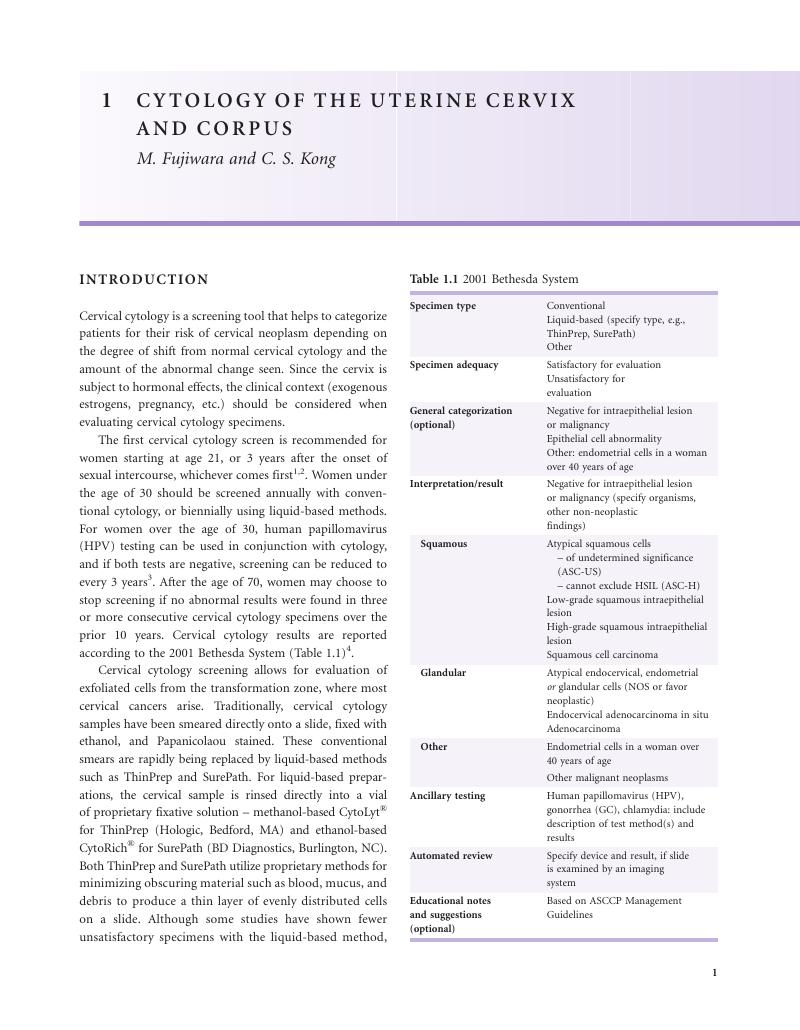
1 - Cytology of the uterine cervix and corpus
Published online by Cambridge University Press: 05 July 2013

Book contents
- Frontmatter
- Contents
- List of contributors
- Preface
- Acknowledgments
- 1 Cytology of the uterine cervix and corpus
- 2 Cervix: squamous cell carcinoma and precursors
- 3 Cervix: adenocarcinoma and precursors, including variants
- 4 Miscellaneous cervical abnormalities
- 5 Non-neoplastic endometrium
- 6 Endometrial carcinoma precursors: hyperplasia and endometrial intraepithelial neoplasia
- 7 Endometrioid adenocarcinoma
- 8 Serous adenocarcinoma
- 9 Clear cell adenocarcinoma and other uterine corpus carcinomas, including unusual variants
- 10 Carcinosarcoma
- 11 Adenofibroma and adenosarcoma
- 12 Uterine smooth muscle tumors
- 13 Endometrial stromal tumors
- 14 Other uterine mesenchymal tumors
- 15 Miscellaneous primary uterine tumors
- 16 Uterine metastases: cervix and corpus
- 17 Gestational trophoblastic disease
- 18 Other pregnancy-related abnormalities
- 19 Lynch syndrome (hereditary non-polyposis colorectal cancer syndrome)
- 20 Cytology of peritoneum and abdominal washings
- Index
- References
Summary
A summary is not available for this content so a preview has been provided. Please use the Get access link above for information on how to access this content.
- Type
- Chapter
- Information
- Uterine Pathology , pp. 1 - 18Publisher: Cambridge University PressPrint publication year: 2012
References
, , , et al. American Cancer Society guideline for the early detection of cervical neoplasia and cancer. J Low Genit Tract Dis 2003;7:67–86.CrossRefGoogle ScholarPubMed
, , , et al. Cancer screening in the United States, 2011: a review of current American Cancer Society guidelines and issues in cancer screening. CA Cancer J Clin 2011;61:8–30.CrossRefGoogle ScholarPubMed
, , , et al. 2006 Consensus guidelines for the management of women with abnormal cervical cancer screening tests. Am J Obstet Gynecol 2007;197:346–55.CrossRefGoogle ScholarPubMed
, , , et al. The 2001 Bethesda System: terminology for reporting results of cervical cytology. Jama 2002;287:2114–19.CrossRefGoogle ScholarPubMed
, , , et al. Accuracy of reading liquid based cytology slides using the ThinPrep Imager compared with conventional cytology: prospective study. BMJ 2007;335:31.CrossRefGoogle ScholarPubMed
, , , et al. Comparison of liquid-based cytology with conventional cytology for detection of cervical cancer precursors: a randomized controlled trial. JAMA 2009;302:1757–64.CrossRefGoogle ScholarPubMed
. ACOG Practice Bulletin no. 109: Cervical cytology screening. Obstet Gynecol 2009;114:1409–20.CrossRefGoogle Scholar
. Cytopathology of false negatives preceding cervical carcinoma. Am J Obstet Gynecol 1996;175:1110–13.CrossRefGoogle ScholarPubMed
, , , , . Chapter 10: New dimensions in cervical cancer screening. Vaccine 2006;24(Suppl 3):S90–7.CrossRefGoogle ScholarPubMed
, , . Prospective follow-up suggests similar risk of subsequent cervical intraepithelial neoplasia grade 2 or 3 among women with cervical intraepithelial neoplasia grade 1 or negative colposcopy and directed biopsy. Am J Obstet Gynecol 2003;188:1406–12.CrossRefGoogle ScholarPubMed
, , . Cervical cytology of atypical squamous cells-cannot exclude high-grade squamous intraepithelial lesion (ASC-H): characteristics and histologic outcomes. Cancer 2006;108:298–305.CrossRefGoogle ScholarPubMed
, , , et al. Distribution of human papillomavirus types in ThinPrep Papanicolaou tests classified according to the Bethesda 2001 terminology and correlations with patient age and biopsy outcomes. Cancer 2006;106:1054–64.CrossRefGoogle ScholarPubMed
, , . Atypical glandular cells: new Bethesda Terminology and Management Guidelines. Obstet Gynecol Surv 2003;58:399–406.CrossRefGoogle ScholarPubMed
, , , et al. Atypical glandular cells and adenocarcinoma in situ according to the Bethesda 2001 classification: cytohistological correlation and clinical implications. Eur J Obstet Gynecol Reprod Biol 2008;139:79–85.CrossRefGoogle ScholarPubMed
, , . How predictive is a cervical smear suggesting glandular neoplasia?Cytopathology 2002;13:83–91.CrossRefGoogle ScholarPubMed
, , , , . Women with atypical glandular cells: a long-term follow-up study in a high-risk population. Am J Clin Pathol 2004;122:575–9.CrossRefGoogle Scholar
, , , . Histologic follow-up results in 662 patients with Pap test findings of atypical glandular cells: results from a large academic womens hospital laboratory employing sensitive screening methods. Gynecol Oncol 2009;114:383–9.CrossRefGoogle ScholarPubMed
. HPV Genotyping Clinical Update; 2011. (Accessed Oct 14 2011, at http://www.asccp.org/ConsensusGuidelines/HPVGenotypingClinicalUpdate/tabid/5963/Default.aspx.)
, , , , . Natural history of cervicovaginal papillomavirus infection in young women. N Engl J Med 1998;338:423–8.CrossRefGoogle ScholarPubMed
, , . Comparison of three management strategies for patients with atypical squamous cells of undetermined significance: baseline results from a randomized trial. J Natl Cancer Inst 2001;93:293–9.CrossRefGoogle ScholarPubMed
, , , et al. Efficiency of the hybrid capture 2 HPV DNA test in cervical cancer screening. A study by the French Society of Clinical Cytology. Am J Clin Pathol 2003;120:492–9.CrossRefGoogle ScholarPubMed
, , , et al. Clinical validation of the Cervista HPV HR and 16/18 genotyping tests for use in women with ASC-US cytology. Gynecol Oncol 2010;118:116–22.CrossRefGoogle ScholarPubMed
, , , et al. Analytical performance of the Investigational Use Only Cervista HPV HR test as determined by a multi-center study. J Clin Virol 2009;45(Suppl 1):S63–72.CrossRefGoogle ScholarPubMed
, , , et al. A population-based clinical trial comparing endocervical high-risk HPV testing using hybrid capture 2 and Cervista from the SHENCCAST II study. Am J Clin Pathol 2011;135:790–5.CrossRefGoogle ScholarPubMed
, . Use of Cervista HPV HR assay for detection of human papillomavirus in samples with hybrid capture borderline negative results. Apmis 2010;118:681–4.CrossRefGoogle ScholarPubMed
, , , . Age-stratified performance of the Cervista HPV 16/18 genotyping test in women with ASC-US cytology. Cancer Epidemiol Biomarkers Prev 2011; 20:1185–9.CrossRefGoogle ScholarPubMed
, , , , . Analytical performance of Cervista HPV 16/18 genotyping test for cervical cytology samples. J Clin Virol 2011;51:38–43.CrossRefGoogle ScholarPubMed
, , . Special commentary: patient safety and the next generation of HPV DNA tests. Am J Clin Pathol 2010;134:193–9.CrossRefGoogle ScholarPubMed
, , . Test performance comparison of inform HPV and hybrid capture 2 high-risk HPV DNA tests using the SurePath liquid-based Pap test as the collection method. Am J Clin Pathol 2005;124:24–30.CrossRefGoogle ScholarPubMed
, , , et al. Comparison of hybrid capture 2 with in situ hybridization for the detection of high-risk human papillomavirus in liquid-based cervical samples. Cancer 2004;102:11–18.CrossRefGoogle ScholarPubMed
, , , , . Comparison of methods trial for high-risk HPV. Diagn Cytopathol 2010;38:104–8.Google ScholarPubMed
, , , . ProEx C immunocytochemistry and high-risk human papillomavirus DNA testing in Papanicolaou tests with atypical squamous cell (ASC-US) cytology: correlation study with histologic biopsy. Arch Pathol Lab Med 2008;132:1648–52.Google ScholarPubMed
, , , et al. Comparison of the Digene HC2 assay and the Roche AMPLICOR human papillomavirus (HPV) test for detection of high-risk HPV genotypes in cervical samples. J Clin Microbiol 2006;44:2141–6.CrossRefGoogle ScholarPubMed
. Liquid-based cytology for cervical cancer screening. Expert Rev Mol Diagn 2005;5:857–71.CrossRefGoogle ScholarPubMed
, , , et al. Evaluation of the FocalPoint GS system performance in an Italian population-based screening of cervical abnormalities. Acta Cytol 2007;51:865–71.CrossRefGoogle Scholar
, , , et al. Introduction of the Thin Prep Imaging System (TIS): experience in a high volume academic practice. Cytojournal 2007;4:6.Google Scholar
, , , . Accuracy of ThinPrep Imaging System in detecting low-grade squamous intraepithelial lesions. Arch Pathol Lab Med 2007;131:773–6.Google ScholarPubMed
, , , . Accuracy of Thinprep Imaging System in detecting atypical glandular cells. Diagn Cytopathol 2009;37:479–82.CrossRefGoogle ScholarPubMed
, , . Effectiveness of the ThinPrep Imaging System: clinical experience in a low risk screening population. Diagn Cytopathol 2008;36:155–60.CrossRefGoogle Scholar
. The FocalPoint System: FocalPoint slide profiler and FocalPoint GS. Cancer 2004;102:334–9.CrossRefGoogle ScholarPubMed
, , . BD FocalPoint slide profiler performance with atypical glandular cells on SurePath Papanicolaou smears. Cancer Cytopathol 2010;118:68–74.CrossRefGoogle ScholarPubMed
, , , et al. The Becton Dickinson FocalPoint GS Imaging System: clinical trials demonstrate significantly improved sensitivity for the detection of important cervical lesions. Am J Clin Pathol 2009;132:767–75.CrossRefGoogle ScholarPubMed
. Cytological features of chronic follicular cervicitis in liquid-based specimens: a potential diagnostic pitfall. Cytopathology 2002;13:364–70.CrossRefGoogle ScholarPubMed
, , , . Pathology correlates of a Papanicolaou diagnosis of low-grade squamous intraepithelial lesion, cannot exclude high-grade squamous intraepithelial lesion. Cancer 2008;114:469–73.CrossRefGoogle ScholarPubMed
, , , . Comparison of the clinical significance of the Papanicolaou test interpretations LSIL cannot rule out HSIL and ASC-H. Diagn Cytopathol 2010;38:313–17.Google ScholarPubMed
, , , . “Low-grade squamous intraepithelial lesion, cannot exclude high-grade squamous intraepithelial lesion” is a distinct cytologic category: histologic outcomes and HPV prevalence. Am J Clin Pathol 2007;128:398–403.CrossRefGoogle ScholarPubMed
, , . The significance of “low-grade squamous intraepithelial lesion, cannot exclude high-grade squamous intraepithelial lesion” as a distinct squamous abnormality category in Papanicolaou tests. Cancer 2006;108:277–81.CrossRefGoogle ScholarPubMed
, , , , . Tubal metaplasia: a cytologic study with comparison to other neoplastic and non-neoplastic conditions of the endocervix. Diagn Cytopathol 1993;9:98–103; discussion –5.CrossRefGoogle ScholarPubMed
, , , . Tubal metaplasia. A frequent potential pitfall in the cytologic diagnosis of endocervical glandular dysplasia on cervical smears. Acta Cytol 1992;36:1–10.Google ScholarPubMed
, , . Superficial endometriosis of the cervix: a source of abnormal glandular cells on cervicovaginal smears. Diagn Cytopathol 2004;30:88–91.CrossRefGoogle ScholarPubMed
, , , . An approach to post-radical trachelectomy vaginal-isthmus cytology. Diagn Cytopathol 2009;37:437–42.CrossRefGoogle ScholarPubMed