Skip to main content Accessibility help
×
Hostname: page-component-848d4c4894-75dct Total loading time: 0 Render date: 2024-04-30T16:55:23.425Z Has data issue: false hasContentIssue false

Chapter 24 - Evaluation of Embryo Quality

Time-Lapse Imaging to Assess Embryo Morphokinesis

from Section 5 - Embryo Selection and Transfer

Published online by Cambridge University Press:  04 January 2019

Gabor Kovacs
Affiliation:
Monash University, Victoria
Anthony Rutherford
Affiliation:
University of Leeds
David K. Gardner
Affiliation:
University of Melbourne
Get access

Summary

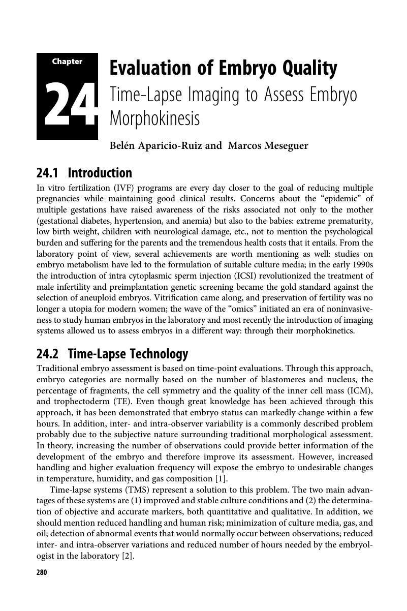
Image of the first page of this content. For PDF version, please use the ‘Save PDF’ preceeding this image.'
Type
Chapter
Information
Publisher: Cambridge University Press
Print publication year: 2019

Access options

Get access to the full version of this content by using one of the access options below. (Log in options will check for institutional or personal access. Content may require purchase if you do not have access.)

References

Aparicio, B, Cruz, M, Meseguer, M. Is morphokinetic analysis the answer? Reproductive Biomedicine Online 2013;27(6): 654–63.Google Scholar
Basile, N, Caiazzo, M, Meseguer, M. What does morphokinetics add to embryo selection and in-vitro fertilization outcomes? Curr Opin Obstet Gynecol 2015 June;27(3):193200.Google Scholar
Wong, CC, Loewke, KE, Bossert, NL et al. Non-invasive imaging of human embryos before embryonic genome activation predicts development to the blastocyst stage. Nat Biotechnol 2010;28(10): 1115–21.CrossRefGoogle Scholar
Meseguer, M, Herrero, J, Tejera, A et al. The use of morphokinetics as a predictor of embryo implantation. Human Reproduction 2011;26(10): 2658–71.Google Scholar
Rubio, I, Galán, A, Larreategui, Z et al. Clinical validation of embryo culture and selection by morphokinetic analysis: a randomized, controlled trial of the EmbryoScope. Fertil Steril 2014;102(5): 1287–94. e5.Google Scholar
Basile, N, Vime, P, Florensa, M et al. The use of morphokinetics as a predictor of implantation: a multicentric study to define and validate an algorithm for embryo selection. Hum Reprod 2015 February;30(2): 276–83.CrossRefGoogle ScholarPubMed
Conaghan, J, Chen, AA, Willman, SP et al. Improving embryo selection using a computer-automated time-lapse image analysis test plus day 3 morphology: results from a prospective multicenter trial. Fertil Steril 2013;100: 412–9.CrossRefGoogle ScholarPubMed
Kirkegaard, K, Campbell, A, Agerholm, I et al. Limitations of a time-lapse blastocyst prediction model: a large multicentre outcome analysis. Reproductive BioMedicine Online 2014;2: 156–8.Google Scholar
Petersen, BM, Boel, M, Montag, M, Gardner, DK. Development of a generally applicable morphokinetic algorithm capable of predicting the implantation potential of embryos transferred on Day 3. Hum Reprod 2016 October;31(10): 2231–44.Google Scholar
Huang, T, Chinn, K, Kosasa, T, Ahn, H, Kessel, B. Morphokinetics of human blastocyst expansion in vitro. Reproductive BioMedicine Online 2016;33: 659–67.Google Scholar
Nogales, MDC, Bronet, F, Basile, N et al. Type of chromosome abnormality affects embryo morphology dynamics. Fertil Steril 2017;107: 229–35.Google Scholar
Milewski, R, Czerniecki, J, Kuczyńska, A, Stankiewicz, B, Kuczyński, W. Morphokinetic parameters as a source of information concerning embryo developmental and implantation potential. Ginekol Pol 2016;87(10): 677–84.CrossRefGoogle ScholarPubMed
Liu, Y, Chapple, V, Feenan, K, Roberts, P, Matson, P. Time-lapse deselection model for human day 3 in vitro fertilization embryos: the combination of qualitative and quantitative measures of embryo growth. Fertil Steril 2016;105(3): 656–62. e1.Google Scholar
Motato, Y, de los Santos, María José, Escriba, MJ et al. Morphokinetic analysis and embryonic prediction for blastocyst formation through an integrated time-lapse system. Fertil Steril 2015.Google Scholar
Kong, X, Yang, S, Gong, F et al. The Relationship between cell number, division behavior and developmental potential of cleavage stage human embryos: a time-lapse study. PloS one 2016;11(4):e0153697.CrossRefGoogle ScholarPubMed
Bodri, D, Sugimoto, T, Serna, JY et al. Blastocyst collapse is not an independent predictor of reduced live birth: a time-lapse study. Fertil Steril 2016;105(6): 1476–83. e3.CrossRefGoogle Scholar
Adamson, GD, Abusief, ME, Palao, L et al. Improved implantation rates of day 3 embryo transfers with the use of an automated time-lapse–enabled test to aid in embryo selection. Fertil Steril 2016;105(2): 369–75. e6.CrossRefGoogle ScholarPubMed
Kieslinger, DC, De Gheselle, S, Lambalk, CB et al. Embryo selection using time-lapse analysis (Early Embryo Viability Assessment) in conjunction with standard morphology: a prospective two-center pilot study. Hum Reprod 2016;31: 2450–57.Google Scholar
Aparicio-Ruiz, B, Basile, N, Albalá, SP et al.Automatic time-lapse instrument is superior to single-point morphology observation for selecting viable embryos: retrospective study in oocyte donation. Fertil Steril 2016;106(6): 1379–85. e10.CrossRefGoogle ScholarPubMed
Goodman, LR, Goldberg, J, Falcone, T, Austin, C, Desai, N. Does the addition of time-lapse morphokinetics in the selection of embryos for transfer improve pregnancy rates? A randomized controlled trial. Fertil Steril 2016;105(2): 275–85. e10.CrossRefGoogle ScholarPubMed
Haikin Herzberger, E, Ghetler, Y, Tamir Yaniv, R et al. Time lapse microscopy is useful for elective single-embryo transfer. Gynecological Endocrinology 2016;32:13.CrossRefGoogle ScholarPubMed
Ahlstrom, A, Park, H, Bergh, C, Selleskog, U, Lundin, K. Conventional morphology performs better than morphokinetics for prediction of live birth after day 2 transfer. Reproductive Biomedicine Online 2016;33:6170.CrossRefGoogle ScholarPubMed
Costa-Borges, N, Bellés, M, Meseguer, M et al.Blastocyst development in single medium with or without renewal on day 3: a prospective cohort study on sibling donor oocytes in a time-lapse incubator. Fertil Steril 2016;105(3): 707–13.Google Scholar
Dal Canto, M, Novara, PV, Coticchio, G et al. Morphokinetics of embryos developed from oocytes matured in vitro. J Assist Reprod Genet 2016;33(2): 247–53.Google Scholar
Tejera, A, Castelló, D, de los Santos, Jose Maria et al. Combination of metabolism measurement and a time-lapse system provides an embryo selection method based on oxygen uptake and chronology of cytokinesis timing. Fertil Steril 2016 July;106(1): 119–26.Google Scholar
Patel, DV, Shah, PB, Kotdawala, AP et al. Morphokinetic behavior of euploid and aneuploid embryos analyzed by time-lapse in embryoscope. Journal of Human Reproductive Sciences 2016;9(2):112.Google Scholar
Balakier, H, Sojecki, A, Motamedi, G, Librach, C. Impact of multinucleated blastomeres on embryo developmental competence, morphokinetics, and aneuploidy. Fertil Steril 2016 July;106: 119–26.CrossRefGoogle ScholarPubMed
Bar-El, L, Kalma, Y, Malcov, M et al. Blastomere biopsy for PGD delays embryo compaction and blastulation: a time-lapse microscopic analysis. J Assist Reprod Genet 2016 November;33:1491457.CrossRefGoogle ScholarPubMed
Bodri, D, Kawachiya, S, Sugimoto, T et al. Time-lapse variables and embryo gender: a retrospective analysis of 81 live births obtained following minimal stimulation and single embryo transfer. J Assist Reprod Genet 2016;33(5): 589–96.CrossRefGoogle Scholar
Wu, Y, Lazzaroni-Tealdi, E, Wang, Q et al. Different effectiveness of closed embryo culture system with time-lapse imaging (EmbryoScope TM) in comparison to standard manual embryology in good and poor prognosis patients: a prospectively randomized pilot study. Reproductive Biology and Endocrinology 2016;14(1):49.CrossRefGoogle ScholarPubMed
Demirel, C, Bastu, E, Aydogdu, S et al. The Presence of endometrioma does not impair time-lapse morphokinetic parameters and quality of embryos: a study on sibling oocytes. Reprod Sci 2016 August;23(8): 1053–57.Google Scholar
Kirkegaard, K, Sundvall, L, Erlandsen, M et al. Timing of human preimplantation embryonic development is confounded by embryo origin. Hum Reprod 2016 February;31(2): 324–31.Google Scholar

Save book to Kindle

To save this book to your Kindle, first ensure coreplatform@cambridge.org is added to your Approved Personal Document E-mail List under your Personal Document Settings on the Manage Your Content and Devices page of your Amazon account. Then enter the ‘name’ part of your Kindle email address below. Find out more about saving to your Kindle.

Note you can select to save to either the @free.kindle.com or @kindle.com variations. ‘@free.kindle.com’ emails are free but can only be saved to your device when it is connected to wi-fi. ‘@kindle.com’ emails can be delivered even when you are not connected to wi-fi, but note that service fees apply.

Find out more about the Kindle Personal Document Service.

Available formats
×

Save book to Dropbox

To save content items to your account, please confirm that you agree to abide by our usage policies. If this is the first time you use this feature, you will be asked to authorise Cambridge Core to connect with your account. Find out more about saving content to Dropbox.

Available formats
×

Save book to Google Drive

To save content items to your account, please confirm that you agree to abide by our usage policies. If this is the first time you use this feature, you will be asked to authorise Cambridge Core to connect with your account. Find out more about saving content to Google Drive.

Available formats
×