Skip to main content Accessibility help
×
  • Cited by 35
Publisher:
Cambridge University Press
Online publication date:
March 2012
Print publication year:
2011
Online ISBN:
9780511921001

Book description

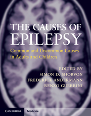
Causation is an aspect of epilepsy neglected in the scientific literature and in the conceptualization of epilepsy at a clinical and experimental level. It was to remedy this deficiency that this book was conceived. The book opens with a draft etiological classification that goes some way to filling the nosological void. The book is divided into four etiological categories: idiopathic, symptomatic, cryptogenic, and provoked epilepsies. Each chapter considers topics in a consistent fashion, dealing with the phenomenon of epilepsy in each etiology, including its epidemiology, clinical features and prognosis, and any specific aspects of treatment. The book is a comprehensive reference work, a catalogue of all important causes of epilepsy, and a clinical tool for all clinicians dealing with patients who have epilepsy. It is aimed at epileptologists and neurologists and provides a distillation of knowledge in a form that is helpful in the clinical setting.

Reviews

'… really very good. I am not aware of anything else that deals with the causes of epilepsy as succinctly and is as well organised and user friendly. This is the sort of book that one would dip into when faced with a particularly interesting or problematic case … should be on the bookshelf of everyone who investigates and manages people with epilepsy.'

Professor Martin J. Brodie - Director, Epilepsy Unit, Western Infirmary, Glasgow

'Most of the authors are world experts on the subjects, and I admire the energy and professional network required to assemble this impressive work … it will have a huge impact.'

Source: The Lancet Neurology

'… extremely comprehensive … fills a significant void in the epilepsy literature … The chapters are well written and address both basic pathogenic mechanisms and clinical diagnosis and management … This book fills a unique niche in the field of epileptology: it is the first to carefully and concisely address the varied etiologies. It is a very readable and comprehensive book that will be valued both by practising clinicians and by trainees and basic neuroscientists. It is an essential resource and should be on the bookshelf of any clinician caring for persons with epilepsy worldwide.'

Source: Epilepsy and Behavior

Refine List

Actions for selected content:

Select all | Deselect all
  • View selected items
  • Export citations
  • Download PDF (zip)
  • Save to Kindle
  • Save to Dropbox
  • Save to Google Drive

Save Search

You can save your searches here and later view and run them again in "My saved searches".

Please provide a title, maximum of 40 characters.
×

Contents


Page 3 of 5


  • Chapter 51 - Arachnoid cysts
    pp 341-345
  • View abstract

    Summary

    The clinical manifestations of Urea cycle disorders are the result of acute or chronic hyperammonemia and include acute neurologic and gastrointestinal symptoms and signs. Chronic hyperammonemia may induce changes in N-methyl-D-aspartate (NMDA) receptor-mediated neurotransmission and induction of astrocytosis. Patients with partial urea cycle enzyme deficiencies may have a much later presentation, usually with hepatogastric, neurologic, or psychiatric symptoms. Seizures in the neonate are a sign of acute hyperammonemia and occur in approximately 50% of severely hyperammonemic neonates. The electroencephalographic (EEG) abnormalities may parallel the clinical course with improvement in the background and resolution of the epileptiform discharges. Measurement of blood ammonia after a protein load may help to establish the diagnosis in patients with normal baseline ammonia levels. Hemodialysis and hemofiltration can be used to lower ammonia levels acutely while measures to reverse the catabolic state are implemented by infusion of glucose and insulin.
  • Chapter 52 - Malformations of human cerebral cortex
    pp 346-362
  • View abstract

    Summary

    Wilson disease should be considered as a possible diagnosis in any child who presents with liver disease particularly if it is sub acute or chronic. The pathogenesis of Wilson disease is an inability to excrete copper via the bile. Studies with radioactive copper have shown that in the early, presymptomatic stage, the liver takes up the metal avidly. Treatment is conventional as for other forms of epilepsy, but treatment of Wilson disease must be vigorously pursued with penicillamine or trientine. Magnetic resonance imaging (MRI) brain scans may show lesions in the basal ganglia, most commonly the putamen, also widening of the cerebral ventricles and cortical atrophy. Wilson disease is best managed at a specialist clinic. The prognosis for most patients with Wilson disease is excellent but the degree of recovery of the neurological deficit inevitably depends upon the amount of damage to the brain before treatment is started.
  • Chapter 53 - Hippocampal sclerosis
    pp 363-372
  • View abstract

    Summary

    Genetically determined and acquired disorders leading to relative cobalamin deficiency result in hematologic changes characterized by megaloblastic anemia. Acquired disorders of cobalamin deficiency occur far more frequently than genetically determined disorders and often result in a variety of neurologic syndromes. Seizures are recognized as an uncommon manifestation of cobalamin deficiency, especially in adults. Early-onset cblC or cblD disorder, which results in both homocystinuria and methylmalonic aciduria, has been described in several reports to feature prominent seizures. Use of valproic acid, carbamazepine, and phenytoin has been shown in epileptic patients to lower serum cobalamin levels. Mutations affecting the proton-coupled folate transporter (PCFT) result in deficient uptake of folate at both the intestinal and choroid plexus levels and causes hereditary folate malabsorption. Neuroimaging studies detect non-specific cerebral atrophy and the intracranial, particularly occipital, calcifications noted especially in folate deficiency.
  • Chapter 54 - Neonatal seizures and postneonatal epilepsy – causes
    pp 373-381
  • View abstract

    Summary

    Epileptic seizures can occur in many inborn errors of metabolism (IEMs), usually as part of a larger clinical spectrum. This chapter considers IEMs, most of which are amenable to specific metabolic treatment. Epilepsy is frequent in Glucose transporter type 1 (GLUT-1) deficiency. Hyperinsulinism/hyperammonemia syndrome (HI/HA) is a rare disorder resulting from missense mutations in glutamate dehydrogenase. Non-ketotic hyperglycinemia (NKH) results from mutations in the subunits of the glycine cleavage enzyme that degrades the neurotransmitter glycine. The congenital disorder of glycosylation (CDG) syndrome covers a group of autosomal recessive diseases, resulting from mutations in the enzymes responsible for protein glycosylation. Epileptic patients, when an IEM is suspected, should have a complete clinical examination, EEG, brain magnetic resonance imaging (MRI) with proton magnetic resonance spectroscopy, ophthalmologic examination, and abdominal ultrasonography. Practical efforts should be made to identify the IEM that can benefit from tailored metabolic therapies.
  • Chapter 55 - Cerebral palsy
    pp 382-387
  • View abstract

    Summary

    Down syndrome (DS) is caused by trisomy 21, and is one of the most common chromosomal disorders and etiology of congenital mental retardation. Down syndrome increases the risk of developing acute megakaryoblastic anemia (AMKL) and acute lymphoblastic leukemia (ALL). Approximately 10% of newborns with DS present with transient myeloproliferative disorder (TMD), and 10-20% with TMD develop AMKL before 4 years of age. People with DS have a greatly increased risk of early-onset Alzheimer disease (AD). Epileptic seizures occur in 5-10% of DS patients and prevalence increases with age. Children with DS may exhibit various seizures types, including myoclonic, atonic, tonic-clonic, and, rarely, partial seizures. There is no etiologic treatment for DS, but supportive care and treatment of associated conditions such as cardiac and gastrointestinal abnormalities are indicated. Approximately one-third of patients die in infancy and 50% during the first 5 years from cardiac and respiratory infections.
  • Chapter 57 - Open head injury
    pp 393-399
  • View abstract

    Summary

    Fragile X syndrome is the most frequent cause of familial mental retardation and the second most common overall cause of mental retardation after Down syndrome. Clinical features of fragile X syndrome vary widely, and include cognitive, behavioral, and morphologic signs and symptoms. Fragile X mental retardation protein (FMRP) plays a number of crucial roles in dendritic maturation and function. The most common seizure type seen in fragile X syndrome is complex partial and the most common electroencephalogram (EEG) pattern is centrotemporal spikes. The definitive diagnosis of fragile X syndrome is made by genetic testing and requires an alteration in the FMR1 gene which can be detected by polymerase chain reaction (PCR) or Southern blot analysis. The treatment of individuals with fragile X syndrome is mainly symptomatic and ideally involves a multidisciplinary team to address common comorbidities including anxiety, behavioral problems, attentional difficulties, and other psychiatric disorders.
  • Chapter 58 - Closed head injury
    pp 400-406
  • View abstract

    Summary

    Wolf-Hirschhorn syndrome (WHS) is a multiple congenital anomalies/intellectual disability disorder due to deletion, or loss of material, of the distal portion of the short arm of chromosome 4. Seizures/epilepsy represents one of the main clinical challenges in 4p WHS. Epilepsy is well controlled in most WHS individuals (80%). Phenobarbital has been reported as the most effective drug against tonic-clonic seizures. About 30-40% of the deletions will not be detected by karyotype. Therefore, fluorescence in situ hybridization (FISH) with cosmid probes from the WHS critical region (WHSCR), or comparative genomic hybridization microarray (aCGH) are necessary to confirm the diagnosis. Brain magnetic resonance imaging (MRI) should be performed in all individuals with seizures, since malformation of the cerebral cortex may modify prognosis and management. A waking/sleeping video-EEG-polygraphic study, electrocardiogram [ECG], surface electromyogram [EMG]) is recommended in infancy and childhood, in order to achieve the best characterization of seizures.
  • Chapter 59 - De novo epilepsy after neurosurgery
    pp 407-412
  • View abstract

    Summary

    Inverted duplicated chromosome 15 (idic (15)) is the most common of the heterogeneous group of the supernumerary marker chromosomes (SMCs). The idic (15) distinct behavior disorder has been described as autistic or autistic-like. Epilepsy represents one of the main clinical challenges in idic (15) individuals. It occurs with a wide variety of seizures, with onset between ages 6 months and 9 years. Infantile spasms associated with an hypsarrhythmic electroencephalogram (EEG) have been reported in several patients. Standard cytogenetics must be associated with fluorescent in situ hybridization (FISH) analysis, using probes both from proximal chromosome 15 and from the Prader-Willi syndrome/Angelman syndrome (PWS/AS) critical region. Although brain neuroimaging (CT/MRI) is reportedly normal in most idic (15) individuals brain MRI should be performed in all those having seizures, since a malformation of the cerebral cortex may modify prognosis and management. Regular follow-up of the seizure status and pharmacotherapy is essential.
  • Chapter 60 - Epilepsy after epilepsy surgery
    pp 413-424
  • View abstract

    Summary

    Ring chromosome 20 is a rare chromosomal abnormality and a rare cause of intractable epilepsy. The most commonly accepted pathophysiological hypothesis is that the deleted regions (p13 and q13) of chromosome 20 contain important genes, and that their loss leads to the development of epilepsy with or without other clinical manifestations. The most consistent clinical feature described is epilepsy. The diagnosis is made by karyotype analysis. Since the majority of patients are mosaic, at least 100 mitoses should be examined when the diagnosis is strongly suspected. It is of note that the mosaicism is typically post-zygotic. The EEG may help in suggesting the diagnosis. Typical EEG features are seen on both the interictal and ictal EEG. Neuroimaging, including high-resolution MRI and other imaging techniques, is typically normal. Occasional cases are reported with focal abnormalities on MRI such as cortical dysplasia or focal atrophy.
  • Chapter 61 - Non-accidental brain injury
    pp 425-432
  • View abstract

    Summary

    Hemimegalencephaly often presents in early infancy with either cerebral hemisphere affected, and occurs in every ethnic group and both genders. Hemimegalencephaly may be the primary etiology of several epilepsy syndromes. In Ohtahara syndrome, tonic seizures, frequently asymmetric, present early in association with a burst suppression pattern. West syndrome is another frequently encountered epilepsy type with infantile spasms present in up to 50% of patients with hemimegalencephaly. The diagnosis of hemimegalencephaly is made definitively by magnetic resonance imaging (MRI) examination; however, early detection using postnatal cranial ultrasound and computed tomography (CT) is possible. Brain single photon emission computed tomography (SPECT) imaging often reveals hypoperfusion of the malformed hemisphere in the interictal state and hyperperfusion during the ictus. The choice of antiepileptic medication rests largely on the seizure type present in each case. Anatomic or functional hemispherectomy have been employed in patients with hemimegalencephaly and both show similar rates of postoperative seizure-freedom.
  • Chapter 63 - Ganglioglioma, dysembryoplastic neuroepithelial tumor, and related tumors
    pp 441-448
  • View abstract

    Summary

    Focal cortical dysplasia (FCD) is a definable disorder among the malformation due to abnormal cortical development (MCD) and as such it should be recognized and diagnosed accordingly. The molecular mechanisms mediating epileptogenesis in FCD are not well understood despite the fact that FCD is one of nature's best models for intrinsic epileptogenicity. The clinical manifestations of patients with cortical FCD are variable. Ictal electroencephalogram (EEG) studies have suggested that FCD is a neural network disorder with secondary ictal zones. Intraictal activation is typical of FCD and can be contiguous or at a distance from the primary epileptogenic area. In transmantle cortical dysplasia (TCD), the imaging abnormality extends from the ventricle to the cerebral cortex. The extent and the localization in Bottom of the sulcus FCD (BOSD) distinguish it from TCD or other FCDs. New surgical treatment options that combine network disconnection and augmentation such as electrical stimulation are being developed.
  • Chapter 64 - Hypothalamic hamartoma and gelastic epilepsy
    pp 449-453
  • View abstract

    Summary

    Lissencephaly (LIS) is a neuronal migration disorder characterized by absent (agyria) or decreased (pachygyria) convolutions, producing a smooth cerebral surface. Subcortical band heterotopia (SBH) is a related disorder in which there are bilateral bands of gray matter interposed in the white matter, between the cortex and the lateral ventricles. This chapter discusses the genetic basis for diagnosis and pathogenesis of LIS. The LIS1 gene encodes a 45-kDa protein (PAFAH1B1), which functions as a regulatory subunit of platelet-activating factor acetylhydrolase (PAF-AH). It has been proposed that LIS1 colocalizes with microtubules and promotes their stabilization in vitro. The DCX gene encodes a 40-kDa microtubule-associated protein (DCX) which is expressed in migrating neuroblasts. X-linked lissencephaly with absent corpus callosum and ambiguous genitalia (XLAG) is a severe malformation syndrome that is only observed in boys. Lissencephaly with cerebellar hypoplasia (LCH) is associated with severe abnormalities of the cerebellum, hippocampus, and brainstem.
  • Chapter 65 - Meningioma
    pp 454-458
  • View abstract

    Summary

    Disrupted formation of the corpus callosum is the second most common central nervous system birth defect, occurring in approximately 1 in 3000 live births. The incidence of epilepsy in patients with agenesis of the corpus callosum (ACC) and its related syndromes ranges from 25% to 62%. Aicardi syndrome is a presumed X-linked syndrome, as it is seen only in females and Klinefelter syndrome (XXY) males. The ARX gene is a homeobox gene that has been shown to be crucial in forebrain, pancreatic, and testicular development. Andermann syndrome, also known as peripheral neuropathy associated with agenesis of the corpus callosum (ACCPN). An autosomal dominant disorder, Mowat-Wilson, is caused by de novo mutations or deletions within the ZFHX1B (ZEB2) gene. Recent work in studying large cohorts of ACC individuals is providing insight into the diversity of causes which lead to ACC, which will provide important insight into mechanisms and outcomes.
  • Chapter 66 - Metastatic disease
    pp 459-466
  • View abstract

    Summary

    Polymicrogyria has been related to mutations of several genes including SRPX2, PAX6, TBR2, KIAA1279, RAB3GAP1, COL18A1 and copy-number variations. The spectrum of clinical manifestations associated with polymicrogyria ranges from normal individuals with only selective impairment of cognitive functions. Generalized polymicrogyria is often accompanied by microcephaly and severe-profound cognitive and motor delay, as well as epilepsy. Both polymicrogyria and schizencephaly have been reported in the same family and both may occur with prenatal cytomegalovirus infection. Clinical findings include focal seizures present in most patients usually beginning before age 3 years if bilateral clefts are present. Epilepsy, motor impairment, and disorders of higher cortical functions are the most common clinical manifestation of polymicrogyria. Specific electroclinical features of Aicardi syndrome include early onset infantile spasms and partial seizures. Electroencephalography in polymicrogyria and schizencephaly shows variable patterns of abnormality. Selected patients with schizencephaly have been treated with surgery for epilepsy.
  • Chapter 67 - Viral encephalitis
    pp 467-474
  • View abstract

    Summary

    Periventricular nodular heterotopia (PNH) is the most common clinically encountered heterotopia and makes up 15-20% of all cortical dysgenesis series. The majority of patients with PNH have seizures or epilepsy. Abnormal cellular representation and connectivity of interneurons in heterotopia may explain or contribute to epileptogenicity of PNH. Several groups have advocated a subclassification of PNH using radiological grounds, based on contiguity and symmetry of nodules, or the clinical and genetic associations. Brain computerized tomography (CT) would usually demonstrate the malformation in cases of diffuse and contiguous PNH but can miss more subtle and focal types. Neuropsychological testing is useful to identify specific domains of deficiency in patients who display signs of neurological developmental delay. Fluoro-deoxy-glucose positron emission tomography (FDGPET) and single positron emission computerized tomography (SPECT) have shown that the metabolic activity and perfusion to the nodules are identical to that of the overlying cortex.
  • Chapter 68 - Bacterial meningitis and focal suppurative intracranial infections in children
    pp 475-481
  • View abstract

    Summary

    Virtually all forms of microcephaly display some degree of simplification of the cortical gyral pattern, with fewer gyri and shallow sulci. Two particular modes of neural progenitor division in the developing cerebral cortex are symmetric and asymmetric. In asymmetric divisions, radial glial progenitors in the ventricular zone (VZ) give rise to a post-mitotic neuron and a radial glial daughter that re-enters the cell cycle. Disruption of neural progenitor proliferation is but one path leading to microcephaly. Seizures are a manifestation of microcephaly that occurs with variable frequency, according to the clinical syndrome and causative gene. The first-line diagnostic test for microcephaly is an accurate measurement of occipital-frontal circumference (OFC) obtained in the delivery room and repeated at each postnatal office visit. The ability to distinguish between congenital microcephaly and postnatal microcephaly will be helpful in prioritizing the possible etiologies.
  • Chapter 69 - Bacterial meningitis and pyogenic abscess in adults
    pp 482-491
  • View abstract

    Summary

    Congenital Arachnoid cysts (AC) have been reported to account for roughly 1% of atraumatic intracranial mass lesions. Sylvian fissure cysts can manifest clinically at any age, but they become symptomatic more frequently in children and adolescents than in adults, and in most series infants and toddlers account for about a quarter of the cases. The incidence of epilepsy in AC patients has been reported to be between 7.5% and 42.4%; conversely MRI screening studies in epileptic patients have showed that AC are incidentally found in up to 2% of the cases. Single photon emission computed tomography (SPECT) studies proved more sensitive than increased intracranial pressure (ICP) monitoring in children with unspecific clinic radiologic correlation. Open cyst marsupialization is considered the preferable surgical procedure. Cyst extrathecal diversions are obviously safer, but are accompanied by a high incidence of additional surgical procedures and the stigma of lifelong shunt dependency.
  • Chapter 70 - Malaria
    pp 492-494
  • View abstract

    Summary

    Fetal studies show that a specific malformation, e.g., polymicrogyria or cobblestone cortex, may be the end point of disruption of one of a number of developmental pathways and may have more than one etiology. Histology of the cerebral hemispheres showed, apart from necrosis, failure of neuronal migration with subcortical heterotopia, polymicrogyria, overmigration into the leptomeninges, transmantle dysplasia, and schizencephaly. Malformations resulting from undermigration may be generalized, as in type 1 lissencephaly and subcortical band (double cortex) syndromes, or focal as represented by subcortical and periventricular heterotopias. Focal cortical dysplasia and tuberous sclerosis are lesions restricted to a small region of the cortex and are thought to result from early abnormalities in cell growth and proliferation. Cobblestone cortex (type 2 lissencephaly), bilateral frontoparietal polymicrogyria, MARCKS deficiency, TUBB2B mutation, polymicrogyria and schizencephaly are some of the over migration syndromes.
  • Chapter 71 - Neurocysticercosis
    pp 495-500
  • View abstract

    Summary

    Hippocampal sclerosis (HS) has been recognized as the most commonly encountered pathological substrate of mesial temporal lobe epilepsy (MTLE). The pathological condition was initially described by early neuropathologists based on postmortem material. Studies in familial MTLE are also important for better understanding of the pathogenesis of HS. HS identified by magnetic resonance imaging (MRI) has been associated with poor medical control of seizures. Neurologic examination is usually normal except for facial asymmetry and memory deficits, which are material-specific for the side of ictal onset. Interictal electroencephalogram (EEG) findings in patients with MTLE typically include unilateral or bilaterally independent mesial temporal spikes, best seen with basal derivations. Treatment should start with a first-line antiepileptic drug (AED) in monotherapy, the dose of which is increased until seizure freedom or the occurrence of side effects such as tiredness, dizziness, diploplia, or gait disturbance.
  • Chapter 73 - Tuberculosis
    pp 511-519
  • View abstract

    Summary

    Neonatal seizures are usually considered of epileptic origin, although some may be generated by non-epileptic mechanisms. Neonatal seizures may be classified by clinical features: focal clonic, focal tonic, myoclonic, spasms, generalized tonic and motor automatisms. The main categories of etiological factors of neonatal seizures include hypoxic- ischemic encephalopathy, central nervous system (CNS) infections, structural brain abnormalities, and metabolic disturbances. Early neuroimaging studies, particularly magnetic resonance imaging (MRI) with diffusion imaging, should show acute diffuse abnormalities consistent with hypoxia/ischemia. Some neonatal seizures result from long-standing disorders. These disorders include cerebral dysgenesis, neurocutaneous syndromes, genetic disorders, or very early onset epilepsy in association with well-defined epileptic syndromes. Clinical examination and seizure characterization are initial points of reference in the evaluation of infants suspected of early myoclonic encephalopathy (EME). Benzodiazepines are frequently used as alternative secondline antiepileptic drugs (AEDs), although more often they are given following phenobarbital and in place of phenytoin.
  • Chapter 74 - HIV infection
    pp 520-527
  • View abstract

    Summary

    Cerebral palsy (CP) affects between 2 and 3 per 1000 live births and is thought to be the most common cause of serious physical disability in childhood. A widely used topographical classification of CP has been formulated by the Surveillance of Cerebral Palsy in Europe group. This divides CP into spastic, dyskinetic, and ataxic subtypes. Children with CP due to central nervous system (CNS) malformation, infection, and gray matter damage were more likely to have epilepsy than those with CP due to white matter damage. The predominant type is focal seizures or focal seizures evolving into secondary generalized tonic-clonic (GTC) seizures. The majority of CP etiologies can be established based on a detailed history, examination, and neuroimaging without the need for other investigations. Magnetic resonance imaging (MRI) is the imaging modality of choice. Epilepsy surgery can have a good outcome particularly for children with hemiplegia.
  • Chapter 75 - Emerging and less common central nervous system viral encephalitides
    pp 528-536
  • View abstract

    Summary

    Epilepsy can be caused by vaccination as a leading symptom of vaccine-induced encephalopathy and febrile seizures can also occur as a result of vaccine-induced fever. Pertussis vaccination is a highly effective means to prevent a disabling and sometimes deadly disease. The live vaccines and the vaccines prepared from infective neural tissue are perhaps the vaccines that carry the greatest risk. The cellular vaccines, and also vaccines with high levels of endotoxin, are said to be generally less safe than the acellular vaccines although evidence on these points is weak especially where seizures are a rare side effect, largely because of the absence of sufficiently powered comparisons. The risks also probably vary according to manufacturer and to the quantity and type of adjuvant which further complicates assessment. Case reports exist of epilepsy, autism, and cerebral palsy developing after measles, mumps, rubella (MMR) and measles vaccines.
  • Chapter 76 - Cerebral hemorrhage
    pp 537-543
  • View abstract

    Summary

    Open head injuries (OHI) may be classified according to the dynamics of trauma into perforating and penetrating: perforating injuries occur when an object enters and exits the skull, while penetrating injuries occur when the object does not exit the cranial vault. Two types of mechanism for brain damages are described in OHIs: primary and secondary. The diagnosis of post-traumatic epilepsy requires at least two seizures after head injury. Post-traumatic seizures are usually partial at onset even if secondary generalization may be rapid enough to simulate generalized seizures from onset. About 25% of seizures are focal, 50% focal with secondary generalization, while 25% are generalized. The number of damaged brain lobes may be a predictive factor to evaluate the volume of brain loss. Antiepileptic drugs (AEDs) should be administered to all patients with OHI, in both early and late post-injury phases, especially if a significant loss of brain tissue has occurred.
  • Chapter 78 - Arteriovenous malformations
    pp 551-558
  • View abstract

    Summary

    The most common measures of traumatic brain injury (TBI) severity include the Glasgow Coma Scale (GCS), the duration of loss of consciousness (LOC), and the duration of post-traumatic amnesia (PTA). Post-traumatic seizures are usually divided into three categories: immediate, early seizures, and late seizures. Early seizures have a different pathogenesis than late seizures; early post-traumatic seizure (PTS) are thought to be due to mechanical damage to neurons, related to extravasated blood, brain swelling, and perioperative events from cerebral manipulation or stress from general anaesthesia and metabolic factors. The relative risks of epilepsy are raised twofold after a mild head injury and sevenfold after severe head injury, risks are slightly greater in women than in men, and are increased with older age at time of injury. Structural imaging has shown promise for improving prediction of PTS risk. Phenytoin has the most evidence to support its use to reduce early post-traumatic seizures.
  • Chapter 79 - Cavernous malformations
    pp 559-564
  • View abstract

    Summary

    The frequency of epilepsy in certain conditions is well known, for example, de novo epilepsy after operative treatment of intracranial abscess is around 70% but this would probably occur independent of the surgical technique used. It seems that craniotomy probably increases the liability of de novo epilepsy by 5-10%. The complexity of the procedure also increases the incidence. Studies of post-traumatic epilepsy with modern imaging techniques have shown the relationship between cortical damage, in particular cortical contusions and post-traumatic epilepsy. The literature on cerebral tumors suggests that late postoperative seizures are more likely to be partial seizures and may be more difficult to control. Antiepileptic drugs (AEDs) are known to be metabolized along established pathways that they may share with other AEDs and also with non-anticonvulsant drugs. Other treatments such as further surgery and adjuvant therapy for intracranial tumors may be useful in treating difficult de novo seizures.
  • Chapter 80 - Other vascular disorders
    pp 565-572
  • View abstract

    Summary

    This chapter provides an overview of biological and psychosocial issues that may be associated with seizure recurrence after epilepsy surgery. It reviews seizure recurrence without introducing the variable constituted by antiepileptic drugs (AED) tapering or discontinuation. Unilateral mesial temporal lobe epilepsy due to hippocampal sclerosis (MTLE/HS) is the prototype of a surgically remediable epilepsy syndrome, in which the chances for medical control are reduced whereas the chances for surgical remediation are elevated. The chapter discusses what could be seen as de novo epilepsy after epilepsy surgery and is subdivided into two independent parts: postoperative seizures possibly related to surgically inflicted cortical damage, and postoperative seizures possibly associated with disinhibition of a potential epileptogenic zone (EZ). Stopping AEDs would be the closest construct for the cure of epilepsy. There is evidence that at least in some pathologies epilepsy may be a release phenomenon following initial resection for epilepsy.

Page 3 of 5


Metrics

Altmetric attention score

Full text views

Total number of HTML views: 0
Total number of PDF views: 0 *
Loading metrics...

Book summary page views

Total views: 0 *
Loading metrics...

* Views captured on Cambridge Core between #date#. This data will be updated every 24 hours.

Usage data cannot currently be displayed.