Skip to main content Accessibility help
×
Hostname: page-component-76fb5796d-vvkck Total loading time: 0 Render date: 2024-04-27T05:49:09.343Z Has data issue: false hasContentIssue false

13 - Intracranial perfusion imaging

Published online by Cambridge University Press:  05 May 2016

László Csiba
Affiliation:
Department of Neurology, Debreceni Egyetem, Hungary
Claudio Baracchini
Affiliation:
Department of Neuroscience, Università degli Studi di Padova, Italy
Get access

Summary

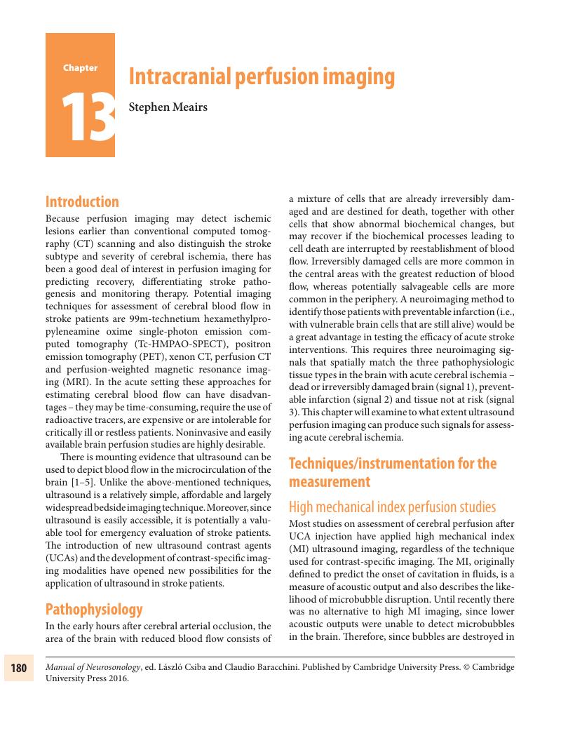
Image of the first page of this content. For PDF version, please use the ‘Save PDF’ preceeding this image.'
Type
Chapter
Information
Publisher: Cambridge University Press
Print publication year: 2016

Access options

Get access to the full version of this content by using one of the access options below. (Log in options will check for institutional or personal access. Content may require purchase if you do not have access.)

References

Seidel, G, Greis, C, Sonne, J, Kaps, M. Harmonic grey scale imaging of the human brain. J Neuroimaging. 1999;9:171174.CrossRefGoogle ScholarPubMed
Postert, T, Muhs, A, Meves, S, et al. Transient response harmonic imaging: an ultrasound technique related to brain perfusion. Stroke. 1998;29:19011907.Google Scholar
Meairs, S, Daffertshofer, M, Neff, W, Eschenfelder, C, Hennerici, M. Pulse-inversion contrast harmonic imaging: ultrasonographic assessment of cerebral perfusion. Lancet. 2000;355:550551.CrossRefGoogle ScholarPubMed
Federlein, J, Postert, T, Meves, S, et al. Ultrasonic evaluation of pathological brain perfusion in acute stroke using second harmonic imaging. J Neurol Neurosurg Psychiatry. 2000;69:616622.Google Scholar
Harrer, JU, Mayfrank, L, Mull, M, Klotzsch, C. Second harmonic imaging: a new ultrasound technique to assess human brain tumour perfusion. J Neurol Neurosurg Psychiatry. 2003;74:333338.Google Scholar
Pohl, C, Tiemann, K, Schlosser, T, Becher, H. Stimulated acoustic emission detected by transcranial color Doppler ultrasound: a contrast-specific phenomenon useful for the detection of cerebral tissue perfusion. Stroke. 2000;31:16611666.Google Scholar
Eyding, J, Krogias, C, Wilkening, W, et al. Parameters of cerebral perfusion in phase-inversion harmonic imaging (PIHI) ultrasound examinations. Ultrasound Med Biol. 2003;29:13791385.CrossRefGoogle ScholarPubMed
Postert, T, Hoppe, P, Federlein, J, et al. Contrast agent specific imaging modes for the ultrasonic assessment of parenchymal cerebral echo contrast enhancement. J Cereb Blood Flow Metab. 2000;20:17091716.CrossRefGoogle ScholarPubMed
Eyding, J, Wilkening, W, Reckhardt, M, et al. Contrast burst depletion imaging (CODIM): a new imaging procedure and analysis method for semiquantitative ultrasonic perfusion imaging. Stroke. 2003;34:7783.CrossRefGoogle ScholarPubMed
Meyer, K, Seidel, G. Transcranial contrast diminution imaging of the human brain: a pilot study in healthy volunteers. Ultrasound Med Biol. 2002;28:14331437.Google Scholar
Seidel, G, Meyer, K, Metzler, V, et al. Human cerebral perfusion analysis with ultrasound contrast agent constant infusion: a pilot study on healthy volunteers. Ultrasound Med Biol. 2002;28:183189.CrossRefGoogle ScholarPubMed
Eyding, J, Wilkening, W, Krogias, C, et al. Validation of the depletion kinetic in semiquantitative ultrasonographic cerebral perfusion imaging using 2 different techniques of data acquisition. J Ultrasound Med. 2004;23:10351040.CrossRefGoogle ScholarPubMed
Harrer, JU, Klotzsch, C. Second harmonic imaging of the human brain: the practicability of coronal insonation planes and alternative perfusion parameters. Stroke. 2002;33:15301535.CrossRefGoogle ScholarPubMed
Meves, SH, Wilkening, W, Thies, T, et al. Comparison between echo contrast agent-specific imaging modes and perfusion-weighted magnetic resonance imaging for the assessment of brain perfusion. Stroke. 2002;33:24332437.Google Scholar
Seidel, G, Algermissen, C, Christoph, A, Katzer, T, Kaps, M. Visualization of brain perfusion with harmonic gray scale and power Doppler technology: an animal pilot study. Stroke. 2000;31:17281734.Google Scholar
Wiesmann, M, Seidel, G. Ultrasound perfusion imaging of the human brain. Stroke. 2000;31:24212425.Google Scholar
Krogias, C, Postert, T, Meves, S, et al. Semiquantitative analysis of ultrasonic cerebral perfusion imaging. Ultrasound Med Biol. 2005;31:10071012.Google Scholar
Kern, R, Perren, F, Kreisel, S, et al. Multiplanar transcranial ultrasound imaging: standards, landmarks and correlation with magnetic resonance imaging. Ultrasound Med Biol. 2005;31:311315.Google Scholar
Wiesmann, M, Meyer, K, Albers, T, Seidel, G. Parametric perfusion imaging with contrast-enhanced ultrasound in acute ischemic stroke. Stroke. 2004;35:508513.Google Scholar
Seidel, G, Meyer-Wiethe, K, Berdien, G, et al. Ultrasound perfusion imaging in acute middle cerebral artery infarction predicts outcome. Stroke. 2004;35: 11071111.CrossRefGoogle ScholarPubMed
Eyding, J, Krogias, C, Schollhammer, M, et al. Contrast-enhanced ultrasonic parametric perfusion imaging detects dysfunctional tissue at risk in acute MCA stroke. J Cereb Blood Flow Metab. 2006;26:576582.CrossRefGoogle ScholarPubMed
Eyding, J, Nolte-Martin, A, Krogias, C, Postert, T. Changes of contrast-specific ultrasonic cerebral perfusion patterns in the course of stroke; reliability of region-wise and parametric imaging analysis. Ultrasound Med Biol. 2007;33:329334.Google Scholar
Holscher, T, Wilkening, W, Draganski, B, et al. Transcranial ultrasound brain perfusion assessment with a contrast agent-specific imaging mode: results of a two-center trial. Stroke. 2005;36:22832285.Google Scholar
Sobesky, J, Zaro, WO, Lehnhardt, FG, et al. Which time-to-peak threshold best identifies penumbral flow? A comparison of perfusion-weighted magnetic resonance imaging and positron emission tomography in acute ischemic stroke. Stroke. 2004;35:28432847.Google Scholar
Kern, R, Perren, F, Schoeneberger, K, et al. Ultrasound microbubble destruction imaging in acute middle cerebral artery stroke. Stroke. 2004;35:16651670.Google Scholar
Meyer-Wiethe, K, Cangur, H, Seidel, GU. Comparison of different mathematical models to analyze diminution kinetics of ultrasound contrast enhancement in a flow phantom. Ultrasound Med Biol. 2005;31:9398.Google Scholar
Lucidarme, O, Kono, Y, Corbeil, J, Choi, SH, Mattrey, RF. Validation of ultrasound contrast destruction imaging for flow quantification. Ultrasound Med Biol. 2003;29:16971704.Google Scholar
Kern, R, Diels, A, Pettenpohl, J, et al. Real-time ultrasound brain perfusion imaging with analysis of microbubble replenishment in acute MCA stroke. J Cereb Blood Flow Metab. 2011;31:17161724.Google Scholar
Harrer, JU, Klotzsch, C, Stracke, CP, Moller-Hartmann, W. [Cerebral perfusion sonography in comparison with perfusion MRT: a study with healthy volunteers]. Ultraschall Med. 2004;25:263269.Google Scholar
Jungehulsing, GJ, Brunecker, P, Nolte, CH, et al. Diagnostic transcranial ultrasound perfusion-imaging at 2.5 MHz does not affect the blood-brain barrier. Ultrasound Med Biol. 2008;34:147150.CrossRefGoogle Scholar
Stroick, M, Alonso, A, Fatar, M, et al. Effects of simultaneous application of ultrasound and microbubbles on intracerebral hemorrhage in an animal model. Ultrasound Med Biol. 2006;32:13771382.Google Scholar
Fatar, M, Stroick, M, Griebe, M, et al. Effect of combined ultrasound and microbubbles treatment in an experimental model of cerebral ischemia. Ultrasound Med Biol. 2008;34:14141420.CrossRefGoogle Scholar
Shiogai, T, Takayasu, N, Mizuno, T, Nakagawa, M, Furuhata, H. Comparison of transcranial brain tissue perfusion images between ultraharmonic, second harmonic, and power harmonic imaging. Stroke. 2004;35:687693.Google Scholar
Postert, T, Federlein, J, Weber, S, Przuntek, H, Buttner, T. Second harmonic imaging in acute middle cerebral artery infarction. Preliminary results. Stroke. 1999;30:17021706.Google Scholar
Schlachetzki, F, Hoelscher, T, Dorenbeck, U, et al. Sonographic parenchymal and brain perfusion imaging: preliminary results in four patients following decompressive surgery for malignant middle cerebral artery infarct. Ultrasound Med Biol. 2001;27:2131.CrossRefGoogle ScholarPubMed
Stolz, E, Allendorfer, J, Jauss, M, Traupe, H, Kaps, M. Sonographic harmonic grey scale imaging of brain perfusion: scope of a new method demonstrated in selected cases. Ultraschall Med. 2002;23:320324.Google Scholar
Meyer, K, Wiesmann, M, Albers, T, Seidel, G. Harmonic imaging in acute stroke: detection of a cerebral perfusion deficit with ultrasound and perfusion MRI. J Neuroimaging. 2003;13:166168.Google Scholar
Seidel, G, Albers, T, Meyer, K, Wiesmann, M. Perfusion harmonic imaging in acute middle cerebral artery infarction. Ultrasound Med Biol. 2003;29:12451251.Google Scholar
Eyding, J, Krogias, C, Wilkening, W, Postert, T. Detection of cerebral perfusion abnormalities in acute stroke using phase inversion harmonic imaging (PIHI): preliminary results. J Neurol Neurosurg Psychiatry. 2004;75:926929.Google Scholar
Meyer-Wiethe, K, Cangur, H, Schindler, A, Koch, C, Seidel, G. Ultrasound perfusion imaging: determination of thresholds for the identification of critically disturbed perfusion in acute ischemic stroke – a pilot study. Ultrasound Med Biol. 2007;33:851856.Google Scholar
Nolte, CH, Gruss, J, Steinbrink, J, et al. Ultrasound perfusion imaging of small stroke involving the thalamus. Ultraschall Med. 2009;30:466470.CrossRefGoogle ScholarPubMed
Niesen, W, Marouf, W, Weiller, C, Hetzel, A. Transcranial perfusion sonography preceding thrombolysis – perfusion deficit correlates with clinical as well as vascular status. Cerebrovasc Dis. 2008;25 (Suppl. 1):27.Google Scholar
Bartels, E, Bittermann, HJ. Transcranial contrast imaging of cerebral perfusion in stroke patients following decompressive craniectomy. Ultraschall Med. 2004;25:206213.Google Scholar
Ay, T, Havaux, X, Van Camp, G, et al. Destruction of contrast microbubbles by ultrasound: effects on myocardial function, coronary perfusion pressure, and microvascular integrity. Circulation. 2001;104: 461466.Google Scholar
Hynynen, K, McDannold, N, Martin, H, Jolesz, FA, Vykhodtseva, N. The threshold for brain damage in rabbits induced by bursts of ultrasound in the presence of an ultrasound contrast agent (Optison). Ultrasound Med Biol. 2003;29:473481.Google Scholar
Hynynen, K, McDannold, N, Sheikov, NA, Jolesz, FA, Vykhodtseva, N. Local and reversible blood-brain barrier disruption by noninvasive focused ultrasound at frequencies suitable for trans-skull sonications. Neuroimage. 2005;24:1220.Google Scholar
Bolognese, M, Artemis, D, Alonso, A, et al. Real-time ultrasound perfusion imaging in acute stroke: assessment of cerebral perfusion deficits related to arterial recanalization. Ultrasound Med Biol. 2013;39:745752.Google Scholar
Pettenpohl, J, Diels, A, Kablau, M, et al. Dynamic microvascular perfusion maps for assessment of brain infarction. Cerebrovasc Dis. 2007; 23:16.Google Scholar
Seidel, G, Roessler, F, Al-Khaled, M. Microvascular imaging in acute ischemic stroke. J Neuroimaging. 2013;23:166169.Google Scholar

Save book to Kindle

To save this book to your Kindle, first ensure coreplatform@cambridge.org is added to your Approved Personal Document E-mail List under your Personal Document Settings on the Manage Your Content and Devices page of your Amazon account. Then enter the ‘name’ part of your Kindle email address below. Find out more about saving to your Kindle.

Note you can select to save to either the @free.kindle.com or @kindle.com variations. ‘@free.kindle.com’ emails are free but can only be saved to your device when it is connected to wi-fi. ‘@kindle.com’ emails can be delivered even when you are not connected to wi-fi, but note that service fees apply.

Find out more about the Kindle Personal Document Service.

Available formats
×

Save book to Dropbox

To save content items to your account, please confirm that you agree to abide by our usage policies. If this is the first time you use this feature, you will be asked to authorise Cambridge Core to connect with your account. Find out more about saving content to Dropbox.

Available formats
×

Save book to Google Drive

To save content items to your account, please confirm that you agree to abide by our usage policies. If this is the first time you use this feature, you will be asked to authorise Cambridge Core to connect with your account. Find out more about saving content to Google Drive.

Available formats
×