
Book contents
- Soft Tissue Sarcomas
- Soft Tissue Sarcomas
- Copyright page
- Dedication
- Contents
- Contributors
- Preface
- Acknowledgments
- Chapter 1 Introduction
- Chapter 2 Imaging of Tumors and Pseudotumors of Soft Tissues
- Chapter 3A Principles of Local Therapy of Soft Tissue Neoplasms
- Chapter 3B Medical Treatment of Adult Soft Tissue Sarcomas and Gastrointestinal Stromal Tumors
- Chapter 4 Intermediate Malignant and Malignant Tumors of Soft Tissue Featuring a Spindle Cell Morphology
- Chapter 5 Soft Tissue Sarcomas with Epithelioid Morphology
- Chapter 6 Round Cell Sarcomas
- Chapter 7 Pleomorphic Sarcomas
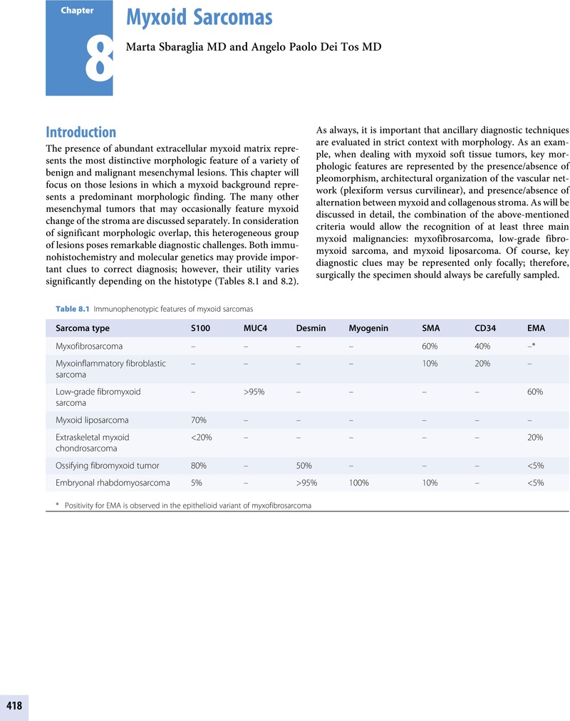
- Chapter 8 Myxoid Sarcomas
- Chapter 9 Intermediate Malignant and Malignant Tumors of Soft Tissue Featuring an Inflammatory Background
- Chapter 10 Intermediate Malignant and Malignant Tumors of Soft Tissue Resembling Normal Tissue
- Index
- References
Chapter 8 - Myxoid Sarcomas
Published online by Cambridge University Press: 10 May 2019

Book contents
- Soft Tissue Sarcomas
- Soft Tissue Sarcomas
- Copyright page
- Dedication
- Contents
- Contributors
- Preface
- Acknowledgments
- Chapter 1 Introduction
- Chapter 2 Imaging of Tumors and Pseudotumors of Soft Tissues
- Chapter 3A Principles of Local Therapy of Soft Tissue Neoplasms
- Chapter 3B Medical Treatment of Adult Soft Tissue Sarcomas and Gastrointestinal Stromal Tumors
- Chapter 4 Intermediate Malignant and Malignant Tumors of Soft Tissue Featuring a Spindle Cell Morphology
- Chapter 5 Soft Tissue Sarcomas with Epithelioid Morphology
- Chapter 6 Round Cell Sarcomas
- Chapter 7 Pleomorphic Sarcomas
- Chapter 8 Myxoid Sarcomas
- Chapter 9 Intermediate Malignant and Malignant Tumors of Soft Tissue Featuring an Inflammatory Background
- Chapter 10 Intermediate Malignant and Malignant Tumors of Soft Tissue Resembling Normal Tissue
- Index
- References
Summary
A summary is not available for this content so a preview has been provided. Please use the Get access link above for information on how to access this content.
- Type
- Chapter
- Information
- Soft Tissue SarcomasA Pattern-Based Approach to Diagnosis, pp. 418 - 492Publisher: Cambridge University PressPrint publication year: 2018
References
Primary Sources
Angervall, L, Kindblom, LG, Merck, C. Myxofibrosarcoma. A study of 30 cases. Acta Pathol Microbiol Scand A. 1977;85A:127–40.Google ScholarPubMed
Busam, KJ, Mentzel, T, Colpaert, C, Barnhill, RL, Fletcher, CD. Atypical or worrisome features in cellular neurothekeoma: a study of 10 cases. Am J Surg Pathol. 1998;22:1067–72.CrossRefGoogle ScholarPubMed
Calonje, E, Guerin, D, McCormick, D, Fletcher, CD. Superficial angiomyxoma: clinicopathologic analysis of a series of distinctive but poorly recognized cutaneous tumors with tendency for recurrence. Am J Surg Pathol. 1999;23:910–17.Google Scholar
Fetsch, JF, Laskin, WB, Lefkowitz, M, Kindblom, LG, Meis-Kindblom, JM. Aggressive angiomyxoma: a clinicopathologic study of 29 female patients. Cancer. 1996;78:79–90.Google Scholar
Fletcher, CD, Chan, JK, McKee, PH. Dermal nerve sheath myxoma: a study of three cases. Histopathology. 1986;10:135–45.CrossRefGoogle ScholarPubMed
Graadt van Roggen, JF, Hogendoorn, PC, Fletcher, CD. Myxoid tumours of soft tissue. Histopathology. 1999;35:291–312.Google Scholar
Graadt van Roggen, JF, McMenamin, ME, Fletcher, CD. Cellular myxoma of soft tissue: a clinicopathological study of 38 cases confirming indolent clinical behaviour. Histopathology. 2001;39:287–97.Google Scholar
Heitzer, E, Sunitsch, S, Gilg, MM, et al. Expanded molecular profiling of myxofibrosarcoma reveals potentially actionable targets. Mod Pathol. 2017;30:1698–1709.Google Scholar
Hollmann, TJ, Bovée, JV, Fletcher, CD. Digital fibromyxoma (superficial acral fibromyxoma): a detailed characterization of 124 cases. Am J Surg Pathol. 2012;36:789–98.Google Scholar
Hornick, JL, Fletcher, CD. Cellular neurothekeoma: detailed characterization in a series of 133 cases. Am J Surg Pathol. 2007;31:329–40.CrossRefGoogle Scholar
Huang, H-Y, Lal, P, Qin, J, et al. Low-grade myxofibrosarcoma: a clinicopathologic analysis of 49 cases treated at a single institution with simultaneous assessment of the efficacy of 3-tier and 4-tier grading systems. Hum Pathol. 2004;35:612–21.CrossRefGoogle Scholar
Liegl, B, Bennett, MW, Fletcher, CD. Microcystic/reticular schwannoma: a distinct variant with predilection for visceral locations. Am J Surg Pathol. 2008;32:1080–7.CrossRefGoogle ScholarPubMed
Meis, JM, Enzinger, FM. Juxta-articular myxoma: a clinical and pathologic study of 65 cases. Hum Pathol. 1992;23:639–46.Google Scholar
Mentzel, T, Brown, LF, Dvorak, HF, et al. The association between tumor progression and vascularity in myxofibrosarcoma and myxoid/round cell liposarcoma. Virchows Arch. 2001;438:13–22.Google Scholar
Mentzel, T, Calonje, E, Wadden, C, et al. Myxofibrosarcoma. Clinicopathologic analysis of 75 cases with emphasis on the low-grade variant. Am J Surg Pathol. 1996;20: 391–405.Google Scholar
Merck, C, Angervall, L, Kindblom, LG, et al. Myxofibrosarcoma. A malignant soft tissue tumor of fibroblastic-histiocytic origin. A clinicopathologic and prognostic study of 110 cases using multivariate analysis. Acta Pathol Microbiol Immunol Scand Suppl. 1983;282:1–40.Google Scholar
Nascimento, AF, Bertoni, F, Fletcher, CDM. Epithelioid variant of myxofibrosarcoma: expanding the clinicomorphologic spectrum of myxofibrosarcoma in a series of 17 cases. Am J Surg Pathol. 2007;31:99–105.Google Scholar
Nielsen, GP, O’Connell, JX, Rosenberg, AE. Intramuscular myxoma: a clinicopathologic study of 51 cases with emphasis on hypercellular and hypervascular variants. Am J Surg Pathol. 1998;22:1222–7.CrossRefGoogle ScholarPubMed
Nucci, MR. Mesenchymal lesions of the lower genital tract. Surg Pathol Clin. 2009;2:603–23.Google Scholar
Nucci, MR, Weremowicz, S, Neskey, DM, et al. Chromosomal translocation t(8;12) induces aberrant HMGIC expression in aggressive angiomyxoma of the vulva. Genes Chromosomes Cancer. 2001;32:172–6.CrossRefGoogle ScholarPubMed
Steeper, TA, Rosai, J. Aggressive angiomyxoma of the female pelvis and perineum. Report of nine cases of a distinctive type of gynecologic soft-tissue neoplasm. Am J Surg Pathol. 1983;7:463–75.Google Scholar
Weiss, SW, Enzinger, FM. Myxoid variant of malignant fibrous histiocytoma. Cancer. 1977;39:1672–85.Google Scholar
Willems, SM, Debiec-Rychter, M, Szuhai, K, Hogendoorn, PC, Sciot, R. Local recurrence of myxofibrosarcoma is associated with increase in tumour grade and cytogenetic aberrations, suggesting a multistep tumour progression model. Mod Pathol. 2006;19:407–16.Google Scholar
Willems, SM, Mohseny, AB, Balog, C, et al. Cellular/intramuscular myxoma and grade I myxofibrosarcoma are characterized by distinct genetic alterations and specific composition of their extracellular matrix. J Cell Mol Med. 2009;13:1291–301.CrossRefGoogle Scholar
Secondary Sources
Antonescu, CR, Zhang, L, Nielsen, GP, et al. Consistent t(1;10) with rearrangements of TGFBR3 and MGEA5 in both myxoinflammatory fibroblastic sarcoma and hemosiderotic fibrolipomatous tumor. Genes Chromosomes Cancer. 2011;50:757–64.CrossRefGoogle ScholarPubMed
Boland, JM, Folpe, AL. Hemosiderotic fibrolipomatous tumor, pleomorphic hyalinizing angiectatic tumor, and myxoinflammatory fibroblastic sarcoma: related or not? Adv Anat Pathol. 2017;24:268–77.CrossRefGoogle ScholarPubMed
Elco, CP, Mariño-Enríquez, A, Abraham, JA, Dal Cin, P, Hornick, JL. Hybrid myxoinflammatory fibroblastic sarcoma/hemosiderotic fibrolipomatous tumor: report of a case providing further evidence for a pathogenetic link. Am J Surg Pathol. 2010;34:1723–7.CrossRefGoogle ScholarPubMed
Enzinger, FM, Dulcey, F. Proliferative myositis. Report of thirty-three cases. Cancer. 1967;20:2213–23.Google Scholar
Hallor, KH, Sciot, R, Staaf, J, et al. Two genetic pathways, t(1;10) and amplification of 3p11-12, in myxoinflammatory fibroblastic sarcoma, haemosiderotic fibrolipomatous tumour, and morphologically similar lesions. J Pathol. 2009;217:716–27.CrossRefGoogle ScholarPubMed
Jurcić, V, Zidar, A, Montiel, MD, et al. Myxoinflammatory fibroblastic sarcoma: a tumor not restricted to acral sites. Ann Diagn Pathol. 2002;6:272–80.CrossRefGoogle Scholar
Kao, YC, Ranucci, V, Zhang, L, et al. Recurrent BRAF gene rearrangements in myxoinflammatory fibroblastic sarcomas, but not hemosiderotic fibrolipomatous tumors. Am J Surg Pathol. 2017;4:1456–65.Google Scholar
Laskin, WB, Fetsch, JF, Miettinen, M. Myxoinflammatory fibroblastic sarcoma: a clinicopathologic analysis of 104 cases, with emphasis on predictors of outcome. Am J Surg Pathol. 2014;38:1–12.CrossRefGoogle ScholarPubMed
Lucas, DR. Myxoinflammatory fibroblastic sarcoma: review and update. Arch Pathol Lab Med. 2017;141:1503–7.Google Scholar
Marshall-Taylor, C, Fanburg-Smith, JC. Hemosiderotic fibrohistiocytic lipomatous lesion: ten cases of a previously undescribed fatty lesion of the foot/ankle. Mod Pathol. 2000;13:1192–9.Google Scholar
Meis-Kindblom, JM, Kindblom, LG. Acral myxoinflammatory fibroblastic sarcoma: a low-grade tumor of the hands and feet. Am J Surg Pathol. 1998;22:911–24.CrossRefGoogle ScholarPubMed
Montgomery, EA, Devaney, KO, Giordano, TJ, Weiss, SW. Inflammatory myxohyaline tumor of distal extremities with virocyte or Reed-Sternberg-like cells: a distinctive lesion with features simulating inflammatory conditions, Hodgkin’s disease, and various sarcomas. Mod Pathol. 1998;11:384–91.Google Scholar
Solomon, DA, Antonescu, CR, Link, TM, et al. Hemosiderotic fibrolipomatous tumor, not an entirely benign entity. Am J Surg Pathol. 2013;37:1627–30.Google Scholar
Allen, PW. The fibromatoses: a clinicopathologic classification based on 140 cases. Am J Surg Pathol. 1977;1:255–70.Google Scholar
Bhattacharya, B, Dilworth, HP, Iacobuzio-Donahue, C, et al. Nuclear beta-catenin expression distinguishes deep fibromatosis from other benign and malignant fibroblastic and myofibroblastic lesions. Am J Surg Pathol. 2005;29:653–9.CrossRefGoogle ScholarPubMed
Burke, AP, Sobin, LH, Shekitka, KM, Federspiel, BH, Helwig, EB. Intra-abdominal fibromatosis. A pathologic analysis of 130 tumors with comparison of clinical subgroups. Am J Surg Pathol. 1990;14:335–41.Google Scholar
Coffin, CM, Davis, JL, Borinstein, SC. Syndrome-associated soft tissue tumours. Histopathology. 2014;64:68–87.Google Scholar
Colombo, C, Bolshakov, S, Hajibashi, S, et al. ‘Difficult to diagnose’ desmoid tumours: a potential role for CTNNB1 mutational analysis. Histopathology. 2011;59:336–40.Google Scholar
de Feraudy, S, Fletcher, CD. Intradermal nodular fasciitis: a rare lesion analyzed in a series of 24 cases. Am J Surg Pathol. 2010;34:1377–81.Google Scholar
de Saint Aubain Somerhausen, N, Rubin, BP, Fletcher, CD. Myxoid solitary fibrous tumor: a study of seven cases with emphasis on differential diagnosis. Mod Pathol. 1999;12:463–71.Google ScholarPubMed
De Wever, I, Dal Cin, P, Fletcher, CD, et al. Cytogenetic, clinical, and morphologic correlations in 78 cases of fibromatosis: a report from the CHAMP Study Group. Mod Pathol. 2000;3: 1080–5.Google Scholar
Doyle, LA, Möller, E, Dal Cin, P, et al. MUC4 is a highly sensitive and specific marker for low-grade fibromyxoid sarcoma. Am J Surg Pathol. 2011;35:733–41.Google Scholar
Erickson-Johnson, MR, Chou, MM, Evers, BR, et al. Nodular fasciitis: a novel model of transient neoplasia induced by MYH9-USP6 gene fusion. Lab Invest. 2011;91:1427–33.Google Scholar
Evans, HL. Low-grade fibromyxoid sarcoma. A report of two metastasizing neoplasms having a deceptively benign appearance. Am J Clin Pathol. 1987;88:615–19.Google Scholar
Evans, HL. Low-grade fibromyxoid sarcoma. A report of 12 cases. Am J Surg Pathol. 1993;17: 595–600.CrossRefGoogle ScholarPubMed
Evans, HL. Low-grade fibromyxoid sarcoma: a clinicopathologic study of 33 cases with long-term follow-up. Am J Surg Pathol. 2011;35:1450–62.Google Scholar
Folpe, AL, Lane, KL, Paull, G, Weiss, SW. Low-grade fibromyxoid sarcoma and hyalinizing spindle cell tumor with giant rosettes: a clinicopathologic study of 73 cases supporting their identity and assessing the impact of high-grade areas. Am J Surg Pathol. 2000;24:1353–60.Google Scholar
Gardner, EJ. Follow-up study of a family group exhibiting dominant inheritance for a syndrome including intestinal polyps, osteomas, fibromas and epidermal cysts. Am J Hum Genet. 1962;14:376–90.Google Scholar
Goodlad, JR, Mentzel, T, Fletcher, CD. Low grade fibromyxoid sarcoma: clinicopathological analysis of eleven new cases in support of a distinct entity. Histopathology. 1995;26:229–37.CrossRefGoogle ScholarPubMed
Gronchi, A, Colombo, C, Le Péchoux, C, et al. Sporadic desmoid-type fibromatosis: a stepwise approach to a non-metastasising neoplasm–a position paper from the Italian and the French Sarcoma Group. Ann Oncol. 2014;25:578–83.Google Scholar
Guillou, L, Benhattar, J, Gengler, C, et al. Translocation-positive low-grade fibromyxoid sarcoma: clinicopathologic and molecular analysis of a series expanding the morphologic spectrum and suggesting potential relationship to sclerosing epithelioid fibrosarcoma: a study from the French Sarcoma Group. Am J Surg Pathol. 2007;31:1387–402.Google Scholar
Honeyman, JN, Theilen, TM, Knowles, MA, et al. Desmoid fibromatosis in children and adolescents: a conservative approach to management. J Pediatr Surg. 2013;48:62–6.Google Scholar
Hornick, JL, Fletcher, CD. Soft tissue perineurioma: clinicopathologic analysis of 81 cases including those with atypical histologic features. Am J Surg Pathol. 2005;29:845–58.Google Scholar
Hornick, JL, Fletcher, CD. Intraarticular nodular fasciitis – a rare lesion: clinicopathologic analysis of a series. Am J Surg Pathol. 2006;30:237–41.Google Scholar
Lane, KL, Shannon, RJ, Weiss, SW. Hyalinizing spindle cell tumor with giant rosettes: a distinctive tumor closely resembling low-grade fibromyxoid sarcoma. Am J Surg Pathol. 1997;21:1481–8.CrossRefGoogle ScholarPubMed
Lau, PP, Lui, PC, Lau, GT, et al. EWSR1-CREB3L1 gene fusion: a novel alternative molecular aberration of low-grade fibromyxoid sarcoma. Am J Surg Pathol. 2013;37:734–8.Google Scholar
Lazar, AJ, Tuvin, D, Hajibashi, S, et al. Specific mutations in the beta-catenin gene (CTNNB1) correlate with local recurrence in sporadic desmoid tumors. Am J Pathol. 2008;173:1518–27.Google Scholar
Le Guellec, S, Soubeyran, I, Rochaix, P, et al. CTNNB1 mutation analysis is a useful tool for the diagnosis of desmoid tumors: a study of 260 desmoid tumors and 191 potential morphologic mimics. Mod Pathol. 2012;25:1551–8.Google Scholar
Linos, K, Bridge, JA, Edgar, MA. MUC 4-negative FUS-CREB3L2 rearranged low-grade fibromyxoid sarcoma. Histopathology. 2014;65:722–4.CrossRefGoogle ScholarPubMed
Matsuyama, A, Hisaoka, M, Shimajiri, S, et al. Molecular detection of FUS-CREB3L2 fusion transcripts in low-grade fibromyxoid sarcoma using formalin-fixed, paraffin-embedded tissue specimens. Am J Surg Pathol. 2006;30:1077–84.Google Scholar
Mentzel, T, Dei Tos, AP, Fletcher, CD. Perineurioma (storiform perineurial fibroma): clinico-pathological analysis of four cases. Histopathology. 1994;25:261–7.Google Scholar
Mertens, F, Fletcher, CD, Antonescu, CR, et al. Clinicopathologic and molecular genetic characterization of low-grade fibromyxoid sarcoma, and cloning of a novel FUS/CREB3L1 fusion gene. Lab Invest. 2005;85:408–15.Google Scholar
Mohamed, M, Fisher, C, Thway, K. Low-grade fibromyxoid sarcoma: clinical, morphologic and genetic features. Ann Diagn Pathol. 2017;28:60–7.Google Scholar
Montgomery, EA, Meis, JM. Nodular fasciitis. Its morphologic spectrum and immunohistochemical profile. Am J Surg Pathol. 1991;15:942–8.Google Scholar
Ng, TL, Gown, AM, Barry, TS, et al. Nuclear beta-catenin in mesenchymal tumors. Mod Pathol. 2005; 18:68–74.Google Scholar
Patel, NR, Chrisinger, JSA, Demicco, EG, et al. USP6 activation in nodular fasciitis by promoter-swapping gene fusions. Mod Pathol. 2017;30:1577–88.Google Scholar
Penel, N, Le Cesne, A, Bonvalot, S, et al. Surgical versus non-surgical approach in primary desmoid-type fibromatosis patients: A nationwide prospective cohort from the French Sarcoma Group. Eur J Cancer. 2017;83:125–31.Google Scholar
Reid, R, de Silva, MV, Paterson, L, Ryan, E, Fisher, C. Low-grade fibromyxoid sarcoma and hyalinizing spindle cell tumor with giant rosettes share a common t(7;16)(q34;p11) translocation. Am J Surg Pathol. 2003;27:1229–36.Google Scholar
Wirman, JA. Nodular fasciitis, a lesion of myofibroblasts: an ultrastructural study. Cancer. 1976;38:2378–89.Google Scholar
Åman, P, Ron, D, Mandahl, N, et al. Rearrangement of the transcript factor gene CHOP in myxoid liposarcomas with t(12;16)(q13;p11). Genes Chromosomes Cancer. 1992;5:278–85.Google Scholar
Antonescu, CR, Tschernyavsky, SJ, Decuseara, R, et al. Prognostic impact of P53 status, TLS-CHOP fusion transcript structure, and histological grade in myxoid liposarcoma: a molecular and clinicopathologic study of 82 cases. Clin Cancer Res. 2001;7:3977–87.Google ScholarPubMed
Bolen, JW, Thorning, D. Benign lipoblastoma and myxoid liposarcoma: a comparative light- and electron-microscopic study. Am J Surg Pathol. 1980;4:163–74.CrossRefGoogle ScholarPubMed
Chen, BJ, Mariño-Enríquez, A, Fletcher, CD, Hornick, JL. Loss of retinoblastoma protein expression in spindle cell/pleomorphic lipomas and cytogenetically related tumors: an immunohistochemical study with diagnostic implications. Am J Surg Pathol. 2012;36:1119–28.Google Scholar
Creytens, D. Lipoblastoma-like tumor of the vulva, an important benign mimic of myxoid liposarcoma. Int J Gynecol Pathol. 2018 Jan 3. [Epub ahead of print]Google Scholar
Dal Cin, P, Sciot, R, Panagopoulos, I, et al. Additional evidence of a variant translocation t(12;22) with EWS/CHOP fusion in myxoid liposarcoma: clinicopathological features. J Pathol. 1997;182:437–41.Google Scholar
Dei Tos, AP. Liposarcomas: diagnostic pitfalls and new insights. Histopathology. 2014;64:38–52.Google Scholar
Dei Tos, AP, Piccinin, S, Doglioni, C, et al. Molecular aberrations of the G1-S checkpoint in myxoid and round cell liposarcoma. Am J Pathol. 1997;151:1531–9.Google Scholar
Demetri, GD, von Mehren, M, Jones, RL, et al. Efficacy and safety of trabectedin or dacarbazine for metastatic liposarcoma or leiomyosarcoma after failure of conventional chemotherapy: results of a phase III randomized multicenter clinical trial. J Clin Oncol. 2016;34:786–93.Google Scholar
de Saint Aubain Somerhausen, N, Coindre, JM, Debiec-Rychter, M, Delplace, J, Sciot, R. Lipoblastoma in adolescents and young adults: report of six cases with FISH analysis. Histopathology. 2008;52:294–8.Google Scholar
de Vreeze, RS, de Jong, D, Tielen, IH, et al. Primary retroperitoneal myxoid/round cell liposarcoma is a nonexisting disease: an immunohistochemical and molecular biological analysis. Mod Pathol. 2009;22:223–31.CrossRefGoogle ScholarPubMed
Fletcher, CD, Martin-Bates, E. Spindle cell lipoma: a clinicopathological study with some original observations. Histopathology. 1987;11:803–17.CrossRefGoogle ScholarPubMed
Fritchie, KJ, Goldblum, JR, Tubbs, RR, et al. The expanded histologic spectrum of myxoid liposarcoma with an emphasis on newly described patterns: implications for diagnosis on small biopsy specimens. Am J Clin Pathol. 2012;137:229–39Google Scholar
Gronchi, A, Ferrari, S, Quagliuolo, V, et al. Histotype-tailored neoadjuvant chemotherapy versus standard chemotherapy in patients with high-risk soft-tissue sarcomas (ISG-STS 1001): an international, open-label, randomised, controlled, phase 3, multicentre trial. Lancet Oncol. 2017;18:812–22.Google Scholar
Grosso, F, Jones, RL, Demetri, GD, et al. Efficacy of trabectedin (ecteinascidin-743) in advanced pretreated myxoid liposarcomas: a retrospective study. Lancet Oncol. 2007;8:595–602.Google Scholar
Hawley, IC, Krausz, T, Evans, DJ, Fletcher, CD. Spindle cell lipoma – a pseudoangiomatous variant. Histopathology. 1994;24(6):565–9.Google Scholar
Hibbard, MK, Kozakewich, HP, Dal Cin, P, et al. PLAG1 fusion oncogenes in lipoblastoma. Cancer Res. 2000;60:4869–72.Google Scholar
Kilpatrick, SE, Doyon, J, Choong, PFM, et al. The clinicopathologic spectrum of myxoid and round cell liposarcoma. A study of 95 cases. Cancer. 1996;77:1450–8.3.0.CO;2-G>CrossRefGoogle ScholarPubMed
Knight, JC, Renwick, PJ, Dal Cin, P, Van den Berghe, H, Fletcher, CD. Translocation t(12;16)(q13;p11) in myxoid liposarcoma and round cell liposarcoma: molecular and cytogenetic analysis. Cancer Res. 1995;55:24–7.Google Scholar
Lee, ATJ, Thway, K, Huang, PH, Jones, RL. Clinical and molecular spectrum of liposarcoma. J Clin Oncol. 2018;10(36):151–9.Google Scholar
Mentzel, T, Calonje, E, Fletcher, CD. Lipoblastoma and lipoblastomatosis: a clinicopathological study of 14 cases. Histopathology. 1993;23:527–33.Google Scholar
Mirkovic, J, Fletcher, CD. Lipoblastoma-like tumor of the vulva: further characterization in 8 new cases. Am J Surg Pathol. 2015;39:1290–5.Google Scholar
Moreau, LC, Turcotte, R, Ferguson, P, et al; Canadian Orthopaedic Oncology Society (CANOOS). Myxoid\round cell liposarcoma (MRCLS) revisited: an analysis of 418 primarily managed cases. Ann Surg Oncol. 2012;19: 1081–8.Google Scholar
Orvieto, E, Furlanetto, A, Laurino, L, Dei Tos, AP. Myxoid and round cell liposarcoma: a spectrum of myxoid adipocytic neoplasia. Semin Diagn Pathol. 2001;18:267–73.Google Scholar
Smith, TA, Easley, KA, Goldblum, JR. Myxoid/round cell liposarcoma of the extremities: a clinicopathologic study of 29 cases with particular attention to extent of round cell liposarcoma. Am J Surg Pathol. 1996;20:171–80.Google Scholar
Tallini, G, Akerman, M, Dal Cin, P, et al. Combined morphologic and karyotypic study of 28 myxoid liposarcomas. Implications for a revised morphologic typing, (a report from the CHAMP Group). Am J Surg Pathol. 1996;20:1047–55.Google Scholar
ten Heuvel, SE, Hoekstra, HJ, van Ginkel, RJ, et al. Clinicopathologic prognostic factors in myxoid liposarcoma: a respective study of 49 patients with long-term follow-up. Ann Surg Oncol. 2007;14:222–9.Google Scholar
Agaram, NP, Zhang, L, Sung, YS, Singer, S, Antonescu, CR. Extraskeletal myxoid chondrosarcoma with non-EWSR1-NR4A3 variant fusions correlate with rhabdoid phenotype and high-grade morphology. Hum Pathol. 2014 45:1084–91.Google Scholar
Antonescu, CR, Argani, P, Erlandson, RA, et al. Skeletal and extraskeletal myxoid chondrosarcoma: a comparative clinicopathologic, ultrastructural, and molecular study. Cancer. 1998 15;83:1504–21.Google Scholar
Attwooll, C, Tariq, M, Harris, M, et al. Identification of a novel fusion gene involving hTAFII68 and CHN from a t(9;17)(q22;q11.2) translocation in an extraskeletal myxoid chondrosarcoma. Oncogene. 1999;18:7599–601.Google Scholar
Broehm, CJ, Wu, J, Gullapalli, RR, Bocklage, T. Extraskeletal myxoid chondrosarcoma with a t(9;16)(q22;p11.2) resulting in a NR4A3-FUS fusion. Cancer Genet. 2014;207:276–80.Google Scholar
Demicco, EG, Wang, WL, Madewell, JE, et al. Osseous myxochondroid sarcoma: a detailed study of 5 cases of extraskeletal myxoid chondrosarcoma of the bone. Am J Surg Pathol. 2013;37:752–62.CrossRefGoogle ScholarPubMed
Enzinger, FM, Shiraki, M. Extraskeletal myxoid chondrosarcoma. An analysis of 34 cases. Hum Pathol. 1972;3:421–35.Google Scholar
Flucke, U, Tops, BB, Verdijk, MA, et al. NR4A3 rearrangement reliably distinguishes between the clinicopathologically overlapping entities myoepithelial carcinoma of soft tissue and cellular extraskeletal myxoid chondrosarcoma. Virchows Arch. 2012;460:621–8.Google Scholar
Hirabayashi, Y, Ishida, T, Yoshida, MA, et al. Translocation (9;22)(q22;q12). A recurrent chromosome abnormality in extraskeletal myxoid chondrosarcoma. Cancer Genet Cytogenet. 1995;81:33–7.Google Scholar
Hisaoka, M, Hashimoto, H. Extraskeletal myxoid chondrosarcoma: updated clinicopathological and molecular genetic characteristics. Pathol Int. 2005;55:453–63.Google Scholar
Hisaoka, M, Ishida, T, Imamura, T, Hashimoto, H. TFG is a novel fusion partner of NOR1 in extraskeletal myxoid chondrosarcoma. Genes Chromosomes Cancer. 2004;40:325–8.Google Scholar
Kohashi, K, Oda, Y, Yamamoto, H, et al. SMARCB1/INI1 protein expression in round cell soft tissue sarcomas associated with chromosomal translocations involving EWS: a special reference to SMARCB1/INI1 negative variant extraskeletal myxoid chondrosarcoma. Am J Surg Pathol. 2008;32:1168–74.CrossRefGoogle ScholarPubMed
Okamoto, S, Hisaoka, M, Ishida, T, et al. Extraskeletal myxoid chondrosarcoma: a clinicopathologic, immunohistochemical, and molecular analysis of 18 cases. Hum Pathol. 2001;32:1116–24.Google Scholar
Panagopoulos, I, Mertens, F, Isaksson, M, et al. Molecular genetic characterization of the EWS/CHN and RBP56/CHN fusion genes in extraskeletal myxoid chondrosarcoma. Genes Chromosomes Cancer. 2002;35:340–52.CrossRefGoogle ScholarPubMed
Smith, MT, Farinacci, CJ, Carpenter, HA, Bannayan, GA. Extraskeletal myxoid chondrosarcoma: a clinicopathological study. Cancer. 1976;37:821–7.Google Scholar
Stacchiotti, S, Pantaleo, MA, Astolfi, A, et al. Activity of sunitinib in extraskeletal myxoid chondrosarcoma. Eur J Cancer. 2014;50:1657–64.Google Scholar
Urbini, M, Astolfi, A, Pantaleo, MA, et al. HSPA8 as a novel fusion partner of NR4A3 in extraskeletal myxoid chondrosarcoma. Genes Chromosomes Cancer. 2017;56:582–6.Google Scholar
Antonescu, CR, Sung, YS, Chen, CL, et al. Novel ZC3H7B-BCOR, MEAF6-PHF1, and EPC1-PHF1 fusions in ossifying fibromyxoid tumors–molecular characterization shows genetic overlap with endometrial stromal sarcoma. Genes Chromosomes Cancer. 2014;53:183–93.CrossRefGoogle ScholarPubMed
Atanaskova Mesinkovska, N, Buehler, D, McClain, CM, et al. Ossifying fibromyxoid tumor: a clinicopathologic analysis of 26 subcutaneous tumors with emphasis on differential diagnosis and prognostic factors. J Cutan Pathol. 2015;42:622–31.Google Scholar
de Silva, MV, Reid, R. Myositis ossificans and fibroosseous pseudotumor of digits: a clinicopathological review of 64 cases with emphasis on diagnostic pitfalls. Int J Surg Pathol. 2003;11:187–95.Google Scholar
Enzinger, FM, Weiss, SW, Liang, CY. Ossifying fibromyxoid tumor of soft parts. A clinicopathological analysis of 59 cases. Am J Surg Pathol. 1989;13: 817–27.Google Scholar
Folpe, AL, Weiss, SW. Ossifying fibromyxoid tumor of soft parts: a clinicopathologic study of 70 cases with emphasis on atypical and malignant variants. Am J Surg Pathol. 2003; 27:421–31.Google Scholar
Gebre-Medhin, S, Nord, KH, Möller, E, et al. Recurrent rearrangement of the PHF1 gene in ossifying fibromyxoid tumors. Am J Pathol. 2012;181:1069–77.Google Scholar
Graham, RP, Dry, S, Li, X, et al. Ossifying fibromyxoid tumor of soft parts: a clinicopathologic, proteomic, and genomic study. Am J Surg Pathol. 2011;35:1615–25.Google Scholar
Kawashima, H, Ogose, A, Umezu, H, et al. Ossifying fibromyxoid tumor of soft parts with clonal chromosomal aberrations. Cancer Genet Cytogenet. 2007; 176:156–60.Google Scholar
Kilpatrick, SE, Ward, WG, Mozes, M, et al. Atypical and malignant variants of ossifying fibromyxoid tumor. Clinicopathologic analysis of six cases. Am J Surg Pathol. 1995; 19:1039–46.Google Scholar
Miettinen, M. Ossifying fibromyxoid tumor of soft parts. Additional observations of a distinctive soft tissue tumor. Am J Clin Pathol. 1991; 95:142–9.Google Scholar
Miettinen, M, Finnell, V, Fetsch, JF. Ossifying fibromyxoid tumor of soft parts–a clinicopathologic and immunohistochemical study of 104 cases with long-term follow-up and a critical review of the literature. Am J Surg Pathol. 2008;32:996–1005.Google Scholar
Min, KW, Seo, IS, Pitha, J. Ossifying fibromyxoid tumor: modified myoepithelial cell tumor? Report of three cases with immunohistochemical and electron microscopic studies. Ultrastruct Pathol. 2005; 29:535–48.Google Scholar
Nishio, J, Iwasaki, H, Ohjimi, Y, et al. Ossifying fibromyxoid tumor of soft parts. Cytogenetic findings. Cancer Genet Cytogenet. 2002; 133:124–8.Google Scholar
Schneider, N, Fisher, C, Thway, K. Ossifying fibromyxoid tumor: morphology, genetics, and differential diagnosis. Ann Diagn Pathol. 2016;20:52–8.Google Scholar
Schofield, JB, Krausz, T, Stamp, GW, et al. Ossifying fibromyxoid tumour of soft parts: immunohistochemical and ultrastructural analysis. Histopathology. 1993; 22:101–12.Google Scholar
Zamecnik, M, Michal, M, Simpson, RH, et al. Ossifying fibromyxoid tumor of soft parts: a report of 17 cases with emphasis on unusual histological features. Ann Diagn Pathol. 1997; 1:73–81.Google Scholar
Asmar, L, Gehan, EA, Newton, WA, et al. Agreement among and within groups of pathologists in the classification of rhabdomyosarcoma and related childhood sarcomas. Report of an international study of four pathology classifications. Cancer. 1994; 74:2579–88,.Google Scholar
Dias, P, Parham, DM, Shapiro, DN, Webber, BL, Houghton, PJ. Myogenic regulatory protein (MyoD1) expression in childhood solid tumors: diagnostic utility in rhabdomyosarcoma. Am J Pathol. 1990; 137:1283–91.Google Scholar
Huang, SC, Alaggio, R, Sung, YS, et al. Frequent HRAS mutations in malignant ectomesenchymoma: overlapping genetic abnormalities with embryonal rhabdomyosarcoma. Am J Surg Pathol. 2016;40:876–85.Google Scholar
Kumar, S, Perlman, E, Harris, CA, Raffeld, M, Tsokos, M. Myogenin is a specific marker for rhabdomyosarcoma: an immunohistochemical study in paraffin-embedded tissues. Mod Pathol. 2000;13:988–93.Google Scholar
Langenau, DM, Keefe, MD, Storer, NY, et al. Effects of RAS on the genesis of embryonal rhabdomyosarcoma. Genes Dev. 2007;21:1382–95.Google Scholar
Newton, WA Jr, Gehan, EA, Webber, B, et al. Classification of rhabdomyosarcomas and related sarcomas. Pathologic aspects and proposal for a new classification–an Intergroup Rhabdomyosarcoma Study. Cancer. 1995;76:1073–85.Google Scholar
Parham, DM, Barr, FG. Classification of rhabdomyosarcoma and its molecular basis. Adv Anat Pathol 2013;20:387–97.Google Scholar
Parham, DM, Ellison, DA. Rhabdomyosarcomas in adults and children. An update. Arch Pathol Lab Med. 2006;130:1454–65.Google Scholar
Parham, DM, Webber, B, Holt, H, Williams, WK, Maurer, H. Immunohistochemical study of childhood rhabdomyosarcomas and related neoplasms. Results of an Intergroup Rhabdomyosarcoma study project. Cancer. 1991; 67:3072–80.Google Scholar
Qualman, S, Lynch, J, Bridge, J, et al. Prevalence and clinical impact of anaplasia in childhood rhabdomyosarcoma : a report from the Soft Tissue Sarcoma Committee of the Children’s Oncology Group. Cancer. 2008;113:3242–7.Google Scholar
Rudzinski, ER, Anderson, JR, Hawkins, DS, et al. The World Health Organization Classification of Skeletal Muscle Tumors in Pediatric Rhabdomyosarcoma: a report from the Children’s Oncology Group. Arch Pathol Lab Med. 2015;139:1281–7.Google Scholar
Seki, M, Nishimura, R, Yoshida, K, et al. Integrated genetic and epigenetic analysis defines novel molecular subgroups in rhabdomyosarcoma. Nat Commun. 2015;6:7557.Google Scholar
Stratton, MR, Fisher, C, Gusterson, BA, Cooper, CS. Detection of point mutations in N-ras and K-ras genes of human embryonal rhabdomyosarcomas using oligonucleotide probes and the polymerase chain reaction. Cancer Res. 1989;49:6324–7.Google Scholar
Tobar, A, Avigad, S, Zoldan, M, et al. Clinical relevance of molecular diagnosis in childhood rhabdomyosarcoma. Diagn Mol Pathol. 2000; 9:9–13.CrossRefGoogle ScholarPubMed
Wijnaendts, LC, van der Linden, JC, van Unnik, AJ, et al. Histopathological classification of childhood rhabdomyosarcomas: relationship with clinical parameters and prognosis. Hum Pathol. 1994; 25:900–7.Google Scholar