Skip to main content Accessibility help
×
Hostname: page-component-848d4c4894-2pzkn Total loading time: 0 Render date: 2024-05-01T18:27:17.398Z Has data issue: false hasContentIssue false

Chapter 9 - Intermediate Malignant and Malignant Tumors of Soft Tissue Featuring an Inflammatory Background

Published online by Cambridge University Press:  10 May 2019

Angelo Paolo Dei Tos
Affiliation:
Università degli Studi di Padova, Italy
Get access

Summary

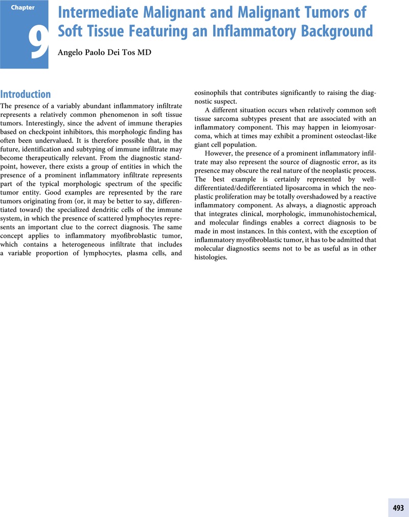
Image of the first page of this content. For PDF version, please use the ‘Save PDF’ preceeding this image.'
Type
Chapter
Information
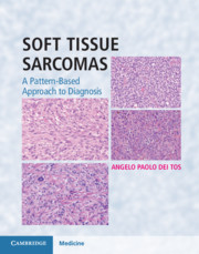
Soft Tissue Sarcomas
A Pattern-Based Approach to Diagnosis
, pp. 493 - 550
Publisher: Cambridge University Press
Print publication year: 2018

Access options

Get access to the full version of this content by using one of the access options below. (Log in options will check for institutional or personal access. Content may require purchase if you do not have access.)

References

Primary Sources

Ehrenstein, V, Andersen, SL, Qazi, I, et al. Tenosynovial giant cell tumor: incidence, prevalence, patient characteristics, and recurrence. A registry-based cohort study in Denmark. J Rheumatol. 2017;44(10):1476–83.Google Scholar
Enzinger, FM, Zhang, RY. Plexiform fibrohistiocytic tumor presenting in children and young adults. An analysis of 65 cases. Am J Surg Pathol. 1988;12:818–26.Google Scholar
Fox, MD, Billings, SD, Gleason, BC, et al. Expression of MiTF may be helpful in differentiating cellular neurothekeoma from plexiform fibrohistiocytic tumor (histiocytoid predominant) in a partial biopsy specimen. Am J Dermatopathol. 2012;34:157–60.CrossRefGoogle Scholar
Hollowood, K, Holley, MP, Fletcher, CD. Plexiform fibrohistiocytic tumour: clinicopathological, immunohistochemical and ultrastructural analysis in favour of a myofibroblastic lesion. Histopathology. 1991;19:503–13.Google Scholar
Jaffer, S, Ambrosini-Spaltro, A, Mancini, AM, Eusebi, V, Rosai, J. Neurothekeoma and plexiform fibrohistiocytic tumor: mere histologic resemblance or histogenetic relationship? Am J Surg Pathol. 2009;33:905–13.Google Scholar
Moosavi, C, Jha, P, Fanburg-Smith, JC. An update on plexiform fibrohistiocytic tumor and addition of 66 new cases from the Armed Forces Institute of Pathology, in honor of Franz M. Enzinger, MD. Ann Diagn Pathol. 2007;11:313–19.Google Scholar
Redlich, GC, Montgomery, KD, Allgood, GA, Joste, NE. Plexiform fibrohistiocytic tumor with a clonal cytogenetic anomaly. Cancer Genet Cytogenet. 1999;108:141–3.Google Scholar
Remstein, ED, Arndt, CA, Nascimento, AG. Plexiform fibrohistiocytic tumor: clinicopathologic analysis of 22 cases. Am J Surg Pathol. 1999;23:662–70.Google Scholar
Salomao, DR, Nascimento, AG. Plexiform fibrohistiocytic tumor with systemic metastases: a case report. Am J Surg Pathol. 1997;21:469–76.CrossRefGoogle ScholarPubMed
Zelger, B, Weinlich, G, Steiner, H, Zelger, BG, Egarter-Vigl, E. Dermal and subcutaneous variants of plexiform fibrohistiocytic tumor. Am J Surg Pathol. 1997;21:235–41.Google Scholar

Secondary Sources

Cupp, JS, Miller, MA, Montgomery, KD, et al. Translocation and expression of CSF1 in pigmented villonodular synovitis, tenosynovial giant cell tumor, rheumatoid arthritis and other reactive synovitides. Am J Surg Pathol. 2007;31:970–6.CrossRefGoogle ScholarPubMed
Folpe, AL, Morris, RJ, Weiss, SW. Soft tissue giant cell tumor of low malignant potential: a proposal for the reclassification of malignant giant cell tumor of soft parts. Mod Pathol. 1999;12:894902.Google ScholarPubMed
Gleason, BC, Fletcher, CD. Deep “benign” fibrous histiocytoma: clinicopathologic analysis of 69 cases of a rare tumor indicating occasional metastatic potential. Am J Surg Pathol. 2008;32:354–62.CrossRefGoogle ScholarPubMed
Guccion, JC, Enzinger, FM. Malignant giant cell tumor of soft parts. An analysis of 32 cases. Cancer. 1972;29:1518–29.Google Scholar
Lee, JC, Liang, CW, Fletcher, CD. Giant cell tumor of soft tissue is genetically distinct from its bone counterpart. Mod Pathol. 2017;30:728–33.Google Scholar
Mancini, I, Righi, A, Gambarotti, M, et al. Phenotypic and molecular differences between giant-cell tumour of soft tissue and its bone counterpart. Histopathology. 2017;71:453–60.CrossRefGoogle ScholarPubMed
Mentzel, T, Calonje, E, Fletcher, CD. Leiomyosarcoma with prominent osteoclast-like giant cells. Analysis of eight cases closely mimicking the so-called giant cell variant of malignant fibrous histiocytoma. Am J Surg Pathol. 1994;18:258–65.Google Scholar
O’Connell, XJ, Wehrli, BM, Nielsen, GP, Rosenberg, AE. Giant cell tumors of soft tissue: a clinicopatologic study of 18 benign and malignant tumors. Am J Surg Pathol. 2000;24:386–95.Google Scholar
Oliveira, AM, Dei Tos, AP, Fletcher, CDM, Nascimento, AG. Primary giant cell tumor of soft tissue: a study of 22 cases. Am J Surg Pathol. 2000;24:248–56.Google Scholar
Rubin, BP. Tenosynovial giant cell tumor and pigmented villonodular synovitis: a proposal for unification of these clinically distinct but histologically and genetically identical lesions. Skeletal Radiol. 2007;6:267–8.Google Scholar
Salm, R, Sissons, HA. Giant-cell tumours of soft tissues. J Pathol. 1972;107:2739.Google Scholar
Sciot, R, Rosai, J, Dal Cin, P, et al. Analysis of 35 cases of localized and diffuse tenosynovial giant cell tumor: a report from the Chromosomes and Morphology (CHAMP) study group. Mod Pathol. 1999;12:576–9.Google Scholar
Tap, WD, Wainberg, ZA, Anthony, SP, et al. Structure-guided blockade of CSF1R kinase in tenosynovial giant-cell tumor. N Engl J Med. 2015;373:428–37.Google Scholar
Akram, S, Pardi, DS, Schaffner, JA, Smyrk, TC. Sclerosing mesenteritis: clinical features, treatment, and outcome in ninety-two patients. Clin Gastroenterol Hepatol. 2007;5:589–96.Google Scholar
Albores-Saavedra, J, Manivel, JC, Essenfeld, H, et al. Pseudosarcomatous myofibroblastic proliferations in the urinary bladder of children. Cancer. 1990;66:1234–41.Google Scholar
Bennett, JA, Nardi, V, Rouzbahman, M, et al. Inflammatory myofibroblastic tumor of the uterus: a clinicopathological, immunohistochemical, and molecular analysis of 13 cases highlighting their broad morphologic spectrum. Mod Pathol. 2017;30:14891503.CrossRefGoogle ScholarPubMed
Bridge, JA, Kanamori, M, Ma, Z, et al. Fusion of the ALK gene to the clathrin heavy chain gene, CLTC, in inflammatory myofibroblastic tumor. Am J Pathol. 2001;159:411–15.Google Scholar
Butrynski, JE, D’Adamo, DR, Hornick, JL, et al. Crizotinib in ALK-rearranged inflammatory myofibroblastic tumor. N Engl J Med. 2010;363:1727–33.CrossRefGoogle ScholarPubMed
Cessna, MH, Zhou, H, Sanger, WG, et al. Expression of ALK1 and p80 in inflammatory myofibroblastic tumor and its mesenchymal mimics: a study of 135 cases. Mod Pathol. 2002;15:931–8.Google Scholar
Cheuk, W, Chan, JK. IgG4-related sclerosing disease: a critical appraisal of an evolving clinicopathologic entity. Adv Anat Pathol. 2010;17:303–32.Google Scholar
Coffin, CM, Hornick, JL, Fletcher, CD. Inflammatory myofibroblastic tumor: comparison of clinicopathologic, histologic, and immunohistochemical features including ALK expression in atypical and aggressive cases. Am J Surg Pathol. 2007;31:509–20.Google Scholar
Coffin, CM, Humphrey, PA, Dehner, LP. Extrapulmonary inflammatory myofibroblastic tumor: a clinical and pathological survey. Semin Diagn Pathol. 1998;15:85101.Google ScholarPubMed
Coffin, CM, Patel, A, Perkins, S, et al. ALK1 and p80 expression and chromosomal rearrangements involving 2p23 in inflammatory myofibroblastic tumor. Mod Pathol. 2001;14:569–76.Google Scholar
Coffin, CM, Watterson, J, Priest, JR, Dehner, LP. Extrapulmonary inflammatory myofibroblastic tumor (inflammatory pseudotumor). A clinicopathologic and immunohistochemical study of 84 cases. Am J Surg Pathol. 1995;19:859–72.CrossRefGoogle ScholarPubMed
Corradi, D, Maestri, R, Palmisano, A, et al. Idiopathic retroperitoneal fibrosis: clinicopathologic features and differential diagnosis. Kidney Int. 2007;72:742–53.Google Scholar
Harik, LR, Merino, C, Coindre, JM, et al. Pseudosarcomatous myofibroblastic proliferations of the bladder: a clinicopathologic study of 42 cases. Am J Surg Pathol. 2006;30:787–94.Google Scholar
Hirsch, MS, Dal Cin, P, Fletcher, CD. ALK expression in pseudosarcomatous myofibroblastic proliferations of the genitourinary tract. Histopathology. 2006;48:569–78.Google Scholar
Hornick, JL, Sholl, LM, Dal Cin, P, Childress, MA, Lovly, CM. Expression of ROS1 predicts ROS1 gene rearrangement in inflammatory myofibroblastic tumors. Mod Pathol. 2015;28:732–9.Google Scholar
Lee, JC, Li, CF, Huang, HY, et al. ALK oncoproteins in atypical inflammatory myofibroblastic tumours: novel RRBP1-ALK fusions in epithelioid inflammatory myofibroblastic sarcoma. J Pathol. 2017;241:316–23.CrossRefGoogle ScholarPubMed
Mariño-Enríquez, A, Wang, WL, Roy, A, et al. Epithelioid inflammatory myofibroblastic sarcoma: An aggressive intra-abdominal variant of inflammatory myofibroblastic tumor with nuclear membrane or perinuclear ALK. Am J Surg Pathol. 2011;35:135–44.CrossRefGoogle ScholarPubMed
Mossé, YP, Voss, SD, Lim, MS, et al. Targeting ALK with crizotinib in pediatric anaplastic large cell lymphoma and inflammatory myofibroblastic tumor: a Children’s Oncology Group study. J Clin Oncol. 2017;35:3215–21.Google Scholar
Nascimento, AF, Ruiz, R, Hornick, JL, Fletcher, CD. Calcifying fibrous ‘pseudotumor’: clinicopathologic study of 15 cases and analysis of its relationship to inflammatory myofibroblastic tumor. Int J Surg Pathol. 2002;10:189–96.CrossRefGoogle ScholarPubMed
Parra-Herran, C, Quick, CM, Howitt, BE, et al. Inflammatory myofibroblastic tumor of the uterus: clinical and pathologic review of 10 cases including a subset with aggressive clinical course. Am J Surg Pathol. 2015;39:157–68.CrossRefGoogle ScholarPubMed
Pettinato, G, Manivel, JC, De Rosa, N, Dehner, LP. Inflammatory myofibroblastic tumor (plasma cell granuloma). Clinicopathologic study of 20 cases with immunohistochemical and ultrastructural observations. Am J Clin Pathol. 1990;94:538–46.Google Scholar
Rossi, GM, Rocco, R, Accorsi Buttini, E, Marvisi, C, Vaglio, A. Idiopathic retroperitoneal fibrosis and its overlap with IgG4-related disease. Intern Emerg Med. 2017;12:287–99.Google Scholar
Sharma, P, Yadav, S, Needham, CM, Feuerstadt, P. Sclerosing mesenteritis: a systematic review of 192 cases. Clin J Gastroenterol. 2017;10:103–11.Google Scholar
Sirvent, N, Hawkins, AL, Moeglin, D, et al. ALK probe rearrangement in a t(2;11;2)(p23;p15;q31) translocation found in a prenatal myofibroblastic fibrous lesion: toward a molecular definition of an inflammatory myofibroblastic tumor family? Genes Chromosomes Cancer. 2001;31:8590.Google Scholar
Sukov, WR, Cheville, JC, Carlson, AW, et al. Utility of ALK-1 protein expression and ALK rearrangements in distinguishing inflammatory myofibroblastic tumor from malignant spindle cell lesions of the urinary bladder. Mod Pathol. 2007;20:592603.Google Scholar
Weaver, J, Goldblum, JR, Turner, S, et al. Detection of MDM2 gene amplification or protein expression distinguishes sclerosing mesenteritis and retroperitoneal fibrosis from inflammatory well-differentiated liposarcoma. Mod Pathol. 2009;22:6670.Google Scholar
Yamamoto, H, Yamaguchi, H, Aishima, S, et al. Inflammatory myofibroblastic tumor versus IgG4-related sclerosing disease and inflammatory pseudotumor: a comparative clinicopathologic study. Am J Surg Pathol. 2009;33:1330–40.Google Scholar
Agaimy, A, Michal, M, Hadravsky, L, Michal, M. Follicular dendritic cell sarcoma: clinicopathologic study of 15 cases with emphasis on novel expression of MDM2, somatostatin receptor 2A, and PD-L1. Ann Diagn Pathol. 2016;23:21–8.Google Scholar
Andersen, EF, Paxton, CN, O’Malley, DP, et al. Genomic analysis of follicular dendritic cell sarcoma by molecular inversion probe array reveals tumor suppressor-driven biology. Mod Pathol. 2017;30:1321–34.Google Scholar
Chan, AC, Chan, KW, Chan, JK, et al. Development of follicular dendritic cell sarcoma in hyaline-vascular Castleman’s disease of the nasopharynx: tracing its evolution by sequential biopsies. Histopathology. 2001;38:510–18.CrossRefGoogle ScholarPubMed
Chan, JK, Fletcher, CD, Nayler, SJ, Cooper, K. Follicular dendritic cell sarcoma. Clinicopathologic analysis of 17 cases suggesting a malignant potential higher than currently recognized. Cancer. 1997;79:294313.3.0.CO;2-W>CrossRefGoogle ScholarPubMed
Chen, T, Gopal, P. Follicular dendritic cell sarcoma. Arch Pathol Lab Med. 2017;141:596–9.Google Scholar
Choe, JY, Go, H, Jeon, YK, et al. Inflammatory pseudotumor-like follicular dendritic cell sarcoma of the spleen: a report of six cases with increased IgG4-positive plasma cells. Pathol Int. 2013;63:245–51.Google Scholar
Griffin, GK, Sholl, LM, Lindeman, NI, Fletcher, CD, Hornick, JL. Targeted genomic sequencing of follicular dendritic cell sarcoma reveals recurrent alterations in NF-κB regulatory genes. Mod Pathol. 2016;29:6774.Google Scholar
Jain, P, Milgrom, SA, Patel, KP, et al. Characteristics, management, and outcomes of patients with follicular dendritic cell sarcoma. Br J Haematol. 2017;178:403–12.CrossRefGoogle ScholarPubMed
Lorenzi, L, Döring, C, Rausch, T, et al. Identification of novel follicular dendritic cell sarcoma markers, FDCSP and SRGN, by whole transcriptome sequencing. Oncotarget. 2017;8:16,463–72.Google Scholar
Pang, J, Mydlarz, WK, Gooi, Z, et al. Follicular dendritic cell sarcoma of the head and neck: Case report, literature review, and pooled analysis of 97 cases. Head Neck. 2016;38 Suppl 1:E22419.Google Scholar
Pileri, SA, Grogan, TM, Harris, NL, et al. Tumors of histiocytes and accessory dendritic cells. An immunohistochemical approach to classification from the International Lymphoma Study Group based on 61 cases. Histopathology. 2002;41:129.Google Scholar
Shek, TW, Ho, FC, Ng, IO, et al. Follicular dendritic cell tumor of the liver. Evidence for an Epstein-Barr virus-related clonal proliferation of follicular dendritic cells. Am J Surg Pathol. 1996;20:313–24.CrossRefGoogle ScholarPubMed
Shia, J, Chen, W, Tang, LH, et al. Extranodal follicular dendritic cell sarcoma: clinical, pathologic, and histogenetic characteristics of an underrecognized disease entity. Virchows Arch. 2006;449:148–58.Google Scholar
Sun, X, Chang, KC, Abruzzo, LV, et al. Epidermal growth factor receptor expression in follicular dendritic cells: a shared feature of follicular dendritic cell sarcoma and Castleman’s disease. Hum Pathol. 2003;34:835–40.Google Scholar
Vermi, W, Lonardi, S, Bosisio, D, et al. Identification of CXCL13 as a new marker for follicular dendritic cell sarcoma. J Pathol. 2008;216:356–64.CrossRefGoogle ScholarPubMed
Gaertner, EM, Tsokos, M, Derringer, GA, et al. Interdigitating dendritic cell sarcoma. A report of four cases and review of the literature. Am J Clin Pathol. 2001;115:589–97.Google Scholar
Magro, CM, Olson, LC, Nuovo, G, Solomon, GJ. Primary cutaneous interdigitating dendritic cell sarcoma is a morphologic and phenotypic simulator of poorly differentiated metastatic melanoma: A report of 2 cases and review of the literature. Ann Diagn Pathol. 2017;30:5965.Google Scholar
Ohtake, H, Yamakawa, M. Interdigitating dendritic cell sarcoma and follicular dendritic cell sarcoma: histopathological findings for differential diagnosis. J Clin Exp Hematop. 2013;53:179–84.Google Scholar
O’Malley, DP, Zuckerberg, L, Smith, LB, et al. The genetics of interdigitating dendritic cell sarcoma share some changes with Langerhans cell histiocytosis in select cases. Ann Diagn Pathol. 2014;18:1820.Google Scholar
Pillay, K, Solomon, R, Daubenton, JD, Sinclair-Smith, CC. Interdigitating dendritic cell sarcoma: a report of four paediatric cases and review of the literature. Histopathology. 2004;44:283–91.Google Scholar
Stowman, AM, Mills, SE, Wick, MR. Spindle cell melanoma and interdigitating dendritic cell sarcoma: do they represent the same process? Am J Surg Pathol. 2016;40:1270–9.CrossRefGoogle ScholarPubMed
Chan, AC, Serrano-Olmo, J, Erlandson, RA, et al. Cytokeratin- positive malignant tumors with reticulum cell morphology: a subtype of fibroblastic reticulum cell neoplasm? Am J Surg Pathol. 2000;24:107–16.CrossRefGoogle ScholarPubMed
Goto, N, Tsurumi, H, Takami, T, et al. Cytokeratin-positive fibroblastic reticular cell tumor with follicular dendritic cell features: a case report and review of the literature. Am J Surg Pathol. 2015;39:573–80.CrossRefGoogle ScholarPubMed
Martel, M, Sarli, D, Colecchia, M, et al. Fibroblastic reticular cell tumor of the spleen: report of a case and review of the entity. Hum Pathol. 2003;34:954–7.Google Scholar
Schuerfeld, K, Lazzi, S, De Santi, MM, et al. Cytokeratin-positive interstitial cell neoplasm: a case report and classification issues. Histopathology. 2003;43:491–4.CrossRefGoogle ScholarPubMed
Ansari, J, Naqash, AR, Munker, R, et al. Histiocytic sarcoma as a secondary malignancy: pathobiology, diagnosis, and treatment. Eur J Haematol. 2016;97:916.Google Scholar
Feldman, AL, Minniti, C, Santi, M, et al. Histiocytic sarcoma after acute lymphoblastic leukaemia: a common clonal origin. Lancet Oncol. 2004;5:248–50.Google Scholar
Foucar, E, Rosai, J, Dorfman, R. Sinus histiocytosis with massive lymphadenopathy (Rosai-Dorfman disease): review of the entity. Semin Diagn Pathol. 1990;7:1973.Google Scholar
Howard, JE, Dwivedi, RC, Masterson, L, Jani, P. Langerhans cell sarcoma: a systematic review. Cancer Treat Rev. 2015;41:320–31.Google Scholar
Kordes, M, Röring, M, Heining, C, et al. Cooperation of BRAF(F595L) and mutant HRAS in histiocytic sarcoma provides new insights into oncogenic BRAF signaling. Leukemia. 2016;30:937–46.Google Scholar
Lauwers, GY, Perez-Atayde, A, Dorfman, RF, Rosai, J. The digestive system manifestations of Rosai-Dorfman disease (sinus histiocytosis with massive lymphadenopathy): review of 11 cases. Hum Pathol. 2000;31:380–5.Google Scholar
Liu, Q, Tomaszewicz, K, Hutchinson, L, et al. Somatic mutations in histiocytic sarcoma identified by next generation sequencing. Virchows Arch. 2016;469:233–41.Google Scholar
Nakamine, H, Yamakawa, M, Yoshino, T, et al. Langerhans cell histiocytosis and Langerhans cell sarcoma: current understanding and differential diagnosis. J Clin Exp Hematop. 2016;56:109–18.Google Scholar
Rosai, J, Dorfman, RF. Sinus histiocytosis with massive lymphadenopathy. A newly recognized benign clinicopathological entity. Arch Pathol. 1969;87:6370.Google Scholar
Sun, W, Nordberg, ML, Fowler, MR. Histiocytic sarcoma involving the central nervous system: clinical, immunohistochemical, and molecular genetic studies of a case with review of the literature. Am J Surg Pathol. 2003;27:258–65.Google Scholar
Thakral, B, Khoury, JD. Histiocytic sarcoma: secondary neoplasm or “transdifferentiation” in the setting of B-acute lymphoblastic leukemia. Blood. 2016;128:2475.Google Scholar
van der Valk, P, Meijer, CJ, Willemze, R, et al. Histiocytic sarcoma (true histiocytic lymphoma): a clinicopathological study of 20 cases. Histopathology. 1984;8:105–23.Google Scholar
Vos, JA, Abbondanzo, SL, Barekman, CL, et al. Histiocytic sarcoma: a study of five cases including the histiocyte marker CD163. Mod Pathol. 2005;18:693704.Google Scholar

Save book to Kindle

To save this book to your Kindle, first ensure coreplatform@cambridge.org is added to your Approved Personal Document E-mail List under your Personal Document Settings on the Manage Your Content and Devices page of your Amazon account. Then enter the ‘name’ part of your Kindle email address below. Find out more about saving to your Kindle.

Note you can select to save to either the @free.kindle.com or @kindle.com variations. ‘@free.kindle.com’ emails are free but can only be saved to your device when it is connected to wi-fi. ‘@kindle.com’ emails can be delivered even when you are not connected to wi-fi, but note that service fees apply.

Find out more about the Kindle Personal Document Service.

Available formats
×

Save book to Dropbox

To save content items to your account, please confirm that you agree to abide by our usage policies. If this is the first time you use this feature, you will be asked to authorise Cambridge Core to connect with your account. Find out more about saving content to Dropbox.

Available formats
×

Save book to Google Drive

To save content items to your account, please confirm that you agree to abide by our usage policies. If this is the first time you use this feature, you will be asked to authorise Cambridge Core to connect with your account. Find out more about saving content to Google Drive.

Available formats
×