


Introduction
Avian haemosporidian parasites comprise species of the genera Haemoproteus, Plasmodium, Leucocytozoon and Fallisia (Valkiūnas, Reference Valkiūnas2005; Perkins, Reference Perkins2014). Haemoproteus spp. are parasites of avian erythrocytes and reticulo-endothelial cells of numerous internal organs. These parasites can be distinguished from other avian haemosporidians by the presence of malarial pigment granules (haemozoin) in blood stages and the absence of asexual reproduction (merogony) in circulating blood cells (Valkiūnas, Reference Valkiūnas2005). Species of this genus are classified into 2 subgenera according to the differences in their sporogony and dipteran vectors. Representatives of the subgenus Haemoproteus are transmitted by louse flies (Hippoboscidae) and produce large-size oocysts containing numerous germinative centres. Representatives of the subgenus Parahaemoproteus are transmitted by biting midges (Ceratopogonidae) and produce tiny oocysts containing only 1 germinative centre (Valkiūnas, Reference Valkiūnas2005). Haemoproteus species are generally presumed to be host-specific. They are not usually transmitted between birds belonging to different orders, and there is limited evidence that some species are host-specific to the family level (Valkiūnas, Reference Valkiūnas2005). However, there are some notable exceptions to this rule, as demonstrated by penguins (Sphenisciformes: Spheniscidae) infected by Haemoproteus larae, which is a common parasite of gulls (Charadriiformes: Laridae) (Inumaru et al., Reference Inumaru, Aratani, Shimizu, Yamamoto, Sato, Murata and Valkiūnas2020).
Charadriiformes is a diverse order of birds, comprising nearly 400 species distributed in 19 families (Gill et al., Reference Gill, Donsker and Rasmussen2022). The suborder Lari comprises 6 families, including aquatic birds in the families Laridae (gulls, terns, noddies, skimmers; 103 species), Alcidae (auklets, murres, puffins; 25 species), Stercorariidae (skuas; 7 species) and Dromadidae (crab-plover; 1 species), as well as terrestrial birds in the families Turnicidae (buttonquails; 18 species) and Glareolidae (coursers, pranticoles; 17 species) (Cracraft et al., Reference Cracraft, Barker, Cibois, Dickinson, Bahr, Dowsett, Pearson, Remsen, Roselaar and Schodde2003; Ericson et al., Reference Ericson, Envall, Irestedt and Norman2003; Gill et al., Reference Gill, Donsker and Rasmussen2022). In spite of the species diversity of the suborder Lari, a relatively small diversity of haemosporidian parasites has been recorded infecting these birds (Valkiūnas, Reference Valkiūnas2005; Quillfeldt et al., Reference Quillfeldt, Arriero, Martínez, Masello and Merino2011; Clark et al., Reference Clark, Clegg and Lima2014). This may be related to a smaller sampling effort compared to other more extensively studied avian groups (e.g. Passeriformes and Galliformes), but it could also relate to the fact that Lari bird species often inhabit either saltwater/brackish or arid environments where the insect vectors of haemosporidian parasites are often less abundant (Jovani et al., Reference Jovani, Tella, Forero, Bertellotti, Blanco, Ceballos and Donázar2001; Martínez-Abraín et al., Reference Martínez-Abraín, Esparza and Oro2004; Quillfeldt et al., Reference Quillfeldt, Arriero, Martínez, Masello and Merino2011).
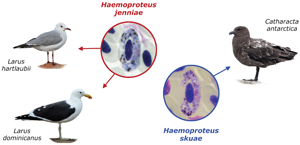
Four morphospecies of Haemoproteus have been detected infecting birds of the suborder Lari, in addition to several unidentified genetic lineages (Table 1). Haemoproteus (Parahaemoproteus) larae is a frequent parasite of several species of gulls (Larus spp.) and terns (Sterna spp.) in the Palaearctic region (Kairullaev, Reference Kairullaev1986; Quillfeldt et al., Reference Quillfeldt, Arriero, Martínez, Masello and Merino2011; Inumaru et al., Reference Inumaru, Murata and Sato2017, Reference Inumaru, Aratani, Shimizu, Yamamoto, Sato, Murata and Valkiūnas2020). Haemoproteus (Haemoproteus) jenniae infects swallow-tailed gulls (Creagrus furcatus) at the Galapagos Islands, Ecuador (Levin et al., Reference Levin, Valkiūnas, Iezhova, O'Brien and Parker2012), and its DNA was detected in the blood of brown noddies (Anous stolidus) at Rocas Atoll, Brazil (Quillfeldt et al., Reference Quillfeldt, Martínez, Bugoni, Mancini and Merino2014), and in the blood of black-headed gulls (Larus ridibundus) in Poland (Włodarczyk et al., Reference Włodarczyk, Bouwhuis, Bichet, Podlaszczuk, Chyb, Indykiewicz, Dulisz, Betleja, Janiszewski and Minias2022). Haemoproteus skuae, for which the subgeneric position has not yet been determined, was described from a brown skua (Catharacta antarctica) sampled while under care at a rehabilitation facility in Cape Town, South Africa (Parsons et al., Reference Parsons, Peirce and Strauss2010, Reference Parsons, Voogt, Schaefer, Peirce and Vanstreels2017), and has not been recorded since its original description. Additionally, DNA from Haemoproteus (Parahaemoproteus) macrovacuolatus was detected in the blood of black skimmers (Rynchops niger) in Brazilian Amazon (Roos et al., Reference Roos, Belo, Silveira and Braga2015); however, considering that H. macrovacuolatus is a frequent parasite of black-bellied whistling-ducks (Dendrocygna autumnalis) (Matta et al., Reference Matta, Pacheco, Escalante, Valkiūnas, Ayerbe-Quiñones and Acevedo-Cendales2014), its occurrence in black skimmers could represent an abortive spill-over infection.
Table 1. Summary of Haemoproteus morphospecies and cytb lineages recorded in birds of the suborder Lari

a Phylogenetic analysis suggests this lineage corresponds to the morphospecies Haemoproteus. (Haemoproteus) multipigmentatus.
b Also recorded in Columbiformes.
c Also recorded in Falconiformes.
d Also recorded in Sphenisciformes.
e Also recorded in Anseriformes.
Although the blood parasites of Lari birds have been extensively studied in Eurasia and the Americas, the African continent remains understudied in this regard (Quillfeldt et al., Reference Quillfeldt, Arriero, Martínez, Masello and Merino2011; Parsons et al., Reference Parsons, Voogt, Schaefer, Peirce and Vanstreels2017). A recent study reported the detection of Haemoproteus sp. in the blood smears from kelp gulls (Larus dominicanus) at various breeding colonies in South Africa, with an average prevalence of 1.6% in chicks (N = 121) and 39.3% in adults (N = 211), however the parasite species was not identified (Reusch et al., Reference Reusch, Ryan and Pichegru2022). In this study, we report on the occurrence of H. jenniae in kelp gulls and Hartlaub's gull (Larus hartlaubii) in South Africa and complement the original description of H. skuae based on its type specimens. Blood stages of H. jenniae and H. skuae have similarities in morphology, and the characters that can be used to distinguish between these parasites are discussed.
Materials and methods
On 15 September 2012, adult kelp gulls were captured and blood samples were sampled at the breeding colony on Robben Island (N = 21; Western Cape, South Africa, 33°47′37″S, 18°21′47″E). The sampled individuals were selected at random and appeared to be healthy (good body condition, no injuries or deformities, normal behaviour and breathing). Additionally, we analysed archival blood samples collected in 2011–2018 from adult kelp gulls under care at rehabilitation facilities of the Southern African Foundation for the Conservation of Coastal Birds (SANCCOB) in Cape Town (N = 13; Western Cape, South Africa, 33°50′01″S, 18°29′29″E) and Port Elizabeth/Gqeberha (N = 2; Eastern Cape, South Africa, 34°00′48″S, 25°41′26″E), and 1 archival blood sample from an adult Hartlaub's gull collected in 2018 at the SANCCOB rehabilitation facility in Cape Town.
For all individuals, blood samples (<1% body mass) were collected from the medial metatarsal or brachial vein. Thin blood smears were freshly prepared, air dried, fixed with absolute methanol, stained with an eosin–methylene blue stain (Kyro-Quick stain set, Kyron Laboratories Pty Ltd, Benrose, South Africa). Blood smears were examined under light microscopy. To estimate parasite prevalence, blood smears from the kelp gulls sampled at Robben Island were screened by examining 200 fields under 1000× magnification (minimum 50 000 erythrocytes).
Blood parasites were morphologically identified using published keys and descriptions (Valkiūnas, Reference Valkiūnas2005; Parsons et al., Reference Parsons, Peirce and Strauss2010; Levin et al., Reference Levin, Valkiūnas, Iezhova, O'Brien and Parker2012; Inumaru et al., Reference Inumaru, Aratani, Shimizu, Yamamoto, Sato, Murata and Valkiūnas2020) and were quantified with the assistance of digital image analysis to count 1000 erythrocytes (Gering and Atkinson, Reference Gering and Atkinson2004). A high-resolution digital camera (AmScope MU1803-HS, United Scope LLC, Irvine, CA, USA) coupled to a light microscope was used to obtain photographs of parasites, and ImageJ 1.53a (Schneider et al., Reference Schneider, Rasband and Eliceiri2012) was used to obtain measurements from uninfected erythrocytes (n = 30), macrogametocytes and their infected cells (n = 30), and microgametocytes and their infected cells (n = 20). The nucleus displacement ratio (NDR) was calculated (Bennett and Campbell, Reference Bennett and Campbell1972). Mann–Whitney tests were conducted to compare the measurements of the parasites from kelp and Hartlaub's gulls. In order to perform proper morphological identification of the parasite found in the gulls, a hapantotype blood smear of H. skuae, deposited at the International Reference Centre for Avian Haematozoa (IRCAH), Queensland Museum (accession number G465379) (Parsons et al., Reference Parsons, Peirce and Strauss2010), was analysed.
One archival sample from a kelp gull (identification number K035/2018, admitted for rehabilitation on 7 March 2018 from Saldanha Bay, 33°04′2″S, 17°56′34″E, and sampled on 26 March 2018 at the Cape Town facility) was selected for parasite measurements and molecular analysis. This sample was selected because the blood smears were of excellent quality and did not present co-infection by other blood parasites (as confirmed through the microscopic examination of 150 000 erythrocytes); a voucher blood smear from this sample was deposited at the collection of IRCAH under accession number G466232.
Total blood was stored at −20°C, and then was transferred to 100% ethanol. Approximately 10 μL of blood was transferred to a 1.5 mL microtube and dried at 37°C for subsequent DNA extraction using the phenol–chloroform method with isopropanol precipitation (Sambrook and Russell, Reference Sambrook and Russell2001). A nested polymerase chain reaction (PCR) test targeting the mitochondrial cytochrome b (cytb) gene of Haemoproteus and Plasmodium was employed (Hellgren et al., Reference Hellgren, Waldenström and Bensch2004). Amplification products of the cytb PCR were purified with polyethylene glycol 8000 (Sambrook and Russell, Reference Sambrook and Russell2001), and sequenced (bi-directional Sanger sequencing with dye-terminator fluorescent labelling). The resulting DNA sequence was deposited in GenBank (accession number OL906299) and MalAvi (lineage code LARDOM01) and compared to publicly available Haemoproteus sequences from the MalAvi database (Bensch et al., Reference Bensch, Hellgren and Pérez-Tris2009).
Phylogenetic relationships were evaluated through a Bayesian phylogenetic tree produced using MrBayes 3.2.7 (Ronquist et al., Reference Ronquist, Teslenko, Van Der Mark, Ayres, Darling, Höhna, Larget, Liu, Suchard and Huelsenbeck2012) with the GTR + I + G model of nucleotide evolution as recommended by jModelTest 2 (Darriba et al., Reference Darriba, Taboada, Doallo and Posada2012) and using reference cytb lineages from public databases (Supplementary File S1). Two Markov chains were run simultaneously for 5 million generations that were sampled every 1000 generations, and the first 1250 trees (25%) were discarded as a burn-in step. The tree was rooted with Leucocytozoon as an outgroup as recommended by multi-gene phylogenetic analyses (Borner et al., Reference Borner, Pick, Thiede, Kolawole, Kingsley, Schulze, Cottontail, Wellinghausen, Schmidt-Chanasit, Bruchhaus and Burmester2016).
Due to the host's small body size, the archival sample from Hartlaub's gull (identification number H230/2018, admitted for rehabilitation on 7 June 2018 from Eden on the Bay, 33°47′35″S, 18°27′22″E, and sampled on 18 June 2018 at the Cape Town facility) did not have a corresponding aliquot of total blood that could be used for molecular analysis. DNA extraction from the blood smear was not considered feasible due to the low parasitaemia. Instead, the blood smear from this sample was deposited at the collection of the IRCAH under accession number G466252.
Results
Haemoproteus sp. infection was detected in the blood smears of 4 of 21 (19.0%) wild kelp gulls sampled at Robben Island. The archived blood smears from 15 kelp gulls and 1 Hartlaub's gull under care at the rehabilitation facilities had been pre-selected for further study on the basis that they had Haemoproteus sp. infections, and therefore prevalence cannot be estimated for this cohort.
The blood smears of all 15 kelp gulls sampled at the rehabilitation facilities showed the presence of a single morphospecies, H. jenniae (Fig. 1). The Hartlaub's gull was also infected by H. jenniae (Fig. 2). The morphological identification of H. jenniae in both hosts was confirmed in blood smears due to the presence of the following diagnostic characteristics: (i) young gametocytes can be seen anywhere in the infected cells, but mainly on erythrocyte poles (Figs 1A and 2A) and can have a wavy to amoeboid outline (Figs 1B–G, Q and 2B, C, E, F); (ii) the presence of unstained spaces in the cytoplasm of young gametocytes (Figs 1D, E and 2D), even though they are less frequently seen in H. jenniae from Hartlaub's gull; (iii) the presence of 1 large vacuole in mature or nearly mature gametocytes (Figs 1D, E, N and 2J, M), also less frequent in H. jenniae from Hartlaub's gull; (iv) growing gametocytes can slightly touch the parasite nucleus in several points (Figs 1I, K and 2C, F) or not touch it, forming thin clefts (Figs 1M, O and 2E); (v) fully grown gametocytes are circumnuclear, they occupy the entire cytoplasm of the infected cells (Figs 1P and 2K, L) and can slightly rotate the erythrocyte nuclei (Figs 1T and 2G, P); (vi) fully grown gametocytes contain predominantly roundish or slightly oval, of approximately uniform size pigment granules (elongate rod-like pigment granules usually are absent) (Figs 1P, T and 2L, O) and (vii) nuclei are diffuse in microgametocytes (Figs 1Q–T and 2M–P).

Fig. 1. Haemoproteus jenniae cytb lineage LARDOM1 from the blood of a kelp gull (Larus dominicanus). All images from the voucher specimen IRCAH accession number G466232. Young gametocytes (A–D), macrogametocytes (E–P) and microgametocytes (Q–T). Simple arrowhead, pigment granules; long simple arrow, parasite nucleus; short simple arrow, vacuole; long simple wide arrow, cleft between parasite and erythrocyte nucleus. Sample K035/2018, eosin–methylene blue stain. Scale bar = 10 μm.

Fig. 2. Haemoproteus jenniae cytb lineage unknown from the blood of a Hartlaub's gull (Larus hartlaubii). All images from the voucher specimen IRCAH accession number G466252. Young gametocytes (A–D), macrogametocytes (E–L) and microgametocytes (M–P). Simple arrowhead, pigment granules; long simple arrow, parasite nucleus; short simple arrow, vacuole; long simple wide arrow, cleft between parasite and erythrocyte nucleus. Sample H230/2018, eosin–methylene blue stain. Scale bar = 10 μm.
A detailed description of macrogametocytes and microgametocytes of H. jenniae (cytb lineage CREFUR01) was provided by Levin et al. (Reference Levin, Valkiūnas, Iezhova, O'Brien and Parker2012) and is not repeated here. However, it is necessary to mention that H. jenniae CREFUR01 induces hypertrophy of the infected erythrocytes in width, which is neither the case for the new H. jenniae LARDOM01 nor for the H. jenniae from Hartlaub's gull (Table 2). It is also noteworthy that the H. jenniae from Hartlaub's gull had generally smaller gametocytes and its macrogametocytes had more numerous pigment granules than those of H. jenniae LARDOM01 and H. jenniae CREFUR01 (Table 2). Mann–Whitney tests detected significant differences (P < 0.05) in the following measurements of H. jenniae LARDOM01 compared to H. jenniae from Hartlaub's gull: macrogametocyte length (W = 703, P = 0.002), area (W = 733, P = 0.007), pigment granules (W = 1137, P = 0.001) and NDR (W = 1245, P < 0.001), and microgametocyte width (W = 284, P < 0.001) and NDR (W = 541, P < 0.001). The slight differences seen between H. jenniae LARDOM01 from kelp gulls and the H. jenniae from the Hartlaub's gull might indicate that they belong to different parasite lineages.
Table 2. Morphometry of host cells and mature gametocytes of H. jenniae (cytb lineage LARDOM01) from a kelp gull (L. dominicanus), H. jenniae (cytb lineage not determined) from a Hartlaub's gull (L. hartlaubii), H. jenniae (cytb lineage CREFUR01) from a swallow-tailed gull (C. furcatus) and H. skuae (cytb lineage not determined) from a brown skua (C. antarctica)

Measurements are provided: mean ± s.d. (minimum–maximum).
a This study.
b Levin et al. (Reference Levin, Valkiūnas, Iezhova, O'Brien and Parker2012).
c Parsons et al. (Reference Parsons, Peirce and Strauss2010).
The morphology of the parasites from the kelp gulls sampled at Robben Island could not be thoroughly characterized due to low parasitaemia (<10 parasites per blood film), but all parasite forms seen were consistent with H. jenniae as seen in the gulls sampled at rehabilitation facilities. One kelp gull (not K035/2018) and the Hartlaub's gull, both sampled at the Cape Town rehabilitation facility, had co-infection by Babesia sp.; morphological and molecular analyses to characterize these parasites are under way and will be presented elsewhere.
Phylogenetic analysis of a partial sequence of the cytb gene (Fig. 3) revealed that the studied parasite is a part of the clade consisting of Haemoproteus subgenus species, and it is most closely related to H. jenniae (CREFUR01). The cytb lineages LARDOM01 and CREFUR01 differed by 3 nucleotides (sequence identity = 476/479 or 99.37%). Both lineages clustered with Haemoproteus iwa (FREMIN01) from frigatebirds (Fregata spp.) (Fig. 3).

Fig. 3. Bayesian phylogenetic tree of a 479 bp fragment of the cytb gene of Haemosporida. Branch lengths are drawn proportionally to the extent of changes (scale bar is shown). Values adjacent to nodes represent posterior probabilities. MalAvi/GenBank accession codes are provided for each lineage. The lineage obtained in this study (red) and other H. (Haemoproteus) spp. lineages previously reported in seagulls (blue) are highlighted. The asterisk indicates lineages attributed to the subgenus Haemoproteus.
Redescription of Haemoproteus skuae Parsons, Peirce and Strauss, Reference Parsons, Peirce and Strauss2010
Type host: Catharacta antarctica (Lesson, 1831).
Type locality: SANCCOB, Bloubergrant, South Africa.
Vector: Unknown.
Description (Fig. 4 and Table 2): The hapantotype of H. skuae was examined, and the following diagnostic characters were observed and complement the original description of this species.

Fig. 4. Haemoproteus skuae from the blood of a brown skua (Catharacta antarctica). Young gametocyte (A), macrogametocytes (B–L) and microgametocytes (M–P). All images from the hapantotype IRCAH accession number G465379. Simple arrowhead, pigment granules; long simple arrow, parasite nucleus; short simple arrow, vacuole; long simple wide arrow, cleft between parasite and erythrocyte nucleus. Sample S02/2006, eosin–methylene blue stain. Scale bar = 10 μm.
Young gametocytes (Fig. 4A): elongated, predominantly located in a sub-polar position in the infected erythrocytes (Fig. 4A); outline is even or slightly amoeboid (Fig. 4A); they neither touch the nucleus nor the envelope of infected erythrocytes (Fig. 4A).
Macrogametocytes (Fig. 4B–L): growing forms have an even or slightly amoeboid outline (Fig. 4D–F); they extend longitudinally along the erythrocyte nuclei touching the envelope of erythrocytes but not the erythrocyte nuclei (Fig. 4B–F), forming thick distinct lateral ‘clefts’ (Fig. 4E, I) – a characteristic feature of this species growth; parasite nucleus is small and frequently seen in a subcentral to subterminal position (Fig. 4C). Readily visible large unfilled spaces (‘clefts’) are usually present between ends of advanced gametocytes and poles of erythrocyte nuclei (Fig. 4F, G, I) – also a distinctive feature of this species development. Fully grown macrogametocytes have a cytoplasm with a granular appearance (Fig. 4H); their outline varies from even to wavy (Fig. 4G, H). Nucleus is usually of central position (Fig. 4J, K), but sometimes in a subcentral position (Fig. 4H, L). Frequently seen touching or slightly touching erythrocyte nucleus (Fig. 4G, J); however, a more or less evident space between the parasites and erythrocyte nuclei was usually visible even in the largest parasites (Fig. 4K, L). Advanced forms grow around infected cell nuclei (Fig. 4J, K) and can enclose it completely (Fig. 4K–L), but often not touching the poles of erythrocytes nucleus (Fig. 4L). A single vacuole (Fig. 4K) was seen occasionally in mature macrogametocytes, but the cytoplasm was not markedly vacuolated in most gametocytes. Pigment granules were mainly of small (<0.5 μm) and medium (0.5–1 μm) size, markedly variable in form; the roundish, oval and elongate (rod-like) granules occur; the later form is common (Fig. 4L).
Microgametocytes (Fig. 4M–P): growing forms extend longitudinally along the erythrocyte nuclei (Fig. 4M), outline is even or slightly amoeboid (Fig. 4M); the parasite does not touch the envelope or the nucleus of the host cell. Pigment granules are small (<0.5 μm) and medium (0.5–1 μm) size, mainly in roundish forms (Fig. 4M). Outline of advanced microgametocytes varies from highly amoeboid (Fig. 4N) to even (Fig. 4O–P); a single vacuole was seen in some microgametocytes (Fig. 4N, P), but the cytoplasm is not markedly vacuolated. Microgametocytes have nuclei with a highly condensed chromatin (Fig. 4M–P). Fully grown gametocytes are closely appressed to the nuclei of erythrocytes, filling the cytoplasm of the infected cells up to their poles and touching the cell envelope (Fig. 4O). Fully grown microgametocytes can encircle the erythrocyte nuclei (Fig. 4P).
Discussion
This is the first record of H. jenniae in Africa, and the first record of this parasite in kelp gull and Hartlaub's gull. There are only 3 previous records of H. jenniae. The species was described from swallow-tailed gulls at Española Island, Galapagos Islands (Levin et al., Reference Levin, Valkiūnas, Iezhova, O'Brien and Parker2012). DNA from H. jenniae was also detected in the blood of brown noddies at Rocas Atoll, off northeast Brazil, but gametocytes were not seen in blood smears (Quillfeldt et al., Reference Quillfeldt, Martínez, Bugoni, Mancini and Merino2014), so it remains unclear if development completes in this avian host. DNA from H. jenniae was also detected in the blood of black-headed gulls in Poland, although blood smears were not examined (Włodarczyk et al., Reference Włodarczyk, Bouwhuis, Bichet, Podlaszczuk, Chyb, Indykiewicz, Dulisz, Betleja, Janiszewski and Minias2022).
Haemoproteus jenniae prevalence in this study was 19% in kelp gulls sampled at Robben Island. This is consistent with a recent study reporting a prevalence of Haemoproteus sp. ranging between 13 and 56% in the blood smears of adult kelp gulls sampled at other breeding colonies in South Africa (Reusch et al., Reference Reusch, Ryan and Pichegru2022) and with the 23% prevalence of H. jenniae in swallow-tailed gulls at Española Island (Levin et al., Reference Levin, Valkiūnas, Iezhova, O'Brien and Parker2012). Włodarczyk et al. (Reference Włodarczyk, Bouwhuis, Bichet, Podlaszczuk, Chyb, Indykiewicz, Dulisz, Betleja, Janiszewski and Minias2022) reported a 44% prevalence of DNA from Haemoproteus spp. in black-headed gulls in Poland; however, cytb sequencing of a subset of individuals revealed that only 4% of these infections corresponded to H. jenniae, therefore species-specific prevalence of H. jenniae in that study may be estimated at 1.8%. In comparison, DNA from H. jenniae was detected in 8% of brown noddies at Rocas Atoll (Quillfeldt et al., Reference Quillfeldt, Martínez, Bugoni, Mancini and Merino2014).
None of the free-ranging gulls or noddies infected with this parasite showed signs of illness (Levin et al., Reference Levin, Valkiūnas, Iezhova, O'Brien and Parker2012; Quillfeldt et al., Reference Quillfeldt, Martínez, Bugoni, Mancini and Merino2014). Furthermore, Haemoproteus sp. infection status does not have a significant effect on the body condition of kelp gulls (Reusch et al., Reference Reusch, Ryan and Pichegru2022), which suggests low pathogenicity at chronic infection. Because the gulls sampled at the South African rehabilitation facility in the current study had other health problems (e.g. malnutrition, botulism, trauma), it is not possible to determine the role that the infections may have played, potentially impairing their health and causing them to be brought for rehabilitation. Studies evaluating subtler signs of disease (e.g. behaviour, breeding success), physiological indicators (e.g. haematology, plasma chemistry) and tissue damage associated with tissue meronts (histopathology) will therefore be necessary to uncover the potential health effects of H. jenniae infections to their hosts. Additionally, it is known that some Haemoproteus species can cause extensive damage to organs, such as Haemoproteus pastoris infecting common starlings (Sturnus vulgaris), in which megalomeronts were reported in the brain and other organs of infected birds (Duc et al., Reference Duc, Ilgūnas, Kubiliūnaitė and Valkiūnas2021). This should be taken into consideration by veterinarians in wildlife rehabilitation centres, since infected birds might present signs of disease due to the presence of Haemoproteus tissue stages, even if they have a low parasitaemia, or the parasitaemia is absent.
Hartlaub's gulls are endemic residents of Namibia and South Africa (BirdLife International and Handbook of the Birds of the World, 2019). Kelp gulls are widely distributed in the Southern Hemisphere, including Namibia and South Africa, and are present at the Galapagos Islands but not at Rocas Atoll (BirdLife International and Handbook of the Birds of the World, 2019). In fact, there are no records of gulls at Rocas Atoll, in contrast to the large numbers of terns and noddies that breed on the atoll (Azevedo Júnior, Reference Azevedo Júnior1992; Schulz-Neto, Reference Schulz-Neto and Branco2004). The detection of DNA from H. jenniae in noddies at Rocas Atoll (Quillfeldt et al., Reference Quillfeldt, Martínez, Bugoni, Mancini and Merino2014) therefore suggests that this parasite is not exclusive to gulls, but probably also infects noddies and terns (which are also members of the family Laridae; Baker et al., Reference Baker, Pereira and Paton2007). To improve our understanding of the ecology of this parasite, additional studies would be valuable to evaluate the occurrence of H. jenniae in other Laridae bird populations. For instance, further research is necessary to investigate whether the Haemoproteus sp. reported by Lowery (Reference Lowery1971) in the blood of a brown noddy sampled at Aldabra Atoll, Indian Ocean, might have corresponded to H. jenniae. Further studies at sites where swallow-tailed gulls (Malpelo Island, off Colombia; BirdLife International and Handbook of the Birds of the World, 2019) and kelp gulls are present (e.g. mainland South America, Australia, New Zealand, Subantarctic islands; BirdLife International and Handbook of the Birds of the World, 2019) are of particular interest, and it would be interesting to sample other sympatric Laridae (including brown noddies) that might share this parasite. Grey-hooded gulls (Larus cirrocephalus) and Sabine's gulls (Xema sabini), whose breeding distribution overlaps with that of kelp gulls and Hartlaub's gulls in southern Africa (BirdLife International and Handbook of the Birds of the World, 2019), should also be considered as potential hosts of H. jenniae.
The dipteran vectors of H. jenniae are unknown, but hippoboscid flies are suspected based on its phylogenetic clustering with other species of the subgenus Haemoproteus (Fig. 3). This is corroborated by the fact that DNA from H. iwa, a parasite of frigatebirds (Fregata spp.) that is closely related to H. jenniae, was detected in Olfersia flies (Levin et al., Reference Levin, Valkiūnas, Santiago-Alarcon, Cruz, Iezhova, O'Brien, Hailer, Dearborn, Schreiber, Fleischer, Ricklefs and Parker2011). Species of Olfersia (O. aenescens, O. fossulata, O. spinifera) have been recorded parasitizing gulls, terns and noddies (Maa, Reference Maa1969) and are plausible vectors of H. jenniae. Other genera of hippoboscid flies also known to parasitize gulls, terns and noddies comprise species of Icosta (I. albipennis, I. americana), Ornithoica (O. pusilla), Ornithomya (O. anchineuria, O. chloropus) and Ornitophila (O. gestroi) (Maa, Reference Maa1969; Nartshuk and Matyukhin, Reference Nartshuk and Matyukhin2019). There is no information about the hippoboscid flies that parasitize gulls in South Africa, but species of Icosta, Ornithoica and Ornithomya have been extensively documented parasitizing South African terrestrial birds (Sychra et al., Reference Sychra, Halajian, Engelbrecht, Symes, Oschadleus, de Swardt and Papousek2020).
It is interesting to note that at least 5 Haemoproteus lineages that have been recorded in Lari birds were also detected in the blood of birds from other orders (Table 1). Haemoproteus larae is a relatively common parasite of gulls and terns (Quillfeldt et al., Reference Quillfeldt, Arriero, Martínez, Masello and Merino2011; Inumaru et al., Reference Inumaru, Murata and Sato2017, Reference Inumaru, Aratani, Shimizu, Yamamoto, Sato, Murata and Valkiūnas2020), and has also been demonstrated to successfully complete life cycle and develop gametocytes in the erythrocytes of penguins at a zoo (Inumaru et al., Reference Inumaru, Aratani, Shimizu, Yamamoto, Sato, Murata and Valkiūnas2020). There are several other instances where DNA from Haemoproteus sp. that infect Lari birds was detected in the blood of birds from other orders and vice versa (Ishtiaq et al., Reference Ishtiaq, Gering, Rappole, Rahmani, Jhala, Dove, Milensky, Olson, Peirce and Fleischer2007; Levin et al., Reference Levin, Valkiūnas, Iezhova, O'Brien and Parker2012; Roos et al., Reference Roos, Belo, Silveira and Braga2015; Inumaru et al., Reference Inumaru, Murata and Sato2017; Spottiswoode et al., Reference Spottiswoode, Bartlett, Conley, Seimon, Griffin and Sykes2020). In these instances, the detection of DNA in the blood without the demonstration of gametocyte development in erythrocytes does not necessarily indicate successful infection, as these could represent abortive infections (Valkiūnas, Reference Valkiūnas2005; Moens et al., Reference Moens, Valkiūnas, Paca, Bonaccorso, Aguirre and Pérez-Tris2016). Nevertheless, these instances of apparent transmission across bird orders suggest that Lari birds may be particularly susceptible to infections from Haemoproteus species of other avian orders and/or that the Haemoproteus lineages that infect Lari birds may have a particularly low host specificity.
In this context, the relationship between H. skuae and H. jenniae merits special consideration. Haemoproteus skuae is only known from 1 brown skua that was sampled at the same rehabilitation facility in Cape Town that was evaluated in the present study (Parsons et al., Reference Parsons, Peirce and Strauss2010); the H. skuae-infected bird was sampled 3 days after admission to the rehabilitation facility (Parsons et al., Reference Parsons, Voogt, Schaefer, Peirce and Vanstreels2017), hence it seems probable that infection occurred in the wild (not while under care). There is morphological similarity between H. skuae and H. jenniae, however, they can be readily distinguished due to several characters of their gametocytes. The following characters are worth mentioning. The cytoplasm of H. skuae macrogametocytes has a granular appearance (Fig. 4H), which is not the case in H. jenniae (Figs 1G–P and 2B–L). Haemoproteus skuae microgametocytes have a highly condensed chromatin (Fig. 4M–P) whereas in H. jenniae the chromatin is not condensed, and nuclei are diffuse (Figs 1Q–T and 2M–P). Haemoproteus jenniae growing gametocytes have clearly visible unstained spaces, resembling vacuoles (Figs 1D and 2A, D), but this feature is not observed in H. skuae (Fig. 4A, B). Additionally, growing macrogametocytes of H. skuae form distinct big unfilled spaces (‘clefts’) between parasite and the poles of erythrocyte nuclei (Fig. 4F). Even though these ‘clefts’ are also present in H. jenniae gametocytes, they are not as prominent as in H. skuae (cf. Figs 1N, O and 2I, K with Fig. 4F, G, I). Morphology of pigment granules is also different, with rod-like granules present in fully grown gametocytes of H. skuae (Fig. 4H–L), but not H. jenniae (Figs 1N–P and 2I–L).
Although skuas are presently considered a separate family (Stercorariidae), they were previously thought to represent a subfamily of Laridae (Olsen and Larsson, Reference Olsen and Larsson1997), and morphological and genetic analyses show that gulls and skuas are closely related (Ericson et al., Reference Ericson, Envall, Irestedt and Norman2003; Baker et al., Reference Baker, Pereira and Paton2007; Chu et al., Reference Chu, Eisenschenk and Zhu2009). The South African coast is home to ~17 500 kelp gulls (Whittington et al., Reference Whittington, Crawford, Martin, Randall, Brown, Ryan, Dyer, Harrison, Huisamen, Makhado, Upfold, Waller and Witteveen2016) and ~16 400 Hartlaub's gulls (Du Toit et al., Reference Du Toit, Boere, Cooper, De Villiers, Kemper, Lenten, Petersen, Simmons, Underhill and Whittington2003) and its coastal waters serve as important wintering grounds for brown skuas (Brooke, Reference Brooke1978; Ryan, Reference Ryan1986; Gartshore et al., Reference Gartshore, Cooper and Hunter1988), therefore, potentially providing opportunities for parasite transmission among these species. Furthermore, the breeding distribution of kelp gulls and brown skuas overlaps extensively in South America and at several Subantarctic islands (BirdLife International and Handbook of the Birds of the World, 2019). It is worth noting that the hippoboscid flies O. aenescens and O. chloropus have been recorded parasitizing gulls, skuas and terns (Maa, Reference Maa1968, Reference Maa1969), and therefore could provide opportunities for transmission of Haemoproteus parasites among these hosts. It is therefore plausible that there are opportunities for cross-transmission of H. skuae to gulls and H. jenniae to skuas. Further studies on the occurrence of blood parasites in wild populations of brown skuas would be particularly valuable to provide better insight on the taxonomy and ecology of these parasites, especially considering how understudied this species is [to date, only 28 free-ranging brown skuas were examined for blood parasites (Quillfeldt et al., Reference Quillfeldt, Arriero, Martínez, Masello and Merino2011)].
In conclusion, H. jenniae is a relatively frequent parasite of kelp gulls in South Africa, and also infects Hartlaub's gulls. The detection of this species in South Africa represents a substantial expansion of its known distribution, and suggests it is a widely distributed parasite of Laridae seabirds. Further research is necessary to improve our knowledge about the host and geographic distribution and health effects of this species, and to clarify the taxonomic relationship between H. jenniae and H. skuae.
Supplementary material
The supplementary material for this article can be found at https://doi.org/10.1017/S003118202300029X.
Data availability
Voucher specimens were deposited at the International Reference Centre for Avian Haematozoa (IRCAH) of the Queensland Museum in Australia (accession codes G466232 and G466252). The cytb sequence produced in this study was deposited in GenBank (accession code OL906299) and MalAvi (lineage LARDOM01).
Acknowledgements
We would like to thank the staff and volunteers of SANCCOB. We also would like to thank Dr Mal Bryant from the Queensland Museum for providing access to the reference slide of Haemoproteus skuae that was used in this study.
Author's contribution
RETV, NJP and PAP conceived and designed the study. RETV, NJP, DGR, AS, RH and KL collected the biological samples and data. RETV, NJP, DGR, AS and RH screened blood smears. NJP, AS and KL curated the archival samples. RETV, CRFC and GV conducted morphological analyses of parasites. CCA and KK conducted molecular analyses of parasites. RETV, CRFC and GV prepared the first draft. All authors reviewed, edited and approved the final version of the manuscript.
Financial support
SANCCOB is supported by a wide range of donors, including ABAX Investments, Bristol Conservation Foundation, Cheyenne Mountain Zoo, Columbus Zoological Park, Georgia Aquarium, Hans Hoheisen Charitable Trust, International Fund for Animal Welfare, Leiden Conservation Foundation, the National Lottery Distribution Trust Fund, Sea Research Foundation (Mystic Aquarium) and SeaWorld and Busch Gardens. K. K. is a CNPq research fellow (309396/2021-2). Additionally, this study was supported by the National Research Foundation (NRF) and the South African National Antarctic Programme (SANAP).
Conflict of interest
The authors declare that they have no conflict of interest in relation to this article.
Ethical standards
This study was conducted under permits from the Department of Environmental Affairs (RES2012/61EXT, RES2016/18, RES2017/56) and CapeNature (AAA007-00047-0056, AAA004-0508-0035, AAA004-000120-0035, AAA007-00040-0035), and under the approval of the University of Cape Town Animal Ethics Committee (2014/V18/SCNP2).


 Open access