
Book contents
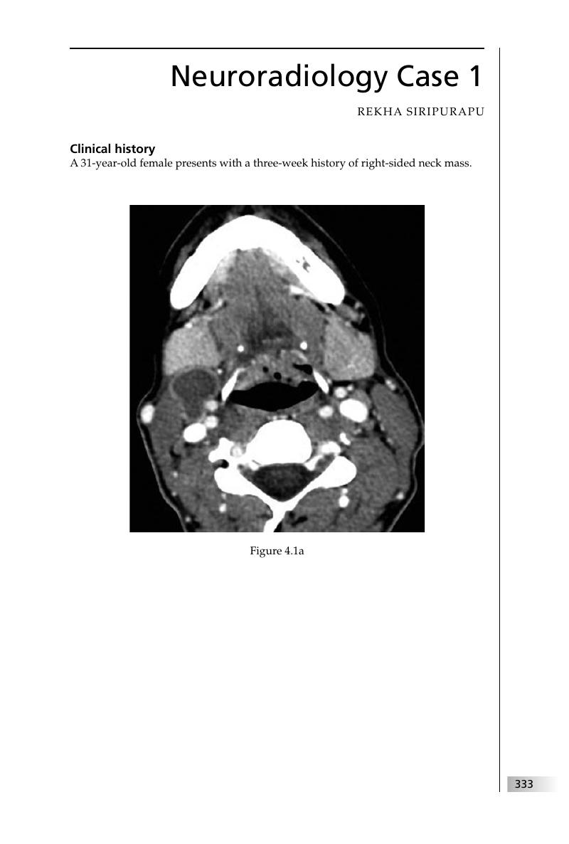
4 - Neuroradiology
Published online by Cambridge University Press: 05 March 2012

Summary
A summary is not available for this content so a preview has been provided. Please use the Get access link above for information on how to access this content.
- Type
- Chapter
- Information
- Final FRCR 2B VivaA Survival Guide, pp. 333 - 452Publisher: Cambridge University PressPrint publication year: 2011
References
, . Second branchial cleft cyst: not!!AJNR Am J Neuroradiol 2009; 30: 1628–9.CrossRefGoogle ScholarPubMed
, , , . Congenital cystic masses of the neck: radiologic–pathologic correlation. Radiographics 1999; 19: 121–46.CrossRefGoogle ScholarPubMed
, . Diagnosis and temporal evolution of signs of intracranial hypotension on MRI of the brain. Neuroradiology 2008; 50: 1025–34.CrossRefGoogle Scholar
, , , . Spinal dural enhancement on magnetic resonance imaging associated with spontaneous intracranial hypotension. J Neurosurg 1998; 88; 912–18.CrossRefGoogle Scholar
, , eds. Handbook of Neuro-oncology Neuroimaging. New York, NY: Academic Press, 2008; 3–4, 13–14, 31.
, , , . Cerebral cavernous malformation: natural history and prognosis after clinical deterioration with or without haemorrhage. J Neurosurg 1997; 87: 190–7.CrossRefGoogle ScholarPubMed
Morphologic characteristics of subcortical heterotopias: MR imaging study. AJNR Am J Neuroradiol 2000; 21: 290–5.Google ScholarPubMed
, , . Sinonasal pathology. In Imaging of the Head and Neck, 2nd edn. Stuttgart: Thieme, 2005; 434–6.Google Scholar
, . Magnetic resonance imaging of sinonasal malignancies. Top Magn Reson Imaging 2007; 18: 259–67.CrossRefGoogle ScholarPubMed
, , , et al. T1 signal hyperintensity in the sellar region: spectrum of findings. Radiographics 2006; 26: 93–113.CrossRefGoogle Scholar
, , , . Imaging of cerebral venous thrombosis: current techniques, spectrum of findings, and diagnostic pitfalls. Radiographics 2006; 26: S19–43.CrossRefGoogle ScholarPubMed
, , , et al. Cerebral venous thrombosis and multidetector CT angiography: tips and tricks. Radiographics 2006; 26: S5–18.CrossRefGoogle ScholarPubMed
, , , et al. The usefulness of MR imaging in the diagnosis of dysembryoplastic neuroepithelial tumour in children: a study of 14 cases. AJNR Am J Neuroradiol 2003; 24: 829–34.Google Scholar
, , , , . Diagnosing idiopathic normal pressure hydrocephalus. Neurosurgery 2005; 57: S4–16.CrossRefGoogle ScholarPubMed
, , , . Psuedo-subarachnoid haemorrhage: a potential imaging pitfall associated with diffuse cerebral oedema. AJNR Am J Neuroradiol 2003; 24: 254–6.Google Scholar
, , , , . CT and MR in non-neonatal hypoxic–ischemic encephalopathy: radiological findings with pathophysiological correlations. Neuroradiology 2010; 52: 949–76.CrossRefGoogle Scholar
, , , . Malignant schwannoma of the trigeminal nerve. AJNR Am J Neuroradiol 2001; 22: 505–7.Google ScholarPubMed
, , , , . Neuropathology for the neuroradiologist: Antoni A and Antoni B tissue patterns. AJNR Am J Neuroradiol 2007; 28: 1633–8.CrossRefGoogle Scholar
. Neurofibromatosis 1 and neurofibromatosis 2: a twenty first century perspective. Lancet Neurol 2007; 6: 340–51.CrossRefGoogle ScholarPubMed
, . Neuroimaging findings in neurofibromatosis type 1 and 2. Neuroimaging Clin N Am 2004; 14: 149–70.CrossRefGoogle ScholarPubMed
, , . Complications of frontal sinusitis and their management. Otolaryngol Clin N Am 2001; 34: 211–25.CrossRefGoogle ScholarPubMed
, . Subdural empyema and other suppurative complications of paranasal sinusitis. Lancet Infect Dis 2007; 7: 62–7.CrossRefGoogle ScholarPubMed
, Donovan . Neuroimaging in the brain in HIV-1-infected patients. Neuroimaging Clin N Am 2008; 18: 93–117.CrossRefGoogle ScholarPubMed
, Primary central nervous system lymphoma. Curr Oncol Rep 2004; 6: 388–95.CrossRefGoogle ScholarPubMed
. Intracranial aneurysms associated with other lesions, disorders or anatomic variations. Neuroimaging Clin N Am 2006; 16: 467–82.CrossRefGoogle ScholarPubMed
, , Carotid cavernous fistulas: anatomy, classification and treatment. Neurosurg Clin N Am 2005; 16: 279–95.CrossRefGoogle ScholarPubMed
, , . Small rosarylike infarctions in the centrum ovale suggest hemodynamic failure. AJNR Am J Neuroradiol 1998; 19: 1479–84.Google ScholarPubMed
, . The pathophysiology of watershed infarction in internal carotid artery disease: review of cerebral perfusion studies. Stroke 2005; 36: 567–77.CrossRefGoogle ScholarPubMed
, . Pilocytic astrocytoma: radiologic–pathologic correlation. Radiographics 2004; 24: 1693–708.CrossRefGoogle ScholarPubMed
, . Neuroimaging findings in neurofibromatosis type 1 and 2. Neuroimaging Clin N Am 2004; 14: 149–70.CrossRefGoogle ScholarPubMed
, . Handbook of Neuroradiology: Brain and Skull. St Louis, MO: Mosby, 1996; 52–3, 568–9.Google Scholar
, , , . The basal ganglia: anatomy, physiology and pharmacology. Psychiatr Clin N Am 2004; 27: 757–99.CrossRefGoogle ScholarPubMed
, , , et al. Early prediction of irreversible brain damage after ischemic stroke at CT. Radiology 2001; 219: 95–100.CrossRefGoogle Scholar
, , , et al. Chiari type I malformation with or without syringomyelia: prevalence and genetics. J Genet Couns 2003; 12: 297–311.CrossRefGoogle ScholarPubMed
, , , , . The pediatric Chiari I malformation: a review. Childs Nerv Syst 2007; 23: 1239–50.CrossRefGoogle ScholarPubMed
, , . Intracranial aneurysms: an overview. Neuroimaging Clin N Am 2006; 16: 371–82.CrossRefGoogle ScholarPubMed
, , , et al. Management of unruptured cerebral aneurysms in patients with polycystic kidney disease. Surg Neurol 2004; 62: 538–45.CrossRefGoogle ScholarPubMed
, , , et al. Colloid cysts of the third ventricle: correlation of MR and CT findings with histology and chemical analysis. AJNR Am J Neuroradiol 1990; 11: 575–81.Google ScholarPubMed
, , , et al. Colloid cysts of the third ventricle: MR findings. J Comput Assist Tomogr 1990; 14: 527–31.CrossRefGoogle ScholarPubMed
Pediatric Neuroimaging, 4th edn. Philadelphia, PA: Lippincott Williams & Wilkins, 2005; 744–52.Google Scholar
TP, . Diastematomyelia: hemicord and meningeal sheaths; single and double arachnoid and dural tubes. AJNR Am J Neuroradiol 1983; 4: 633–6.Google ScholarPubMed
, , , Craniofacial mucormycosis: assessment with CT. Radiology 1986; 160: 207–12.CrossRefGoogle ScholarPubMed
, , , , . Computed tomography and magnetic resonance diagnosis of allergic fungal sinusitis. Laryngoscope 1997; 107: 170–6.CrossRefGoogle ScholarPubMed
, Figueroa RE, Ginsberg LE, et al. Allergic fungal sinusitis: CT findings. Radiology 1998; 207: 417–22.CrossRefGoogle ScholarPubMed
, , . Aneurysmal subarachnoid hemorrhage. N Engl J Med 2006; 354: 387–96.CrossRefGoogle ScholarPubMed
, , , , . Cerebral venous thrombosis: a pictorial review. Eur J Radiol 2010; 74: 110–16.CrossRefGoogle ScholarPubMed
, , , et al. Noncontrast CT in deep cerebral venous thrombosis and sinus thrombosis: comparison of its diagnostic value for both entities. AJNR Am J Neuroradiol 2009; 30: 728–35.CrossRefGoogle ScholarPubMed
, . Radiological diagnosis of cerebral venous thrombosis. Front Neurol Neurosci 2008; 23: 96–111.Google ScholarPubMed
Pediatric Neuroimaging, 4th edn. Philadelphia, PA: Lippincott Williams & Wilkins, 2005; 710–31.Google Scholar
, , , . MR imaging in the tethered spinal cord syndrome. AJR Am J Roentgenol 1989; 152: 843–52.CrossRefGoogle ScholarPubMed
Reppucci VS, Movshovich A. Current concepts in the treatment of traumatic injury to the posterior segment. Ophthalmol Clin N Am 1999; 12: 465–74.
, , , , . Principles, techniques, and applications of T2*-based MR imaging and its special applications. Radiographics 2009; 29: 1433–49.CrossRef
, . Diffuse axonal injury: pathophysiology and imaging. Neuroimaging Clin N Am 2002; 12: 205–16.CrossRefGoogle Scholar
, . Imaging of pediatric head trauma. Neuroimaging Clin N Am 2002; 12: 271–94.CrossRefGoogle ScholarPubMed
, How to simplify the CT diagnosis of Le Fort fractures. AJR Am J Roentgenol 2005; 184: 1700–5.CrossRefGoogle ScholarPubMed
, , , et al. Diffusion-weighted MR imaging of cholesteatoma in pediatric and adult patients who have undergone middle ear surgery. AJR Am J Roentgenol 2003; 181: 261–5.CrossRefGoogle ScholarPubMed
, . The pathophysiology of cholesteatoma. Otolaryngol Clin N Am 2006: 39; 1143–59.CrossRefGoogle ScholarPubMed
, . The management of pediatric cholesteatoma. Otolaryngol Clin N Am 2002; 35: 841–51.CrossRefGoogle ScholarPubMed
, . Complications of chronic otitis media and cholesteatoma. Otolaryngol Clin N Am 2006; 39: 1237–55.CrossRefGoogle ScholarPubMed
, , , , . Benign brain tumors: sellar/parasellar tumors. Neurol Clin 2007; 25: 1231–49.CrossRefGoogle ScholarPubMed
. Nonfunctioning pituitary tumors and pituitary incidentalomas. Endocrinol Metab Clin N Am 2008; 37: 151–71.CrossRefGoogle ScholarPubMed
, . Imaging for functional endoscopic sinus surgery. Otolaryngol Clin N Am 2006; 39: 403–16.CrossRefGoogle ScholarPubMed
. Surgical management of polyps in the treatment of nasal airway obstruction. Otolaryngol Clin N Am 2009; 42: 377–85.CrossRefGoogle ScholarPubMed
. Contemporary management of nasal polyps. Otolaryngol Clin N Am 2004; 37: 327–37.CrossRefGoogle ScholarPubMed
, , , . Oral cavity and oropharynx tumors. Radiol Clin North Am 2007; 45: 1–20.CrossRefGoogle ScholarPubMed
, , , . Retinoblastoma and simulating lesions: role of imaging. Neuroimaging Clin N Am 2005; 15: 49–67.CrossRefGoogle ScholarPubMed
, Imaging for functional endoscopic sinus surgery. Otolaryngol Clin N Am 2006; 39: 403–16.CrossRefGoogle ScholarPubMed
Transnasal endoscopic management of frontal mucoceles. Otolaryngol Clin N Am 2001; 34: 243–51.CrossRefGoogle ScholarPubMed
, . Imaging of chemosensory loss. Otolaryngol Clin N Am 2004; 37: 1255–80.CrossRefGoogle ScholarPubMed
, . Sarcomas of the head and neck. Surg Oncol Clin N Am 2003; 12: 379–417.CrossRefGoogle ScholarPubMed
, , . Nasopharynx: clinical, pathologic, and radiologic assessment. Neuroimaging Clin N Am 2003; 13: 465–83.CrossRefGoogle ScholarPubMed
, , . Best cases from the AFIP: glioblastoma multiforme. Radiographics 2007; 27: 883–8.CrossRefGoogle ScholarPubMed
, . Spinal-cord MRI in multiple sclerosis: conventional and nonconventional MR techniques. Neuroimaging Clin N Am 2009; 19: 81–99.CrossRefGoogle ScholarPubMed
, . Defining the clinical course of multiple sclerosis: results of an international survey. Neurology 1996; 46: 907–11.CrossRefGoogle Scholar
, . Variants of multiple sclerosis. Neuroimaging Clin N Am 2008; 18: 703–16.CrossRefGoogle ScholarPubMed
Pediatric Neuroimaging, 4th edn. Philadelphia, PA: Lippincott Williams & Wilkins, 2005; 205–6.Google Scholar
, . Hypoxic–ischemic brain injury: imaging findings from birth to adulthood. Radiographics 2008; 28: 417–39.Google Scholar
, , , et al. Pictorial review of tuberous sclerosis in various organs. Radiographics 2008; 28: e32.CrossRefGoogle ScholarPubMed