
Book contents
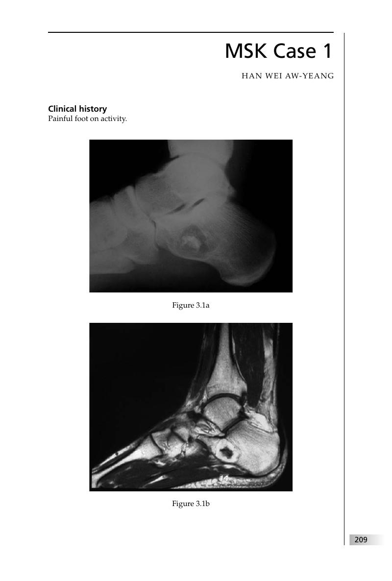
3 - Musculoskeletal radiology
Published online by Cambridge University Press: 05 March 2012

Summary
A summary is not available for this content so a preview has been provided. Please use the Get access link above for information on how to access this content.
- Type
- Chapter
- Information
- Final FRCR 2B VivaA Survival Guide, pp. 209 - 332Publisher: Cambridge University PressPrint publication year: 2011
References
, , . Intraosseous lipoma of the calcaneus. Langenbecks Arch Surg 2001; 386: 313–17.CrossRefGoogle ScholarPubMed
, , , . MR findings of calcaneal intraosseous lipoma with hemorrhage. AJR Am J Roentgenol 2005; 185: 1378–9.CrossRefGoogle ScholarPubMed
, , , et al. From the archives of the AFIP. Benign musculoskeletal lipomatous lesions. Radiographics 2004; 24: 1433–66.CrossRefGoogle ScholarPubMed
. Freiberg's infraction. In Pediatric Orthopedics, 2nd edn. Philadelphia, PA:Saunders, 1990; 1006–10.Google Scholar
, . Problems of the second metatarsophalangeal joint. Orthopedics 1987; 10: 83–9.Google ScholarPubMed
, , , et al. From the archives of the AFIP. Radiologic spectrum of Paget disease of bone and its complications with pathologic correlation. Radiographics 2002; 22: 1191–216.CrossRefGoogle ScholarPubMed
, , , , . Paget's disease of the patella. Skeletal Radiol 1990; 19: 407–10.CrossRefGoogle ScholarPubMed
. The meniscus: recent advances in MR imaging of the knee. AJR Am J Roentgenol 2002; 179: 1115–22.CrossRefGoogle Scholar
, , . MR imaging of tears of discoid lateral menisci. AJR Am J Roentgenol 1998; 171: 963–7.CrossRefGoogle ScholarPubMed
, , , et al. Prevalence, pattern, and spectrum of glenoid bone loss in anterior shoulder dislocation: CT analysis of 218 patients. AJR Am J Roentgenol 2008; 190: 1247–54.CrossRefGoogle ScholarPubMed
, , , et al. Acute traumatic posterior shoulder dislocation: MR findings. Radiology 2008; 248: 185–93.CrossRefGoogle ScholarPubMed
, , Osteomyelitis originating in and around bone infarcts: giant sequestrum phenomena. AJR Am J Roentgenol 2001; 176: 387–91.CrossRefGoogle ScholarPubMed
, , , et al. Magnetic resonance imaging of medullary bone infarction in the early stage. Clin Imaging 2008; 32: 147–51.CrossRefGoogle ScholarPubMed
, , , et al. Efficacy of linezolid versus vancomycin in the treatment of methicillin-resistant Staphylococcus aureus discitis: a controlled animal model. Spine 2006; 31: E830–2.CrossRefGoogle ScholarPubMed
, . Tumoral calcinosis: pearls, polemics, and alternative possibilities. Radiographics 2006; 26: 871–85.CrossRefGoogle ScholarPubMed
, , , . Radiographic evaluation of arthritis: degenerative joint disease and variations. Radiology 2008; 248; 737–47.CrossRefGoogle ScholarPubMed
, , , et al. From the archives of the AFIP: benign musculoskeletal lipomatous lesions. Radiographics 2004; 24: 1433–66.CrossRefGoogle ScholarPubMed
, . Radiologic aspects of hemophilic pseudotumors in bone. Am J Roentgenol Radium Ther Nucl Med 1972; 115: 525–39.Google ScholarPubMed
, , , . Hemophilic pseudotumor: radiologic–pathologic correlation. Radiographics 2003; 23: 852–6.Google Scholar
, . Hemophilic pseudotumor involving the musculoskeletal system: spectrum of radiologic findings. AJR Am J Roentgenol 2004; 183: 55–61.CrossRefGoogle ScholarPubMed
, , , et al. Magnetic resonance imaging of children with Duchenne muscular dystrophy. Pediatric Radiology 1987; 17: 495–7.CrossRefGoogle ScholarPubMed
. Bone tumors and tumorlike conditions: analysis with conventional radiography. Radiology 2008: 246; 662–74.CrossRefGoogle ScholarPubMed
, , , . Radiographic evaluation of arthritis: degenerative joint disease and variations. Radiology 2008: 248: 737–47.CrossRefGoogle ScholarPubMed
, , . A pictorial review of primary synovial osteochondromatosis. Eur Radiol 2008; 18: 2662–9.CrossRefGoogle ScholarPubMed
, , , et al. Pigmented villonodular synovitis: radiologic–pathologic correlation. Radiographics 2008; 28: 1493–518.CrossRefGoogle ScholarPubMed
Imaging and differential diagnosis of primary bone tumors and tumor-like lesions of the spine. Eur J Radiol 2006; 58: 48–67.CrossRefGoogle ScholarPubMed
, , , , Palestro CJ. Radionuclide bone imaging: an illustrative review. Radiographics 2003; 23: 341–58.CrossRefGoogle Scholar
, , , et al. From the archives of the AFIP. Radiologic spectrum of Paget disease of bone and its complications with pathologic correlation. Radiographics 2002; 22: 1191–216.CrossRefGoogle ScholarPubMed
, , , , . Musculoskeletal manifestations of sickle cell disease. Radiographics 2007; 27: 1005–21.CrossRefGoogle ScholarPubMed
, , , et al. The radiological manifestations of sickle cell disease. Clin Radiol 2007; 62: 528–38.CrossRefGoogle ScholarPubMed
, , . 2_148 The Musculoskeletal Manifestations of the Haemoglobinopathies. Radiology Integrated Training Initiative. Royal College of Radiologists, 2010.
. Hemoglobinopathies and other anemias. In , ed., Diagnosis of Bone and Joint Disorders, 4th edn. Philadelphia, PA: Saunders, 2002; 2146–87.Google Scholar
, , , , . Multiple myeloma: clinical review and diagnostic imaging. Radiology 2004; 231: 11–23.CrossRefGoogle ScholarPubMed
, . Common causes of low back pain in children. Radiographics 1991; 11: 269–91.CrossRefGoogle ScholarPubMed
, , , . Skeletal findings in progressive systemic sclerosis (scleroderma). AJR Am J Roentgenol 1981; 136: 1121–6.CrossRefGoogle Scholar
, . Radiology at your fingertips: lesions of the terminal phalanx. Clin Radiol 1988; 39: 478–85.CrossRefGoogle ScholarPubMed
, , . Intravenous urography: technique and interpretation. Radiographics 2001; 21: 799–824.CrossRefGoogle ScholarPubMed
, , , , . Renal papillary necrosis: review and comparison of findings at multi-detector row CT and intravenous urography. Radiographics 2006; 26: 1827–36.CrossRefGoogle ScholarPubMed
. Renal bone disease: radiological investigation. Kidney Int Suppl 1999; 56: S38–41.CrossRefGoogle Scholar
. Radiology of rickets and osteomalacia. In , , , eds., Vitamin D, 2nd edn. New York, NY: Elsevier, 2005; 967–94.Google Scholar
. Rickets and osteomalacia. In , , eds., Bone and Joint Imaging, 3rd edn. Richmond, VA: Elsevier-Saunders, 2005; 563–75.Google Scholar
. Carpal injuries: analytic approach and case exercises. AJR Am J Roentgenol 1979; 133: 503–17.CrossRefGoogle ScholarPubMed
, , , , . Wrist fractures: what the clinician wants to know. Radiology 2001; 219: 11–28.CrossRefGoogle Scholar
, , , et al. Multidetector CT of carpal injuries: anatomy, fractures, and fracture-dislocations. Radiographics 2008; 28: 1771–84.CrossRefGoogle ScholarPubMed
, . Imaging of wrist injuries. In Vanhoenacker FM, Maas M, Gielen JL, eds., Imaging of Orthopedic Sports Injuries. Berlin: Springer, 2007; 201–24.Google Scholar
, , , . Pediatric ribs: a spectrum of abnormalities. Radiographics 2002; 22: 87–104.CrossRefGoogle ScholarPubMed
, . Keep your eyes on the ribs: the spectrum of normal variants and diseases that involves the ribs. Radiographics 1999; 19: 1125–42.CrossRefGoogle Scholar
, , , et al. Radiologic features of eosinophilic granuloma of bone. AJR Am J Roentgenol 1989; 153: 1021–6.CrossRefGoogle ScholarPubMed
, , , et al. Tuberculosis: a radiologic review. Radiographics 2007; 27: 1255–73.CrossRefGoogle ScholarPubMed
, , , . Imaging of extrapulmonary tuberculosis. Radiographics 2000; 20: 471–88.CrossRefGoogle ScholarPubMed
, , , et al. Tuberculosis from head to toe. Radiographics 2000; 20: 449–70.CrossRefGoogle ScholarPubMed
, , , , . Discrimination of tuberculous spondylitis from pyogenic spondylitis on MRI. AJR Am J Roentgenol 2004; 182: 1405–10.CrossRefGoogle ScholarPubMed
, , , . A compartmental approach to the radiographic evaluation of soft-tissue calcifications. Semin Roentgenol 2005; 40: 391–407.CrossRefGoogle ScholarPubMed
, . Tumoral calcinosis: pearls, polemics, and alternative possibilities. Radiographics 2006; 26: 871–85.CrossRefGoogle ScholarPubMed
, , . Maffucci syndrome: radiologic and pathologic findings. Radiographics 2001; 21: 1311–16.Google Scholar
, , , et al. Enchondroma versus chondrosarcoma in the appendicular skeleton: differentiating features. Radiographics 1998; 18: 1213–37.CrossRefGoogle ScholarPubMed
, , . Cortical lesions of the tibia: characteristic appearances at conventional radiography. Radiographics 2003; 23: 157–77.CrossRefGoogle ScholarPubMed
, , , , . Imaging of osteochondroma: variants and complications with radiologic-pathologic correlation. Radiographics 2000; 20: 1407–34.CrossRefGoogle ScholarPubMed
, , , . Neuropathic osteoarthropathy: diagnostic dilemmas and differential diagnosis. Radiographics 2000; 20: S279–93.CrossRefGoogle ScholarPubMed
, , , et al. Pathogenesis of the Segond fracture: anatomic and MR imaging evidence of an iliotibial tract or anterior oblique band avulsion. Radiology 2001; 219: 381–6.CrossRefGoogle ScholarPubMed
, , , . Magnetic resonance imaging of trauma patterns in the knee. Emerg Radiol 1998; 4: 237–44.CrossRefGoogle Scholar
, , . The Segond fracture of the proximal tibia: a small avulsion that reflects major ligamentous damage. AJR Am J Roentgenol 1988; 151: 1163–7.CrossRefGoogle ScholarPubMed
, , , . Avulsions around the knee portend instability. Emerg Radiol 2005; 11: 213–18.CrossRefGoogle ScholarPubMed
, . Radiographic indicators of acute ligament injuries of the knee: a mechanistic approach. Emerg Radiol 2010; 17: 435–44.CrossRefGoogle ScholarPubMed
. Bone tumors and tumorlike conditions: analysis with conventional radiography. Radiology 2008; 246: 662–74.CrossRefGoogle ScholarPubMed
. Diagnosis of Bone and Joint Disorders, 4th edn. Philadelphia, PA: Saunders, 2002; 3757, 3922–4.Google Scholar
, . Musculoskeletal Imaging: a Teaching File, 2nd edn. Philadelphia, PA:Lippincott Williams & Wilkins, 2006.Google Scholar
, . Classification and diagnostic criteria for psoriatic arthritis. Ann Rheum Dis 2005; 64: ii3–8.CrossRefGoogle ScholarPubMed
, . Radiology at your fingertips: lesions of the terminal phalanx. Clin Radiol 1988; 39: 478–85.CrossRefGoogle ScholarPubMed
, , . Best cases from the AFIP: primary hyperparathyroidism due to parathyroid adenoma. Radiographics 2005; 25: 829–34.CrossRefGoogle ScholarPubMed
. Bone tumors and tumorlike conditions: analysis with conventional radiography. Radiology 2008; 246: 662–74.CrossRefGoogle ScholarPubMed
, , , . Osteodystrophy: imaging findings that mimic those of other diseases,AJR Am J Roentgenol 1995; 165: 143–8.CrossRefGoogle Scholar
. Radiology of the hands: review and self-assessment module. AJR Am J Roentgenol 2005; 184: S157–68.CrossRefGoogle ScholarPubMed
, , , et al. Radiologic manifestations of sarcoidosis in various organs. Radiographics 2004; 24: 87–104.CrossRefGoogle ScholarPubMed
, . Musculoskeletal sarcoidosis: spectrum of appearances at mr imaging. Radiographics 2003; 23: 1389–99.CrossRefGoogle ScholarPubMed
, Sarcoidosis. In , , eds., Diagnosis of Bone and Joint Disorders, 3rd edn. Philadelphia, PA: Saunders, 1995; 4333–52.Google Scholar
. Radiographic, angiographic and radionuclide manifestations of osseous sarcoidosis. Radiographics 1983; 3: 375–96.CrossRefGoogle Scholar
, , , et al. Radiographic, CT, and MR imaging features of dedifferentiated chondrosarcomas: a retrospective review of 174 de novo cases. Radiographics 2004; 24: 1397–409.CrossRefGoogle Scholar
, , , et al. From the archives of the AFIP. Imaging of primary chondrosarcoma: radiologic–pathologic correlation. Radiographics 2003; 23: 1245–78.CrossRefGoogle ScholarPubMed
. Bone tumors and tumorlike conditions: analysis with conventional radiography. Radiology 2008; 246: 662–74.CrossRefGoogle ScholarPubMed
, , , et al. From the archives of the AFIP. Imaging of giant cell tumor and giant cell reparative granuloma of bone: radiologic– pathologic correlation. Radiographics 2001; 21: 1283–309.CrossRefGoogle ScholarPubMed
, . Monteggia fracture–dislocation in children. J Accid Emerg Med 1994; 11: 192–4.CrossRefGoogle ScholarPubMed
, , Radiologic history exhibit. Musculoskeletal eponyms: who are those guys?Radiographics 2000; 20: 819–36.CrossRefGoogle ScholarPubMed
, , . Improving detection of pediatric elbow fractures by understanding their mechanics. Radiographics 1996; 16: 1443–60.CrossRefGoogle ScholarPubMed
, , , , . Best cases from the AFIP: adamantinoma of the tibia and fibula with cytogenetic analysis. Radiographics 2008; 28: 1215–20.Google ScholarPubMed
, Cortical lesions of the tibia: characteristic appearances at conventional radiography. Radiographics 2003; 23: 157–77.CrossRefGoogle ScholarPubMed
. Bone tumors and tumorlike conditions: analysis with conventional radiography. Radiology 2008; 246: 862–74.CrossRefGoogle ScholarPubMed
Evaluation of focal bone lesions: basic principles and clinical scenarios. Imaging 2003; 15: 298–323.CrossRefGoogle Scholar
, , , et al. Radiologic diagnosis of osteoid osteoma: from simple to challenging findings. Radiographics 2010; 30: 737–49.CrossRefGoogle ScholarPubMed
, , , . From the archives of the AFIP: osteoid osteoma. Radiographics 1991; 11: 671–96.CrossRefGoogle Scholar
, , . Improving detection of pediatric elbow fractures by understanding their mechanics. Radiographics 1996; 16: 1443–60.CrossRefGoogle ScholarPubMed
, , , . Radiologic and pathologic characteristics of benign and malignant lesions of the mandible. Radiographics 2006; 26: 1751–68.CrossRefGoogle ScholarPubMed
, , . Pattern of the month: periosteal reaction. AJR Am J Roentgenol 2009; 193: W259–72.CrossRefGoogle Scholar
, . Cross-sectional imaging of peripheral nerve sheath tumors: characteristic signs on CT, MR imaging, and sonography. AJR Am J Roentgenol 2001; 176: 75–82.CrossRefGoogle ScholarPubMed
, , , et al. Chest wall tumors: radiologic findings and pathologic correlation. Part 1. Benign tumors. Radiographics 2003; 23: 1477–90.CrossRefGoogle ScholarPubMed