Skip to main content Accessibility help
×
  • Cited by 1
Publisher:
Cambridge University Press
Online publication date:
May 2011
Print publication year:
2011
Online ISBN:
9780511997426

Book description

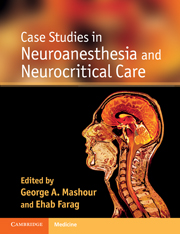
The anesthetic considerations and procedures involved in the perioperative care of the neurosurgical patient are among the most complex in anesthesiology. The practice of neurosurgery and neuroanesthesiology encompasses a wide range of cases, from major spine surgery, to aneurysm clipping and awake craniotomy. Case Studies in Neuroanesthesia and Neurocritical Care provides a comprehensive view of real-world clinical practice. It contains over 90 case presentations with accompanying focussed discussions, covering the broad range of procedures and monitoring protocols involved in the care of the neurosurgical patient, including preoperative and postoperative care. The book is illustrated throughout with practical algorithms, useful tables and examples of neuroimaging. Written by leading neuroanesthesiologists, neurologists, neuroradiologists and neurosurgeons from the University of Michigan Medical School and the Cleveland Clinic, these clear, concise cases are an excellent way to prepare for specific surgical cases or to aid study for both written and oral board examinations.

Reviews

'This textbook is very useful for teaching … we would wholeheartedly recommend this book to anaesthetic trainees and all clinicians involved in neuroanaesthesia or intensive care. It complements traditional neuroanaesthesia textbooks and would be an excellent portable book to dip in and out of during a busy clinical day.'

Source: British Journal of Anesthesia

Refine List

Actions for selected content:

Select all | Deselect all
  • View selected items
  • Export citations
  • Download PDF (zip)
  • Save to Kindle
  • Save to Dropbox
  • Save to Google Drive

Save Search

You can save your searches here and later view and run them again in "My saved searches".

Please provide a title, maximum of 40 characters.
×

Contents


Page 1 of 4


  • Case 8 - Delayedemergence after posterior fossa surgery
    pp 27-29
  • View abstract

    Summary

    Supratentorial craniotomy is a common case in neurosurgery during which both the neurosurgeon and anesthesiologist are modulating the same organ. This chapter presents a case study of a 61-year-old female who presented with new-onset seizures, which were preceded by several weeks of bilateral frontal headaches described as dull and achy. The patient was scheduled for tumor excision via stealth-guided craniotomy. The patient's anxiety, increased blood pressure, and bronchospasm were addressed immediately. The patient emerged within several minutes of the head dressing being applied, without bucking or coughing. Normally, a patient presenting for supratentorial craniotomy is best served by an organized and systematic approach. The chapter presents one strategy for the preoperative evaluation of these neurosurgical patients. Supratentorial craniotomy is a common case in neurosurgery during which both the neurosurgeon and anesthesiologist are modulating the same organ. Thoughtful planning and clear communication between the teams is required for optimal patient care.
  • Case 10 - Trigeminocardiacreflex
    pp 33-35
  • View abstract

    Summary

    Intracranial pressure (ICP) is of paramount importance because the cranial vault is nondistensible and within it is contained three noncompressible substances: brain, blood, and cerebrospinal fluid (CSF). This chapter presents a case study of a 75-year-old male who presented for emergent subdural hematoma evacuation. The immediate anesthetic goal was to minimize the rise in ICP while at the same time maintaining adequate cerebral perfusion pressure until the neurosurgeons could provide definitive treatment. Definitive correction of intracranial hypertension was achieved with hematoma evacuation. Intracranial hematomas, blood-filled space-occupying lesions, are classified by their location relative to the meningeal layers. Numerous therapeutic maneuvers exist for lowering ICP, each with the common mechanism of decreasing the volume of one or more intracranial components. increased ICP can be a life-threatening condition, the definitive treatment of which is often in the hands of the neurosurgeon.
  • Case 11 - Sittingcraniotomy
    pp 36-37
  • View abstract

    Summary

    The injury spectrum in traumatic brain injury (TBI) encompasses not only the initial insult, but includes the cascade of systemic responses and pathophysiology that occur after the focal or diffuse brain injury. This chapter presents a case study of a 49-year-old male motorcyclist who was evaluated in the emergency room after a motor vehicle collision with an articulated truck. Around the time of computed tomography (CT) scanning, the patient's right pupil became dilated and unreactive. Hypoxic cerebral damage, a common postmortem finding in TBI, is associated with arterial hypoxemia, decreased mean arterial pressure, or cerebral hypoperfusion, occurring as a consequence of shock, intracranial hypertension, or cerebral vasospasm. Although a definitively effective neuroprotective therapy in central nervous system (CNS) trauma remains elusive, through the skilled interactions of prehospital, emergency department, anesthesiology, and surgical personnel, the lives of many critically injured individuals can be saved and their neurologic function preserved.
  • Case 12 - Cerebellarhemorrhage
    pp 38-40
  • View abstract

    Summary

    Postoperative seizures can have physiologic, pharmacologic, and pathologic causes. This chapter presents a case study of a 56-year-old female presented for resection of a 2 cm by 2.5 cm mass in the right temporal lobe. The patient was brought to the operating room for a scheduled resection of the tumor under general endotracheal anesthesia. Midazolam was administered and the seizure was terminated, but she was now lethargic and combative. Citrate is added to stored blood to bind calcium and prevent coagulation of the stored blood. Some advocate a remifentanil infusion be used intraoperatively as it can provide a smooth emergence and because of its short half life a quick return to neurologic baseline. Immediate termination of the seizure should be followed by a rapid assessment of reversible physiologic causes and then amore extensive differential to identify the underlying source.
  • Case 13 - Preoperativeevaluation
    pp 41-44
  • View abstract

    Summary

    The perioperative pain management for craniotomies can be extremely challenging. This chapter presents a common clinical scenario and offers options for perioperative pain management. It presents a case study of a 52-year-old female American Society of Anesthesiologists class 3 patient presented for clipping of a cerebral aneurysm. The case described is a common example of the complexity frequently associated with neurosurgical patients. The combined regimen provided for analgesia and hemodynamic control, while allowing for an adequate neurologic examination. In addition, opioids were limited, thereby decreasing the risk of postoperative nausea and vomiting. Opioids are a key component of intraoperative and postoperative pain management for craniotomies. Morphine can cause histamine release, which can lead to venodilation and subsequent hypotension. A combination of intravenous analgesics and regional anesthesia can provide excellent pain relief and decrease the wide hemodynamic changes that can accompany anesthesia and surgery.
  • Case 14 - Intracranialaneurysm clipping with intraoperative rupture
    pp 45-47
  • View abstract

    Summary

    The posterior fossa is an intracranial compartment that houses the cerebellum and the brainstem. Mass lesions and increased intracranial pressure (ICP) in this area can have profound consequences for neurologic, cardiac, and respiratory functions. This chapter presents a case study of a 54-year-old female with complaints of headaches, progressive hearing loss and episodes of aspiration that were increasing in frequency. A diagnosis of neurofibromatosis type 2 was made and a posterior fossa craniotomy was scheduled. Immediate concerns for the anesthetic team included (1) a depressed level of consciousness, (2) the potential for increased ICP, and (3) aspiration on induction. Neurosurgical procedures involving the posterior fossa can be challenging for both the surgical and anesthetic teams. Preoperative assessment and preparation of these patients should focus on the consequences of posterior fossa abnormalities as well as the potentially life-threatening complications that may occur intraoperatively.
  • Case 15 - Awakefiberoptic intubation
    pp 48-50
  • View abstract

    Summary

    A venous air embolism (VAE) is a potentially life-threatening event caused by air in the vascular system. The entrainment of air from an operative site into the venous vasculature produces a wide array of systemic effects. This chapter presents a case study of a 37-year-old female with a right-sided acoustic neuroma presenting for a suboccipital approach to tumor resection. VAE was historically most often associated with craniotomies performed in the sitting position. Clinical presentation depends on the severity of the air embolus. There are several monitors that are capable of detecting venous air emboli. The most sensitive is transesophageal echocardiography (TEE). The presence of TEE also enables direct visualization of air aspiration through a central catheter if a VAE should occur. Monitors for high-risk cases should be chosen depending on the expertise of the anesthesiologist, the surgery being performed, and the position of the patient.
  • Case 16 - Patientwith coronary artery stent
    pp 51-54
  • View abstract

    Summary

    Immediate emergence after neurosurgery is desirable to facilitate neurologic examination and early identification of complications. Awakening is determined by many factors including preoperative status, type of surgery, and intraoperative events. This chapter presents a case study of a 58-year-old female with a body mass index of 32 who complained about gradual hearing loss, increasing frequency of headaches and vertigo and subsequently was diagnosed with an acoustic neuroma. In order to facilitate early detection of neurologic complications, anesthesiologists usually aim for an early emergence following intracranial surgery. The most common causes of delayed postoperative emergence include residual drug effects, respiratory failure, metabolic derangements, and neurologic complications. Cerebral swelling can occur intraoperatively or preexist. In the context of posterior fossa surgery this is of particular concern. Multiple factors contribute to a delayed emergence from anesthesia and a systematic approach to rule out all possible causes is necessary.
  • Case 17 - Deephypothermic circulatory arrest for intracranial aneurysm clipping
    pp 55-58
  • View abstract

    Summary

    Trigeminal neuralgia is multifactorial in etiology: it may be due to vascular compression, tumor, demyelination, or idiopathic causes. The treatment options are either medical (carbamazepine, oxacarbazepine, lamotrigine, neurontin, clonazepam, pregabalin, phenytoin, and baclofen) or surgical. Surgical management includes microsurgical exploration/decompression, percutaneous procedures (rhizotomies) on the Gasserian ganglion (radiofrequency lesioning, glycerol injection, and balloon compression), radiosurgery, and neurectomy. Bradycardia and even asystole as a result of the trigemino cardiac reflex can complicate this procedure. The noxious stimulus on the trigeminal nerve during needle insertion and balloon inflation is thought to induce the reflex. This reflex can also be activated without surgery; the mere pain of trigeminal neuralgia has reportedly resulted in bradycardia that was severe enough to cause syncope and in other instances degenerated to complete cardiac arrest. Events such as these demonstrate the potentially debilitating nature of trigeminal neuralgias.
  • Case 18 - Neuroprotectionduring surgical clip ligation of cerebral aneurysms
    pp 59-62
  • View abstract

    Summary

    Bradycardia and even asystole may occur suddenly during posterior fossa surgery and requires immediate evaluation and treatment in order to prevent potential ischemia and major neurologic complications. Trigeminocardiac reflex (TCR) commonly manifests as bradycardia and hypotension in response to mechanical stimulation of any of the branches of the trigeminal nerve. This chapter presents a case study of a 53-year-old female with a history of progressive headaches and a syncopal episode was found to have a right-sided tentorial mass consistent with a falcine meningioma. The tentorial nerves arise from the intracranial portions of ophthalmic branch (V1) and course into the dura of the parieto-occipital region and the posterior third of the falx, where there is a converging and bilaterally overlapping innervation at its midpoint. When stimulation of the falx results in the TCR, cessation of the surgical manipulation in that area is the first step in correcting the hemodynamic instability.
  • Case 19 - Dexmedetomidineand nitrous oxide for cerebral aneurysm clipping
    pp 63-65
  • View abstract

    Summary

    Sitting craniotomies pose a unique set of problems for perioperative care of the neurosurgical patient. Although there are benefits to neurosurgery in this position, a number of potentially catastrophic complications may also result. This chapter presents a case study of a 20-year-old morbidly obese male presented for resection of a pineocytoma via a supracerebellar approach in the sitting position. The sitting position for craniotomies offers several surgical advantages. Exposure to posterior cervical and posterior fossa structures is improved. It has been suggested that a ventriculo-atrial shunt, pulmonary hypertension, a patent foramen ovale, and symptomatic cerebral ischemia may be absolute contraindications to this procedure. Performing surgery in the sitting position for patients with uncontrolled hypertension, significant chronic obstructive airway disease, or at the extremes of age should also be done with caution. However, there are few data to support appropriate patient selection.
  • Case 20 - Anaphylaxisassociated with indocyanine green administration for intraoperative fluorescence angiography
    pp 66-69
  • View abstract

    Summary

    This chapter explores the management issues surrounding a hemorrhagic stroke of the cerebellum, one of the most common sites for intracerebral hemorrhage, and one where proper management can have a profound impact on outcome. It presents a case study of a 75-year-old female with a history of hypertension and end-stage renal disease requiring dialysis. Examination consistently revealed appropriate, symmetric limb movements and limited cranial nerve exams. Computed tomography scans showed satisfactory decompression of the posterior fossa and absence of hydrocephalus. Intracerebral hemorrhage is most commonly associated with chronic hypertension, amyloid angiopathy, anticoagulation, trauma or underlying pathology such as tumor or vascular malformation. As ventricular obstruction may occur when the patient is positioned, prepared or opened, allowing access for an emergency external ventricular drainage device is desirable in preparing and draping the patient.
  • Case 21 - Subarachnoidhemorrhage during aneurysm coiling
    pp 70-72
  • View abstract

    Summary

    Subarachnoid hemorrhage (SAH) is a complex disease with high morbidity and mortality. Management of patients with SAH requires a multisystem approach. This chapter presents a case study of a 45-year-old female who had presented to an outside hospital with a 1-month history of progressive right-sided facial and body numbness that had worsened acutely over the week prior to her admission. The patient underwent definitive correction of the aneurysm the following day. Aneurysmal SAH is a neurologic emergency, resulting from blood extravasation into the subarachnoid space normally filled with cerebrospinal fluid (CSF), that requires complex treatment and monitoring. Patients present for elective clipping of an unruptured aneurysm or emergent surgery following SAH. Thorough assessment of the patient, effective organ support and correction of pathophysiology are vital prior to leaving the intensive care unit (ICU) for what may be a challenging case in the operating room.
  • Case 23 - Postoperativeretroperitoneal bleed
    pp 77-79
  • View abstract

    Summary

    Intraoperative management of subarachnoid hemorrhage (SAH) is high-risk anesthesia with the potential for severe consequences. This chapter presents a case study of a 56-year-old African-American female smoker who presented with sudden onset of frontal headache, vomiting, and neck stiffness. Anesthesia for patients with SAH is challenging. The maintenance of an adequate mean arterial blood pressure, and hence cerebral perfusion pressure (CPP), during the induction of anesthesia is key to prevent ischemic secondary injury. CPP and transmural pressure are essentially influenced by the same variables and are equal to the mean arterial pressure minus intracranial pressure (ICP). Aneurysm rupture during laryngoscopy is an uncommon but life-threatening complication, which should be suspected if severe hypertension and bradycardia develop. Constant vigilance regarding hemodynamic control and preparedness for the possibility of intraoperative aneurysmal rupture are essential for good outcomes.

Page 1 of 4


Metrics

Altmetric attention score

Full text views

Total number of HTML views: 0
Total number of PDF views: 0 *
Loading metrics...

Book summary page views

Total views: 0 *
Loading metrics...

* Views captured on Cambridge Core between #date#. This data will be updated every 24 hours.

Usage data cannot currently be displayed.