Skip to main content Accessibility help
×
  • Cited by 1
Publisher:
Cambridge University Press
Online publication date:
May 2011
Print publication year:
2011
Online ISBN:
9780511997426

Book description

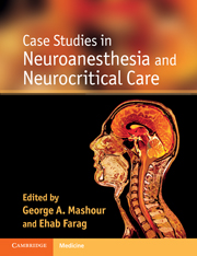
The anesthetic considerations and procedures involved in the perioperative care of the neurosurgical patient are among the most complex in anesthesiology. The practice of neurosurgery and neuroanesthesiology encompasses a wide range of cases, from major spine surgery, to aneurysm clipping and awake craniotomy. Case Studies in Neuroanesthesia and Neurocritical Care provides a comprehensive view of real-world clinical practice. It contains over 90 case presentations with accompanying focussed discussions, covering the broad range of procedures and monitoring protocols involved in the care of the neurosurgical patient, including preoperative and postoperative care. The book is illustrated throughout with practical algorithms, useful tables and examples of neuroimaging. Written by leading neuroanesthesiologists, neurologists, neuroradiologists and neurosurgeons from the University of Michigan Medical School and the Cleveland Clinic, these clear, concise cases are an excellent way to prepare for specific surgical cases or to aid study for both written and oral board examinations.

Reviews

'This textbook is very useful for teaching … we would wholeheartedly recommend this book to anaesthetic trainees and all clinicians involved in neuroanaesthesia or intensive care. It complements traditional neuroanaesthesia textbooks and would be an excellent portable book to dip in and out of during a busy clinical day.'

Source: British Journal of Anesthesia

Refine List

Actions for selected content:

Select all | Deselect all
  • View selected items
  • Export citations
  • Download PDF (zip)
  • Save to Kindle
  • Save to Dropbox
  • Save to Google Drive

Save Search

You can save your searches here and later view and run them again in "My saved searches".

Please provide a title, maximum of 40 characters.
×

Contents


Page 2 of 4


  • Case 24 - Angiographyin the patient with kidney failure
    pp 80-82
  • View abstract

    Summary

    Awake fiberoptic intubation (AFOI) has the potential to trigger hypertension, tachycardia and hypoxia or hypercarbia. Patients who require fiberoptic-guided endotracheal intubation for the clipping or coiling of an intracranial aneurysm pose particular challenges for the safe completion of both procedures. This chapter presents a case study of a 56-year-old female with a poorly documented history of difficult intubation presented for elective clipping of a middle cerebral artery aneurysm. The case discussion highlights the considerations for awake endotracheal intubation in the patient with an unsecured aneurysm. The indications for AFOI in this case are essentially the same as for any difficult airway: concern for the ability to visualize the glottic opening via direct laryngoscopy combined with concern for the ability to mask ventilate. Hypercarbia or hypoxia during an awake fiberoptic intubation are frequently due to loss of respiratory efforts in a narcotized patient.
  • Case 25 - Postoperativenormal perfusion pressure breakthrough
    pp 83-84
  • View abstract

    Summary

    The increasing use of coronary artery angioplasty with deployment of stents for treatment of coronary artery disease poses several dilemmas for perioperative management. These conflicting requirements are manifested most acutely in the management of patients with neurovascular disease. This chapter presents a case study of a 51-year-old female with a past medical history of ischemic heart disease, hypertension, and undifferentiated autoimmune disease with interstitial lung involvement, as an example. The presence of coronary stents in patients undergoing neurosurgical procedures warrants specific consideration prior to anesthesia. It is necessary to balance the risks of stent thrombosis, and the subsequent risk of myocardial infarction, arrhythmia, or cardiac arrest, against the risks of hemorrhage during or after a neurosurgical procedure. There is currently an irresolvable conflict between the risks of with holding and continuing antiplatelet agents in the perioperative period.
  • Case 26 - Preoperativeevaluation
    pp 85-87
  • View abstract

    Summary

    This chapter presents a case study of a 62-year-old female presented to the operating room with a diagnosis of two large intracranial aneurysms. The case is a discussion of the application of deep hypothermic circulatory arrest (DHCA) for patients undergoing large and/or complex intracranial aneurysm clipping and repair. The patient was brought to the operating room, noninvasive monitors placed, and under local anesthesia a right radial arterial line was established. The rationale behind the technique of DHCA stems from the significant advantage the surgeon has once blood flow has stopped circulating to the aneurysm. The margin for error is small, and success depends upon an experienced and knowledgeable team. The safe practice and management of DHCA requires an extensive understanding of cardiac and neurosurgical anesthetic practice, cardiopulmonary bypass (CPB), as well as careful consideration and proper planning.
  • Case 27 - Monitoringmodalities
    pp 88-90
  • View abstract

    Summary

    This chapter discusses the different strategies of neuroprotection applied to prevent ischemic damage to the brain during surgical clip ligation of cerebral aneurysms. It presents a case study of a 44-year-old male presented to hospital with acute subarachnoid hemorrhage (SAH); diagnostic cerebral angiogram was performed emergently that demonstrated an anterior communicating artery (ACOM) aneurysm. The ACOM artery aneurysm was deemed to be the cause of the SAH based on its morphology and the distribution of blood in the subarachnoid space. An interdisciplinary approach to the management of cerebrovascular disease is necessary for optimal patient outcome. Another method that has been reported to have some benefit in case of an unforeseen hemorrhage during clip ligation of a cerebral aneurysm is the use of adenosine to induce temporary cardiac arrest. Larger multicenter studies and more translational research are needed to demonstrate the efficacy of currently employed cerebral protective measures.
  • Case 28 - Neurologicdecline after carotid surgery
    pp 91-93
  • View abstract

    Summary

    This chapter presents a case study of a 58-year-old female with multiple intact unsecured aneurysms, who presented for clipping of one paraclinoid aneurysm. Dexmedetomidine and nitrous oxide are commonly used as adjuvants to volatile agents or other intravenous anestheticsm. The case describes these agents in combination with each other for anesthetic maintenance of a cerebral aneurysm clipping. Hemodynamic stability was achieved both intraoperatively and postoperatively, preserving cerebral perfusion pressure and avoiding hypertension. Dexmedetomidine is unique in its ability to produce sedation, anxiolysis and analgesia with little respiratory depression. The hemodynamic stability associated with dexmedetomidine may also be cardioprotective. It becomes especially important when discussing an anesthetic agent in neurosurgery to examine its neurophysiologic profile. The combination of nitrous oxide and dexmedetomidine provided a complete anesthetic while maintaining hemodynamic stability, enabling neurophysiologic monitoring, and facilitating a prompt emergence in a patient with a history of delayed recovery from general anesthesia.
  • Case 29 - Postoperativehematoma and airway compromise after carotid endarterectomy
    pp 94-95
  • View abstract

    Summary

    Indocyanine green (ICG) is a tricarbocyanine organic dye that has diverse clinical uses including cardiac dye-dilution studies, liver function and blood flow determination, and ophthalmic angiography. This chapter presents a case study of a 67-year-old American Society of Anesthesiologists Class III female scheduled to undergo elective left pterional craniotomy for clipping of intracranial aneurysms. Adverse reactions to ICG dye vary both in system involvement and severity. Treatment in case reports has included intravenous crystalloid and colloids, airway management if necessary, corticosteroids, epinephrine, diphenhydramine, beta-agonist nebulizers, and theophylline. It was initially proposed that patients with iodine sensitivity were susceptible because of the solubilizing iodine component of the pharmaceutical product, but this has been refuted by a large case series. Both anaphylactoid and nonallergic reactions have been proposed as possible mechanisms for ICG dye reactions. Awareness of adverse reactions associated with ICG dye is imperative given its increasing use in neurosurgery.
  • Case 30 - Postoperativestroke after carotid endarterectomy
    pp 96-98
  • View abstract

    Summary

    Intraprocedural rupture of an intracranial aneurysm is a potentially catastrophic complication, but can be even more devastating in a heparinized patient in an offsite interventional suite. This chapter presents a case study of a 67-year old woman with hypertension presented to the emergency department with the sudden onset of the worst headache of her life. A noncontrast head computed tomography scan was obtained, which showed expected diffuse subarachnoid hemorrhage (SAH) and a small hypo-attenuating spherical abnormality in the basilar artery tip. The intracranial pressure (ICP) measurement from the ventriculostomy precipitously increased to 40 mm Hg. An injection of contrast dye demonstrated extravasation from the aneurysm. Intraprocedural rupture can occur both in elective procedures, as well as in the treatment of previously-ruptured aneurysms. The anesthesiology team must therefore always be prepared for expeditious transport to the operating room in the event that an endovascular procedure fails.
  • Case 31 - Postoperativemyocardial infarction
    pp 99-102
  • View abstract

    Summary

    There are significant cardiac abnormalities observed following subarachnoid hemorrhage (SAH) that varies depending upon the grade of SAH, but correlate with the degree of elevation of cardiac troponin I (cTnI). These effects are likely mitigated through sympathetic and parasympathetic dysfunction that results from global cerebral dysfunction following SAH. This chapter presents a case study of a 54-year-old male with no significant past medical history who suddenly developed a thunderclap headache. The patient underwent an uneventful ventriculoperitoneal shunt placement and after 2 more weeks in the neurosurgical intensive care unit (ICU) was transferred to the general care ward. The histopathology of neurogenic cardiac lesions is distinct from the coagulation necrosis observed following myocardial infarction. Both sympathetic overactivity and parasympathetic dysfunction result in a pro-arrhythmogenic state as well that worsens electrocardiogram (ECG) changes associated with myocardial necrosis.
  • Case 32 - Preoperativeevaluation for deep brain stimulator surgery
    pp 103-105
  • View abstract

    Summary

    Retroperitoneal hematoma is an uncommon clinical entity that may be encountered more frequently as iatrogenic injuries occur during interventional procedures. This chapter presents a case study of a 71-year-old male who presented to the emergency room with new onset headache, stiff neck, and slight confusion. The clinical manifestations of retroperitoneal hematoma are vague and thus the clinician must have a high index of suspicion to make the diagnosis. If the hematoma develops near or within the iliopsoas muscle, patients will present with a femoral neuropathy. Diagnosis of retroperitoneal hematoma is made either via computed tomography (CT) or angiography. However, if the patient is unstable or has ongoing bleeding, endovascular therapy with stent-grafting across the injured vessel is a treatment option if interventional radiology is available. Surgery may also be indicated to decompress the retroperitoneal space if nerve or ureteral compression exists.
  • Case 33 - Airwaycrisis during deep brain stimulator placement
    pp 106-107
  • View abstract

    Summary

    Contrast-induced nephropathy (CIN) from iodine contrast media during radiologic procedures is one of the most common causes of acute kidney injury. This chapter presents a case study of a 65-year-old, 88-kg female presented for cerebral angiography and planned coiling of a large middle cerebral artery aneurysm. It presents a case that necessitated continued perioperative assessment of renal function during cerebral aneurysm coiling in a patient with chronic renal insufficiency. Initial management included a noncontrast computed tomography (CT) scan, which diagnosed extensive subarachnoid hemorrhage suggestive of aneurismal rupture. General anesthesia with endotracheal intubation, central venous pressure monitoring, and careful blood pressure control was planned for the aneurysm coiling procedure. The best plan for preventing acute renal failure secondary to CIN includes hydration with normal saline, intravenous sodium bicarbonate infusion prior to the procedure, minimization of dye exposure, and consideration of postprocedure hemodialysis.
  • Case 34 - Postoperativemanagement of Parkinson's medications
    pp 108-109
  • View abstract

    Summary

    Normal perfusion pressure breakthrough is a potentially catastrophic event after arteriovenous malformation (AVM) surgery. Anesthesia providers should strive for tight perioperative blood pressure control and should be vigilant for signs of postoperative neurologic deterioration. This chapter presents a case study of a 37-year-old female with a 2-month history of generalized tonic-clonic seizures. The patient underwent a successful left craniotomy for clipping and resection of an AVM located in the left parietooccipital lobe. Emergent computed tomography (CT) scan of the brain showed massive cerebral edema, as well as enlarged vascular enhancement suggesting hyperperfusion and a small intracerebral hemorrhage. One month after surgery, examination demonstrated no neurologic deficit and a cerebral angiography showed normalization of flows and diameter of the left posterior cerebral artery. Neuroimaging evidence of normal pattern of cerebral vasoreactivity along with neurologic status improvement may warrant barbiturate withdrawal and careful liberalization of the blood pressure control.
  • Case 35 - Epilepsysurgery: intraoperative seizure
    pp 110-112
  • View abstract

    Summary

    The role of the anesthesiologist as a perioperative consultant requires a thorough knowledge of current guidelines, a deep understanding of perioperative risk associated with various noncardiac surgeries in order to follow an evidence-based approach to perioperative management. This chapter presents a case study of a 76-year-old male was scheduled for a right carotid endarterectomies (CEA) under general anesthesia after an episode of transient monocular blindness (amaurosis fugax) prompted a duplex ultrasound of the carotid vessels. Preoperative laboratory work-up was significant for a hematocrit of 33 and a serum creatinine of 1.6 mg/dL. The vascular surgeon consulted the anesthesia team for preoperative evaluation. The classification of cardiac risk in noncardiac surgery is based on the incidence of cardiac death and nonfatal myocardial infarction. Patients undergoing vascular surgery have a high incidence of concomitant coronary artery disease and cardiac causes are the most common causes of morbidity and mortality after CEA.
  • Case 36 - Awakecraniotomy and intraoperative neurologic decline
    pp 113-116
  • View abstract

    Summary

    Intraoperative neurologic monitoring has been the subject of intense research for many years, the goal being to accurately identify intraoperative cerebral ischemia, and predict which patients may benefit from intraoperative shunting. This chapter presents a case study of a 73-year-old male scheduled for a right carotid endarterectomy (CEA) under general anesthesia. The patient experienced delayed emergence and displayed signs of a left hemiparesis. A diffusion-weighted magnetic resonance imaging confirmed the presence of a right-sided ischemic stroke in the middle cerebral artery (MCA) territory. For patients undergoing CEA under general anesthesia a number of monitoring modalities exist: monitors of cerebral hemodynamics, monitors of cerebral oxygenation and metabolism, and monitors of electrophysiologic parameters. None of the monitoring modalities commonly used for CEA under general anesthesia have been shown to either reliably identify or prevent cerebral ischemia or stroke, nor predict which patients may benefit from shunt placement.
  • Case 37 - Epilepsysurgery and awake craniotomy
    pp 117-120
  • View abstract

    Summary

    The perioperative management of patients undergoing carotid endarterectomy (CEA), along with attempts to prevent its associated neurologic decline, continues to be a challenge to clinicians. Due to the marked changes in cerebral perfusion, there is a spectrum of neurologic sequelae directly attributable to this procedure, ranging from intraoperative stroke and death to more subtle perioperative neurocognitive deterioration. This chapter presents a case study of a 68-year-old male with demonstrated occlusive cerebrovascular disease with a history of a right- internal carotid artery (ICA) territory transient ischemic attack (TIA) 3 months previously. Blood pressure control has long been recognized as crucial during and after CEA to prevent critical hypo- or hyperperfusion states. The performance of CEA under local anesthesia has the potential to demonstrate immediate neurologic decline. Further investigation is warranted to ensure the best neurologic outcomes in patients undergoing this common procedure.
  • Case 38 - Acutesurgery: spinal and neurogenic shock
    pp 121-124
  • View abstract

    Summary

    This chapter presents a case study of a 76-year-old female arrived in the postanesthesia care unit (PACU) after an uneventful left carotid endarterectomy (CEA) for a severe left internal carotid artery stenosis. On emergence from anesthesia, she developed severe hypertension requiring intravenous nitroglycerin boluses as well as intravenous labetalol. Airway management is often challenging in the PACU or critical care unit. Postoperative bleeding after CEA is particularly hazardous because bleeding into a closed space can quickly result in an expanding neck hematoma that can cause impingement on laryngeal structures and airway compromise. In cases of progressive expansion of the neck hematoma, even in the absence of airway compromise, awake intubation may be prudent, followed by surgical exploration of the wound and drainage of the hematoma. If ventilation is unsuccessful or becomes inadequate despite drainage of the neck hematoma, invasive airway access should proceed.
  • Case 39 - Returningpatient with autonomic hyperreflexia
    pp 125-127
  • View abstract

    Summary

    Perioperative stroke can be a devastating complication of surgery and has a particularly high incidence in association with vascular procedures such as carotid endarterectomy (CEA). This chapter presents a case study of a 75-year-old right-handed male with a history of a previous stroke 1 month before admission, which manifested as left upper extremity weakness that steadily improved over about a week. The patient was taken for arterial thrombolysis with a mechanical clot retrieval device. Postprocedure, he was awake with slightly improved arm and leg strength on the left side. A number of studies have shown that the risk of subsequent stroke is decreased in correctly selected patients who have CEA. Improvement in stroke outcome has been shown to correlate with early and aggressive physical therapy and rehabilitation. This is an important and potentially overlooked aspect of care in patients with stroke after any surgery.
  • Case 40 - Preoperativeevaluation
    pp 128-132
  • View abstract

    Summary

    Patients with atherosclerotic carotid disease have a high incidence of concomitant coronary artery disease. This chapter presents a case study of a 59-year-old male presented for a right carotid endarterectomy (CEA) under regional anesthesia. A cardiac catheterization was followed by percutaneous coronary intervention with two bare metal stents to the left anterior descending and obtuse marginal vessels. The patient was instructed to hold his clopidogrel 5 days prior to his scheduled carotid surgery and to continue his aspirin perioperatively. After a stable intraoperative course, the patient was transferred to the recovery room in a stable condition. Patients with S-T segment elevation myocardial infarction (STEMI) of sufficient size have a reduction in left ventricle (LV) function resulting in a reduced stroke volume, reduced systemic blood pressure, and a consequent reduction in coronary perfusion pressure. Surgical patients with postoperative STEMI are poor candidates for fibrinolytic therapy.
  • Case 41 - Lossof evoked potentials
    pp 133-136
  • View abstract

    Summary

    Deep brain stimulation surgery for Parkinson's disease requires a systematic approach to preoperative assessment. The success of deep brain stimulation (DBS) surgery depends on proper patient selection, proper placement of the DBS electrode in the intended nucleus and proper programming. In view of this, the preoperative evaluation of the DBS patient assumes added importance. This chapter presents a case study of a 65-year-old male who was to be evaluated for DBS surgery. An awake intubation was performed and intraoperative electrophysiologic monitoring was employed during the surgery. This case highlights the following important preoperative evaluation issues in DBS patients: general surgical patient evaluation, disease-specific evaluation, neuropsychological evaluation, evaluation of associated medical conditions and airway evaluation. The neuropsychological assessment should include assessment of cognition, neuropsychiatric symptoms, social support, and goals for surgery. Understanding the medical and neuropsychological considerations of Parkinson's disease is essential for proper perioperative care.
  • Case 43 - Neurofibromatosistype 1 and spinal deformity
    pp 140-142
  • View abstract

    Summary

    Deep brain stimulation (DBS) is the ultimate therapy for motor disease disorders like Parkinson's disease (PD) and dystonia. This chapter presents a case study of a 70-year-old male with a past medical history of hypertension, hyperlipidemia, obstructive sleep apnea, and PD. The patient was scheduled for unilateral DBS electrode insertion in the subthalamic nucleus under sedation. The DBS procedure requires fixation of the patient's head to the stereotactic apparatus for accurate electrode placement. Patient selection and preparation for the procedure are very important in order to minimize the perioperative complications. Propofol can induce respiratory depression and airway obstruction, as was seen in the case. Placement of a laryngeal mask airway (LMA) is the ideal method for managing the upper airway in emergency situations during DBS procedures. Dexmedetomidine creates an environment in which the patient feels comfortable and relaxed during the procedure.
  • Case 44 - Majorvascular complication during spine surgery
    pp 143-145
  • View abstract

    Summary

    Deep brain stimulation (DBS) for Parkinson's disease (PD) has now become a routine therapeutic option. The success of surgery depends on proper patient selection, proper placement of electrodes, and postoperative programming. This chapter presents a case study of a 62-year-old female with a 15-year history of PD who was scheduled to have bilateral subthalamic nucleus (STN) DBS implantation. In this case the chapter highlights the importance of restarting PD medications soon after surgery, interactions between the STN DBS and levodopa, effect of postoperative neurologic changes and medication, and interactions of other medication and Parkinsonism. The best antinausea medication for PD patients in the postoperative period is ondansetron, lorazepam in small doses works very well for confusion. STN DBS has been demonstrated to be effective in alleviating the symptoms of medically refractory PD across multiple reports in the literature. These results were confirmed by prospective series with double-blinded assessments.
  • Case 45 - Complexspine surgery for a Jehovah's Witness
    pp 146-148
  • View abstract

    Summary

    Epilepsy is a complex disease that imposes great disability on those affected. Epilepsy surgery is an underutilized treatment option and an awake craniotomy is sometimes warranted to allow precise cortical mapping. This chapter presents a case study of a 20-year-old healthy male with a history of seizure disorder who was found to have a right frontal brain tumor. The primary concerns of the anesthesiology team were: (1) management of intraoperative pain, particularly during Mayfield cranial pin placement, (2) management of the airway, (3) the potential for nausea and vomiting, and (4) intraoperative seizures during cortical stimulation. The patient was brought to the operating room and brief sedation was induced using a propofol and alfentanil infusion. There are several pitfalls for which anesthesiologists should be aware, close monitoring, team communication, the safe and judicious use of long-acting local analgesia and prompt management of intraoperative seizures make for safe perioperative care.
  • Case 46 - Diplopiafollowing spine surgery
    pp 149-149
  • View abstract

    Summary

    Awake craniotomy is routinely used in patients undergoing epilepsy surgery or surgery on eloquent areas of brain. Awake craniotomy allows for optimal lesion resection with minimal postoperative neurologic dysfunction. This chapter presents a case study of a 27-year-old right-handed male with a history of psychotic depression who worked as a baggage handler at a local airline. The primary concerns of the anesthesiology team were (1) preoperative airway assessment and management in the event of intraoperative airway obstruction, (2) intraoperative pain management, and (3) close monitoring for signs of seizure or neurologic decline. Modern use of awake craniotomies began with the introduction of propofol and subsequently dexmedetomidine. Careful patient selection and preoperative consideration of potential contraindications, the use of scalp blocks, improved anesthetic agents, and clear communication among members of the patient's care team will minimize many potential complications and improve patient outcome and satisfaction.
  • Case 47 - Postoperativevisual loss in spine patients
    pp 150-153
  • View abstract

    Summary

    This chapter presents a case study of a 60-year-old right-handed male with a WHO Grade II astrocytoma diagnosed and treated with gross total resection at that time. This case demonstrates some common adversities faced in epilepsy surgery during awake craniotomy. Gross total resection of the tumor was adequately achieved and the lesion was sent for frozen and permanent pathology. At this point, the patient was sedated and the wound was closed in the normal anatomic layers. The addition of dexmedetomidine to propofol decreases the amount of propofol needed for sedation and allows the maintenance of spontaneous respiration. The other benefit of dexmedetomidine is its inhibitory effect on hypercarbia-induced cerebral vasodilation and consequently intracranial hypertension. Patient education and a thorough discussion of the risks and benefits of such a procedure are important prior to surgical intervention being offered because of the potential complications that can be encountered during this procedure.
  • Case 48 - Pronecardiopulmonary resuscitation
    pp 154-156
  • View abstract

    Summary

    Spinal cord injury (SCI) is a devastating, life-threatening condition that produces a number of physiologic and anatomical derangements that must be acutely managed by the anesthetic team. This chapter presents a case study of a 26-year-old male with a loss of sensation and motor control from the neck down. The patient was scheduled for an immediate posterior cervical decompression and stabilization by the neurosurgical service. The patient was evaluated in the emergency room for other associated injuries and high-dose methylprednisolone was started. Maintenance of anesthesia included propofol and remifentanil infusions, in order to facilitate spinal cord monitoring with somatosensory and motor evoked potentials. The postoperative care of these patients might be extensive requiring multiple further anesthetics. Anesthesiologists must be familiar with the unique long-term complications of SCI such as spasticity, autonomic hyperreflexia, and chronic ventilator support that may alter anesthetic management.
  • Case 50 - Extubatingthe trachea after prolonged prone surgery
    pp 160-162
  • View abstract

    Summary

    The number of patients with autonomic hyperreflexia returning for various surgeries is increasing due to improved medical management of urinary tract and respiratory tract complications in patients with spinal cord injury. This chapter presents a case study of a 44-year-old male who sustained a motor vehicle accident 6 years previously that resulted in paraplegia secondary to T6 spinal cord injury. The surgery was performed under the block and light sedation using midazolam without any complications or significant hemodynamic perturbations. Spinal cord injuries can be caused by either traumatic or nontraumatic causes. Most of these patients survive to return for elective surgeries, most commonly urological and orthopedic procedures. General anesthesia can be employed while maintaining adequate depth using potent volatile anesthetic, narcotics, and systemic sympatholytics to decrease the afferent sensory discharge. Perioperative management of these patients requires knowledge of the risks associated with this phenomenon as well as the pathophysiology.
  • Case 51 - Perioperativeperipheral nerve injury
    pp 163-164
  • View abstract

    Summary

    The preoperative evaluation provides the opportunity for assessment of patient, surgical, and anesthetic risks in order to formulate an appropriate anesthetic plan. This chapter presents a case study of a 27-year-old female, who presented with new onset of bilateral upper extremity weakness and sensory changes in neck and upper back pain. Co-morbidities associated with spinal disease range from acute traumatic spinal column instability or spinal shock to decreased cardiopulmonary function from chronic thoracic cage deformity. A comprehensive history and physical examination is a crucial component of preparation for complex spine surgery. The airway examination may be notable for limited cervical spine range of motion as well as instability increasing the risk of spinal cord injury. In an effort to achieve a successful outcome, preoperative evaluation should be thorough and consists of careful assessment of the risks associated with patient pathophysiology, anesthetic requirements, and the surgery itself.
  • Case 52 - Unstablecervical spine
    pp 165-167
  • View abstract

    Summary

    This chapter presents a case study of a 56-year-old male, American Society of Anesthesiologists Class III, scheduled to undergo T9-T12 laminectomy and microsurgical correction of a T10-T12 dural ateriovenous fistula. Central nervous system (CNS) function was monitored using somatosensory evoked potentials (SSEP), electromyography (EMG) and transcranial motor evoked potentials (MEP). The neurophysiologic monitors used in this case were monitors of CNS function, and can be described as follows: sensory evoked potentials (SEPs); motor evoked potentials; electromyography; and intraoperative changes. Sensory evoked potentials (SEPs) are measured electrophysiologic responses to somatosensory, visual, or auditory stimulation. Electromyography consists of monitoring muscle activity in response to either spontaneous or active nerve stimulation. In the immediate postoperative period a brief neurologic exam completed by the anesthetic team should be documented in the anesthetic record. In these ways morbidity and mortality in complex spine surgery using neurophysiologic monitoring can be reduced.
  • Case 53 - Cervicalspine limitations
    pp 168-171
  • View abstract

    Summary

    The primary objective in intraoperative neurophysiologic monitoring is to identify and prevent the development of new neurologic deficits or worsening of a preexisting neurologic injury to a patient who is undergoing surgery. This chapter presents a case study of a 24-year-old right-handed female diagnosed with neurofibromatosis type I at age 3 years, presented with progressive right upper extremity weakness, new onset left upper extremity weakness and difficulty walking. The agents used for anesthesia were fentanyl, sevoflurane, nitrous oxide, and remifentanil. The chapter provides an overview of the anesthetic agents such as inhalational agents, intravenous anesthetic agents and neuromuscular blocking agents and their effects on neurophysiologic monitoring. Almost all of the anesthetic agents can cause depression of the evoked potentials if given at sufficiently high doses and therefore a suitable combination of anesthetic agents should be chosen in discussion with the surgeon, anesthetist, and the monitoring team.

Page 2 of 4


Metrics

Altmetric attention score

Full text views

Total number of HTML views: 0
Total number of PDF views: 0 *
Loading metrics...

Book summary page views

Total views: 0 *
Loading metrics...

* Views captured on Cambridge Core between #date#. This data will be updated every 24 hours.

Usage data cannot currently be displayed.