Hostname: page-component-76fb5796d-2lccl Total loading time: 0 Render date: 2024-04-26T13:01:46.528Z Has data issue: false hasContentIssue false

Nanoscale Imaging of Biomolecules Using Molecule Anchorable Gel-enabled Nanoscale In-situ Fluorescence Microscopy

Published online by Cambridge University Press:  22 July 2022

Aleksandra Klimas
Affiliation:
Department of Biological Sciences, Carnegie Mellon University, Pittsburgh, Pennsylvania, USA
Brendan R. Gallagher
Affiliation:
Department of Biological Sciences, Carnegie Mellon University, Pittsburgh, Pennsylvania, USA
Piyumi Wijesekara
Affiliation:
Department of Biomedical Engineering, Carnegie Mellon University, Pittsburgh, Pennsylvania, USA
Sinda Fekir
Affiliation:
Department of Neuroscience, Brown University, Providence, Rhode Island, USA Carney Institute for Brain Science, Brown University, Providence, Rhode Island, USA
Donna B. Stolz
Affiliation:
Department of Cell Biology, Center for Biologic Imaging, University of Pittsburgh, Pittsburgh, Pennsylvania
Simon Watkins
Affiliation:
Department of Cell Biology, Center for Biologic Imaging, University of Pittsburgh, Pittsburgh, Pennsylvania
Alison L. Barth
Affiliation:
Department of Biological Sciences, Carnegie Mellon University, Pittsburgh, Pennsylvania, USA
Christopher I. Moore
Affiliation:
Department of Neuroscience, Brown University, Providence, Rhode Island, USA Carney Institute for Brain Science, Brown University, Providence, Rhode Island, USA
Xi Ren
Affiliation:
Department of Biomedical Engineering, Carnegie Mellon University, Pittsburgh, Pennsylvania, USA
Yongxin Zhao*
Affiliation:
Department of Biological Sciences, Carnegie Mellon University, Pittsburgh, Pennsylvania, USA
*
*Correspondence to yongxinz@andrew.cmu.edu

Abstract

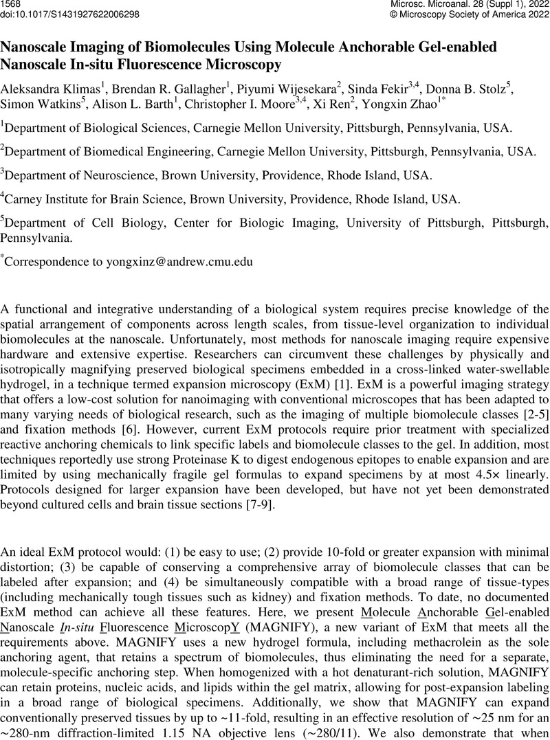
Image of the first page of this content. For PDF version, please use the ‘Save PDF’ preceeding this image.'
Type
Development, Challenges and Biomedical Applications of Tissue Clearing, Expansion Microscopy and Volumetric Imaging
Copyright
Copyright © Microscopy Society of America 2022

References

Chen, F., Tillberg, P. W. and Boyden, E. S. Science 347 (2015), 543548.CrossRefGoogle Scholar
Tillberg, P. W. et al. Nat. Biotechnol. 34 (2016), 987992.CrossRefGoogle Scholar
Chozinski, T. J. et al. Nat. Methods 13 (2016), 17.CrossRefGoogle Scholar
Chen, F. et al. Nat. Methods 13 (2016), 679–84.CrossRefGoogle Scholar
Sun, D. et al. bioRxiv (2020) 2020.03.19.998039. doi:10.1101/2020.03.19.998039Google Scholar
Zhao, Y. et al. Nat. Biotechnol. 35 (2017), 757764.CrossRefGoogle Scholar
Truckenbrodt, S. et al. EMBO Rep. 19 (2018), e45836.CrossRefGoogle Scholar
Damstra, H. G., et al. bioRxiv (2021) 2021.02.03.428837. doi:10.1101/2021.02.03.428837Google Scholar
Chang, J.-B. et al. Nat. Meth 14 (2017), 593599.CrossRefGoogle Scholar
Dertinger, T. et al. Proc. Natl. Acad. Sci. U. S. A. 106 (2009), 2228722292.CrossRefGoogle Scholar
This work was supported by Carnegie Mellon University and DSF Charitable Foundation (Y.Z. and X.R.), U.S. Department of Defense DoD VR190139 (Y.Z.), National Institutes of Health (NIH) Director's New Innovator Award DP2 OD025926-01 (Y.Z), T32 pre-doctoral training grant (Biomechanics in Regenerative Medicine, BiRM) from the National Institute of Biomedical Imaging and Bioengineering of NIH (P.W.), NIH RF1 MH114103 (A.L.B), Air Force Office of Scientific Research AFOSR FA9550-19-1-13022629 (A.L.B.), NeuroNex GR5260228.1001 (C.I.M.), and the Training Program for Interactionist Cognitive Neuroscience (ICoN) T32MH115895 (C.I.M). We thank A. Gittis and J. Schwenk at Carnegie Mellon University for the donation and preparation of mouse brain tissue.Google Scholar
These authors contributed equally to this work: Aleksandra Klimas, Brendan R. GallagherGoogle Scholar