Hostname: page-component-76fb5796d-zzh7m Total loading time: 0 Render date: 2024-04-26T03:52:54.898Z Has data issue: false hasContentIssue false

Time-dependent mechanical properties of rat femoral cortical bone by nanoindentation: An age-related study

Published online by Cambridge University Press:  04 June 2014

Sebastián Jaramillo Isaza*
Affiliation:
Department of Biological Engineering, Laboratoire de Biomécanique et Bioingénierie, UMR CNRS-UTC 7338, Université de Technologie de Compiègne, Compiègne Cedex 60205, France
Pierre-Emmanuel Mazeran
Affiliation:
Department of Mechanical Systems, Laboratoire Roberval Unité de Recherche en Mécanique, UMR CRNS-UTC 7337, Université de Technologie de Compiègne, Compiègne Cedex 60205, France
Karim El Kirat
Affiliation:
Department of Biological Engineering, Laboratoire de Biomécanique et Bioingénierie, UMR CNRS-UTC 7338, Université de Technologie de Compiègne, Compiègne Cedex 60205, France
Marie-Christine Ho Ba Tho*
Affiliation:
Department of Biological Engineering, Laboratoire de Biomécanique et Bioingénierie, UMR CNRS-UTC 7338, Université de Technologie de Compiègne, Compiègne Cedex 60205, France
*
a)Address all correspondence to this author. e-mail: marie-christine.hobatho@utc.fr
Get access

Abstract

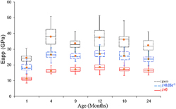
The aim of this study is to assess the time-dependent mechanical properties of rat femoral cortical bone in a lifespan model from growth to senescence. New nanoindentation protocol was performed to assess the time-dependent mechanical behavior. The experimental data were fitted with an elastic–viscoelastic–plastic–viscoplastic mechanical model allowing the calculus of the mechanical properties. Variation of mechanical response of bone as a function of the strain rate and age were highlighted. The most representative variations of the mechanical properties with age were found to be statistically significant (P < 0.001) from 1 to 4 months for elastic properties, from 1 to 9 months for viscoelastic properties and during all lifespan for plastic and viscoplastic properties, highlighting different maturation ages for elastic, viscoelastic, plastic and viscoplastic behaviors. These results suggest that different physical–chemical and structural processes occur at different ages reflecting bone modeling and remodeling activities in the rat's whole lifespan.

Type
Articles
Copyright
Copyright © Materials Research Society 2014 

Access options

Get access to the full version of this content by using one of the access options below. (Log in options will check for institutional or personal access. Content may require purchase if you do not have access.)

References

REFERENCES

Ashman, R.B., Cowin, S.C., Van Buskirk, W.C., and Rice, J.C.: A continuous wave technique for the measurement of the elastic properties of cortical bone. J. Biomech. 17, 349 (1984).CrossRefGoogle ScholarPubMed
Ho Ba Tho, M.C., Rho, J.Y., and Ashman, R.B.: Atlas of mechanical properties of human cortical and cancellous bone. In Vivo Assessment of Bone Quality by Vibration and Wave propagation Techniques. Part II, Van der Perre, G., Lowet, G., Borgwardt, A. eds.; ACCO Publishing: Leuven, (1991); pp. 732.Google Scholar
Thurner, P.J.: Atomic force microscopy and indentation force measurement of bone. WIREs Nanomed. Nanobiotechnol. 1(6), 624649 (2009).Google Scholar
Ho Ba Tho, M.C., Mazeran, P.E., El Kirat, K., and Bensamoun, S.: Multiscale characterization of human cortical bone. CMES-Comput. Model. Eng. 87, 557 (2012).Google Scholar
Currey, J., Brear, K., and Zioupos, P.: The effect of ageing and changes in mineral content in degrading toughness of human femora. J. Biomech. 29, 257 (1996).CrossRefGoogle ScholarPubMed
Currey, J.: Changes in the impact energy absorption of bone with age. J. Biomech. 12, 459 (1979).CrossRefGoogle ScholarPubMed
Currey, J.: Effects of differences in mineralization on the mechanical properties of bone. Philos. Trans. R. Soc. Lond. B. Biol. Sci. 304, 509 (1984).Google Scholar
Akkus, O., Fadar, F., and Schaffer, M.: Age-related changes in physicochemical properties of mineral crystals are related to impaired mechanical function of cortical bone. Bone 34, 443 (2004).Google Scholar
Indrekvam, K., Husby, O.S., Gjerdet, N.R., Engester, L.B., and Langeland, N.: Age-dependent mechanical properties of rat femur. Measured in vivo and in vitro. Acta Orthop. Scand. 62, 248 (1991).Google Scholar
Danielsen, C., Mosekilde, L., and Svenstrup, B.: Cortical bone mass, composition, and mechanical properties in female rats in relation to age, long-term ovariectomy, and estrogen substitution. Calcif. Tissue Int. 52, 26 (1993).CrossRefGoogle ScholarPubMed
Iida, H. and Fukuda, S.: Age-related changes in bone mineral density, cross-sectional area and strength at different skeletal sites in male rats. J. Vet. Med. Sci. 64, 29 (2002).Google Scholar
Willinghamm, M.D., Brodt, M.D., Lee, K.L., Stephens, A.L., Ye, J., and Silva, M.J.: Age-related changes in bone structure and strength in female and male BALB/c mice. Calcif. Tissue Int. 86, 470 (2010).CrossRefGoogle ScholarPubMed
Vanleene, M., Rey, C., and Ho Ba Tho, M.C.: Relationships between density and Young’s modulus with microporosity and physico-chemical properties of Wistar rat cortical bone from growth to senescence. Med. Eng. Phys. 30, 1049 (2008).Google Scholar
Busa, B., Miller, L.M., Rubin, C., Qin, Y-X., and Judex, S.: Rapid establishment of chemical and mechanical properties during lamellar bone formation. Calcif. Tissue Int. 77, 386394 (2005).Google Scholar
Isaksson, H., Malkiewicz, M., Nowak, R., Helminen, H.J., and Jurvelin, J.S.: Rabbit cortical bone tissue increases its elastic stiffness but becomes less viscoelastic with age. Bone 47, 1030 (2010a).Google Scholar
Donnelly, E., Boskey, A.L., Baker, S.P., and Van der Meulen, M.C.H.: Effects of tissue age on bone tissue material composition and nanomechanical properties in the rat cortex. J. Biomed. Mater. Res. 92A, 1048 (2010).CrossRefGoogle Scholar
Burket, J., Gourion-Arsiquaud, S., Havill, L.M., Baker, S.P., Boskey, A.L., and van der Meulen, M.C.H.: Microstructure and nanomechanical properties in osteons relate to tissue and animal age. J. Biomech. 44, 277 (2011).Google Scholar
Oliver, W.C. and Pharr, G.M.: An improved technique for determining hardness and elastic modulus using load and displacement sensing indentation experiments. J. Mater. Res. 7, 1564 (1992).Google Scholar
Lucca, D., Herrmann, K., and Klopfstein, M.J.: Nanoindentation: Measuring methods and applications. CIRP Annals - Manuf. Technol. 59, 803 (2010).Google Scholar
Rho, J.Y., Tsui, T.Y., and Pharr, G.M.: Elastic properties of human cortical and trabecular lamellar bone measured by nanoindentation. Biomaterials 18, 1325 (1997).Google Scholar
Rho, J.Y., Roy, M.E., Tsui, T.Y., and Pharr, G.M.: Elastic properties of microstructural components of human bone tissue as measured by nanoindentation. J. Biomed. Mater. Res. 45, 48 (1999).Google Scholar
Ebenstein, D. and Pruitt, L.: Nanoindentation of biological materials. Nano Today 1, 26 (2006).Google Scholar
Fan, Z. and Rho, J.Y.: Effects of viscoelasticity and time-dependent plasticity on nanoindentation measurements of human cortical bone. J. Biomed. Mater. Res. 67A, 208 (2003).Google Scholar
Vanleene, M., Mazeran, P.E., and Ho Ba Tho, M.C.: Influence of strain rate on the mechanical behavior of cortical bone interstitial lamellae at the micrometer scale. J. Mater. Res. 21, 2093 (2006).Google Scholar
Oyen, M.L. and Cook, R.F.: Load–displacement behavior during sharp indentation of viscous–elastic–plastic materials. J. Mater. Res. 18, 139 (2003).CrossRefGoogle Scholar
Oyen, M.L. and Ko, C.: Examination of local variations in viscous, elastic, and plastic indentation responses in healing bone. J. Mater. Sci.: Mater Med. 18, 623 (2007).Google Scholar
Oyen, M.L.: Nanoindentation hardness of mineralized tissues. J. Biomech. 39, 2699 (2006).CrossRefGoogle ScholarPubMed
Olesiak, S.E., Oyen, M.L., and Ferguson, V.L.: Viscous-elastic-plastic behavior of bone using Berkovich nanoindentation. Mech. Time-Depend Mater. 14, 111 (2010).Google Scholar
Rodriguez-Florez, N., Oyen, M.L., and Shefelbine, S.J.: Insight into differences in nanoindentation properties of bone. J. Mech. Behav. Biomed. Mater. 18, 90 (2013).CrossRefGoogle ScholarPubMed
Isaksson, H., Malkiewicz, M., Nowak, R., Helminen, H.J., and Jurvelin, J.S.: Precision of nanoindentation protocols for measurement of viscoelasticity in cortical and trabecular bone. J. Biomech. 43, 2410 (2010b).CrossRefGoogle ScholarPubMed
Sasaki, N. and Yoshikawa, M.: Stress relaxation in native and EDTA-treated bone as a function of mineral content. J. Biomech. 26, 77 (1993).CrossRefGoogle ScholarPubMed
Mazeran, P-E., Beyaoui, M., Bigerelle, M., and Guigon, M.: Determination of mechanical properties by nanoindentation in the case of viscous materials. Int. J. Mater. Res. 103, 715 (2012).Google Scholar
Lucas, B.N., Oliver, W.C., Pharr, G.M., and Loubet, J.L.: Time-dependent deformation during indentation testing. Mat. Res. Soc. Symp. Proc. 436, 233 (1997).Google Scholar
Hochstetter, G., Jimenez, A., and Loubet, J.L.: Strain-rate effects on hardness of glassy polymers in the nanoscale range. Comparison between quasistatic and continuous stiffness measurements. J. Macromol. Sci. Part B 38, 681 (1999).CrossRefGoogle Scholar
Chen, X., Ogasawara, N., Zhao, M., and Chiba, N.: On the uniqueness of measuring elastoplastic properties from indentation: The indistinguishable mystical materials. J. Mech. Phys. Solids 55, 1618 (2007).CrossRefGoogle Scholar
Reilly, D.T., Burstein, A.H., and Frankel, V.H.: The elastic modulus for bone. J. Biomech. 7, 271 (1974).Google Scholar
Winsor, C.P.: The Gompertz curve as a growth curve. Proc. Natl. Acad. Sci. U S A 18(1), 1 (1932).CrossRefGoogle ScholarPubMed
Liebschner, M.A.K.: Biomechanical considerations of animal models used in tissue engineering of bone. Biomaterials 25, 1697 (2004).CrossRefGoogle ScholarPubMed
Rho, J.Y. and Pharr, G.M.: Effects of drying on the mechanical properties of bovine femur measured by nanoindentation. J. Mater. Sci.: Mater. Med. 10, 485 (1999).Google Scholar
Hodgskinson, R. and Currey, J.D.: Young’s modulus, density and material properties in cancellous bone over a large density range. J. Mater. Sci.: Mater. Med. 3, 377 (1992).Google Scholar
Eberhardsteiner, L., Hellmich, C., and Scheiner, S.: Layered water in crystal interfaces as source for bone viscoelasticity: Arguments from a multiscale approach. Comput. Methods Biomech. Biomed. Engin. 17, 48 (2014).Google Scholar