Skip to main content Accessibility help
×
Hostname: page-component-76fb5796d-vfjqv Total loading time: 0 Render date: 2024-04-25T10:52:18.688Z Has data issue: false hasContentIssue false

9 - Nanostructure Growth, Interactions, and Assembly in the Liquid Phase

from Part II - Applications

Published online by Cambridge University Press:  22 December 2016

Frances M. Ross
Affiliation:
IBM T. J. Watson Research Center, New York
Get access

Summary

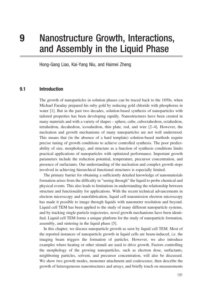
Image of the first page of this content. For PDF version, please use the ‘Save PDF’ preceeding this image.'
Type
Chapter
Information
Publisher: Cambridge University Press
Print publication year: 2016

Access options

Get access to the full version of this content by using one of the access options below. (Log in options will check for institutional or personal access. Content may require purchase if you do not have access.)

References

Faraday, M., The Bakerian lecture: experimental relations of gold (and other metals) to light. Phil. Trans. R. Soc. Lond., 147 (1857), 145181.Google Scholar
Daniel, M. C. and Astruc, D., Gold nanoparticles: assembly, supramolecular chemistry, quantum-size-related properties, and applications toward biology, catalysis, and nanotechnology. Chem. Rev., 104 (2004), 293346.CrossRefGoogle Scholar
Murphy, C. J. Sau, T. P., Gole, A. M., et al., Anisotropic metal nanoparticles: synthesis, assembly, and optical applications. J. Phys. Chem. B, 109 (2005), 1385713870.Google Scholar
Xia, Y., Xiong, Y., Lim, B. and Skrabalak, S. E., Shape-controlled synthesis of metal nanocrystals: simple chemistry meets complex physics? Angew. Chem. Int. Ed., 48 (2009), 60103.CrossRefGoogle ScholarPubMed
Liao, H.-G., Niu, K. and Zheng, H., Observation of growth of metal nanoparticles. Chem. Commun., 49 (2013), 1172011727.CrossRefGoogle ScholarPubMed
Niu, K.-Y., Park, J., Zheng, H. and Alivisatos, A. P., Revealing bismuth oxide hollow nanoparticle formation by the Kirkendall effect. Nano Lett., 13 (2013), 57155719.CrossRefGoogle ScholarPubMed
Xin, H. L. and Zheng, H., In situ observation of oscillatory growth of bismuth nanoparticles. Nano Lett., 12 (2012), 14701474.CrossRefGoogle ScholarPubMed
Zheng, H., Smith, R. K., Jun, Y.-W., et al., Observation of single colloidal platinum nanocrystal growth trajectories. Science, 324 (2009), 13091312.CrossRefGoogle ScholarPubMed
Grogan, J. M., Schneider, N. M., Ross, F. M. and Bau, H. H., Bubble and pattern formation in liquid induced by an electron beam. Nano Lett., 14 (2013), 359364.CrossRefGoogle ScholarPubMed
den Heijer, M., Shao, I., Radisic, A., Reuter, M. C. and Ross, F. M., Patterned electrochemical deposition of copper using an electron beam. APL Materials, 2 (2014), 022101.Google Scholar
Liu, Y., Lin, X.-M., Sun, Y. and Rajh, T., In situ visualization of self-assembly of charged gold nanoparticles. J. Am. Chem. Soc., 135 (2013), 37643767.Google Scholar
Woehl, T. J., Park, C., Evans, J. E., et al., Direct observation of aggregative nanoparticle growth: kinetic modeling of the size distribution and growth rate. Nano Lett., 14 (2013), 373378.Google Scholar
Liao, H.-G., Cui, L., Whitelam, S. and Zheng, H., Real-time imaging of Pt3Fe nanorod growth in solution. Science, 336 (2012), 10111014.Google Scholar
Zhu, G., Jiang, Y., Lin, F., et al., In situ study of the growth of two-dimensional palladium dendritic nanostructures using liquid-cell electron microscopy. Chem. Commun., 50 (2014), 94479450.CrossRefGoogle ScholarPubMed
Woehl, T. J., Evans, J. E., Arslan, I., Ristenpart, W. D. and Browning, N. D., Direct in situ determination of the mechanisms controlling nanoparticle nucleation and growth. ACS Nano, 6 (2012), 85998610.Google Scholar
Evans, J. E., Jungjohann, K. L., Browning, N. D. and Arslan, I., Controlled growth of nanoparticles from solution with in situ liquid transmission electron microscopy. Nano Lett., 11 (2011), 28092813.Google Scholar
Niu, K.-Y., Liao, H.-G. and Zheng, H., Visualization of the coalescence of bismuth nanoparticles. Microsc. Microanal., 20 (2014), 416424.CrossRefGoogle ScholarPubMed
Li, D., Nelson, M. H., Lee, J. R., et al., Direction-specific interactions control crystal growth by oriented attachment. Science, 336 (2012), 10141018.CrossRefGoogle ScholarPubMed
Yuk, J. M., Park, J., Ercius, P., et al., High-resolution EM of colloidal nanocrystal growth using graphene liquid cells. Science, 336 (2012), 6164.Google Scholar
Wulff, G., On the question of speed of growth and dissolution of crystal surfaces. Z. Krystallogr. Mineral., 34 (1901), 449530.Google Scholar
Gibbs, J. W., Bumstead, H. A., Van Name, R. G. and Longley, W. R., The Collected Works of J. Willard Gibbs (London: Longmans, Green and Co., 1902).Google Scholar
Tian, N., Zhou, Z.-Y., Sun, S.-G., Ding, Y. and Wang, Z. L., Synthesis of tetrahexahedral platinum nanocrystals with high-index facets and high electro-oxidation activity. Science, 316 (2007), 732735.CrossRefGoogle ScholarPubMed
Ringe, E., Van Duyne, R. P. and Marks, L. D., Wulff construction for alloy nanoparticles. Nano Lett., 11 (2011), 33993403.Google Scholar
Bealing, C. R., Baumgardner, W. J., Choi, J. J., Hanrath, T. and Hennig, R. G., Predicting nanocrystal shape through consideration of surface-ligand interactions. ACS Nano, 6 (2012), 21182127.Google Scholar
Liao, H.-G., Zherebetskyy, D., Xin, H., et al., Facet development during platinum nanocube growth. Science, 345 (2014), 916919.Google Scholar
Liao, H.-G. and Zheng, H., Liquid cell transmission electron microscopy study of platinum iron nanocrystal growth and shape evolution. J. Am. Chem. Soc., 135 (2013), 50385043.CrossRefGoogle ScholarPubMed
Kimura, Y., Niinomi, H., Tsukamoto, K. and García-Ruiz, J. M., In situ live observation of nucleation and dissolution of sodium chlorate nanoparticles by transmission electron microscopy. J. Am. Chem. Soc., 136 (2014), 17621765.Google Scholar
Sutter, E., Jungjohann, K., Bliznakov, S. et al., In situ liquid-cell electron microscopy of silver-palladium galvanic replacement reactions on silver nanoparticles. Nat. Commun., 5 (2014), 4946.Google Scholar
Jungjohann, K., Bliznakov, S., Sutter, P., Stach, E. A. and Sutter, E., In situ liquid cell electron microscopy of the solution growth of Au–Pd core–shell nanostructures. Nano Lett., 13 (2013), 29642970.Google Scholar
Lewis, E. A., Haigh, S. J., Slater, T. J. A., et al., Real-time imaging and local elemental analysis of nanostructures in liquids. Chem. Commun., 50 (2014), 1001910022.Google Scholar
Wu, J., Gao, W., Wen, J. et al., Growth of Au on Pt icosahedral nanoparticles revealed by low-dose in situ TEM. Nano Lett., 15 (2015), 27112715.Google Scholar
De Clercq, A., Dachraoui, W., Margeat, O., et al., Growth of Pt–Pd nanoparticles studied in situ by HRTEM in a liquid cell. J. Phys. Chem. Lett., 5 (2014), 21262130.CrossRefGoogle ScholarPubMed
Kraus, T. and de Jonge, N., Dendritic gold nanowire growth observed in liquid with transmission electron microscopy. Langmuir, 29 (2013), 84278432.Google Scholar
Liao, H.-G., Shao, Y., Wang, C. M., et al., TEM study of fivefold twinned gold nanocrystal formation mechanism. Mater. Lett., 116 (2014), 299303.Google Scholar
Alloyeau, D., Dachraoui, W., Javed, Y., et al., Unravelling kinetic and thermodynamic effects on the growth of gold nanoplates by liquid transmission electron microscopy. Nano Lett., 15 (2015), 25742581.Google Scholar
Parent, L. R., Robinson, D. B., Woehl, T. J., et al., Direct in situ observation of nanoparticle synthesis in a liquid crystal surfactant template. ACS Nano, 6 (2012), 35893596.Google Scholar
Parent, L. R., Robinson, D. B., Cappillino, P. J., et al., In situ observation of directed nanoparticle aggregation during the synthesis of ordered nanoporous metal in soft templates. Chem. Mater., 26 (2014), 14261433.CrossRefGoogle Scholar
Chen, X. and Wen, J., In situ wet-cell TEM observation of gold nanoparticle motion in an aqueous solution. Nanoscale Res. Lett., 7 (2012), 16.Google Scholar
Ring, E. A. and de Jonge, N., Microfluidic system for transmission electron microscopy. Microsc. Microanal., 16 (2010), 622629.Google Scholar
Zheng, H., Claridge, S. A., Minor, A. M., Alivisatos, A. P. and Dahmen, U., Nanocrystal diffusion in a liquid thin film observed by in situ transmission electron microscopy. Nano Lett., 9 (2009), 24602465.Google Scholar
Chen, Q., Smith, J. M., Park, J., et al., 3D motion of DNA-Au nanoconjugates in graphene liquid cell electron microscopy. Nano Lett., 13 (2013), 45564561.Google Scholar
de Jonge, N., Poirier-Demers, N., Demers, H., Peckys, D. B. and Drouin, D., Nanometer-resolution electron microscopy through micrometers-thick water layers. Ultramicroscopy, 110 (2010), 11141119.Google Scholar
White, E. R., Mecklenburg, M., Shevitski, B., Singer, S. B. and Regan, B. C., Charged nanoparticle dynamics in water induced by scanning transmission electron microscopy. Langmuir, 28 (2012), 36953698.Google Scholar
Mueller, C., Harb, M., Dwyer, J. R. and Miller, R. D., Nanofluidic cells with controlled pathlength and liquid flow for rapid, high-resolution in situ imaging with electrons. J. Phys. Chem. Lett., 4 (2013), 23392347.Google Scholar
Li, F., Josephson, D. P. and Stein, A., Colloidal assembly: the road from particles to colloidal molecules and crystals. Angew. Chem. Int. Ed., 50 (2011), 360388.Google Scholar
Baker, J. L., Widmer-Cooper, A., Toney, M. F., Geissler, P. L. and Alivisatos, A. P., Device-scale perpendicular alignment of colloidal nanorods. Nano Lett., 10 (2009), 195201.Google Scholar
Park, J., Zheng, H., Lee, W. C., et al., Direct observation of nanoparticle superlattice formation by using liquid cell transmission electron microscopy. ACS Nano, 6 (2012), 20782085.CrossRefGoogle ScholarPubMed
Grogan, J. M., Rotkina, L. and Bau, H. H., In situ liquid-cell electron microscopy of colloid aggregation and growth dynamics. Phys. Rev. E, 83 (2011), 061405.Google Scholar
Oleshko, V. P. and Howe, J. M., Are electron tweezers possible? Ultramicroscopy, 111 (2011), 15991606.Google Scholar
Batson, P. E., Reyes-Coronado, A., Barrera, R. G., et al., Nanoparticle movement: plasmonic forces and physical constraints. Ultramicroscopy, 123 (2012), 5058.Google Scholar
Batson, P. E., Reyes-Coronado, A., Barrera, R. G., et al., Plasmonic nanobilliards: controlling nanoparticle movement using forces induced by swift electrons. Nano Lett., 11 (2011), 33883393.Google Scholar
Zheng, H., Mirsaidov, U. M., Wang, L.-W. and Matsudaira, P., Electron beam manipulation of nanoparticles. Nano Lett., 12 (2012), 56445648.CrossRefGoogle ScholarPubMed
Zheng, H., Using molecular tweezers to move and image nanoparticles. Nanoscale, 5 (2013), 40704078.Google Scholar
Chen, Y.-T., Wang, C.-Y., Hong, Y.-J., et al., Electron beam manipulation of gold nanoparticles external to the beam. RSC Adv., 4 (2014), 3165231656.Google Scholar
Jiang, Y., Zhu, G., Lin, F., et al., In situ study of oxidative etching of palladium nanocrystals by liquid cell electron microscopy. Nano Lett., 14 (2014), 37613765.Google Scholar

Save book to Kindle

To save this book to your Kindle, first ensure coreplatform@cambridge.org is added to your Approved Personal Document E-mail List under your Personal Document Settings on the Manage Your Content and Devices page of your Amazon account. Then enter the ‘name’ part of your Kindle email address below. Find out more about saving to your Kindle.

Note you can select to save to either the @free.kindle.com or @kindle.com variations. ‘@free.kindle.com’ emails are free but can only be saved to your device when it is connected to wi-fi. ‘@kindle.com’ emails can be delivered even when you are not connected to wi-fi, but note that service fees apply.

Find out more about the Kindle Personal Document Service.

Available formats
×

Save book to Dropbox

To save content items to your account, please confirm that you agree to abide by our usage policies. If this is the first time you use this feature, you will be asked to authorise Cambridge Core to connect with your account. Find out more about saving content to Dropbox.

Available formats
×

Save book to Google Drive

To save content items to your account, please confirm that you agree to abide by our usage policies. If this is the first time you use this feature, you will be asked to authorise Cambridge Core to connect with your account. Find out more about saving content to Google Drive.

Available formats
×