Skip to main content Accessibility help
×
Hostname: page-component-848d4c4894-x5gtn Total loading time: 0 Render date: 2024-05-01T05:47:09.963Z Has data issue: false hasContentIssue false

16 - Liquid STEM for Studying Biological Function in Whole Cells

from Part II - Applications

Published online by Cambridge University Press:  22 December 2016

Frances M. Ross
Affiliation:
IBM T. J. Watson Research Center, New York
Get access

Summary

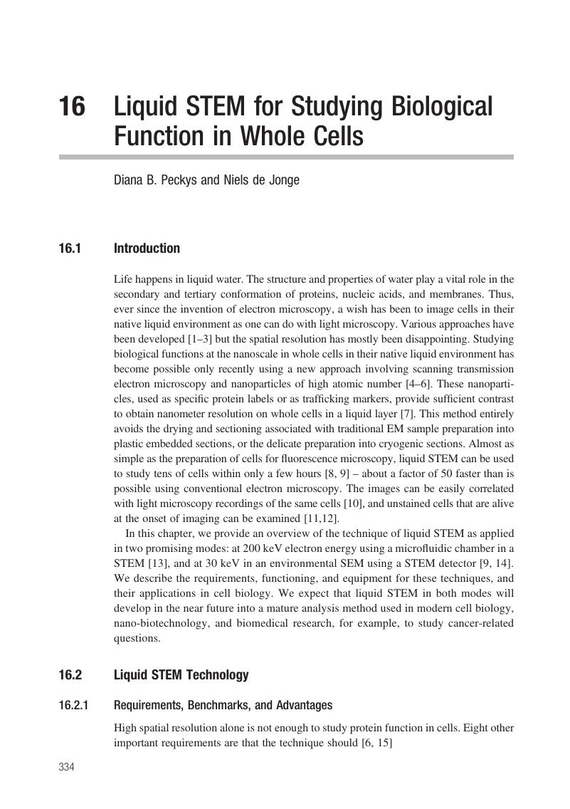
Image of the first page of this content. For PDF version, please use the ‘Save PDF’ preceeding this image.'
Type
Chapter
Information
Publisher: Cambridge University Press
Print publication year: 2016

Access options

Get access to the full version of this content by using one of the access options below. (Log in options will check for institutional or personal access. Content may require purchase if you do not have access.)

References

Parsons, D. F., Matricardi, V. R., Moretz, R. C. and Turner, J. N., Electron microscopy and diffraction of wet unstained and unfixed biological objects. Adv. Biol. Med. Phys., 15 (1974), 161270.Google Scholar
Parsons, D. F., Structure of wet specimens in electron microscopy. Science, 186 (1974), 407414.CrossRefGoogle ScholarPubMed
de Jonge, N. and Ross, F. M., Electron microscopy of specimens in liquid. Nat. Nanotechnol., 6 (2011), 695704.Google Scholar
de Jonge, N., Peckys, D. B., Kremers, G. J. and Piston, D. W., Electron microscopy of whole cells in liquid with nanometer resolution. Proc. Natl. Acad. Sci. USA, 106 (2009), 21592164.Google Scholar
Peckys, D. B., Veith, G. M., Joy, D. C. and de Jonge, N., Nanoscale imaging of whole cells using a liquid enclosure and a scanning transmission electron microscope. PLoS One, 4 (2009), e8214.Google Scholar
Peckys, D. B. and de Jonge, N., Liquid scanning transmission electron microscopy: imaging protein complexes in their native environment in whole eukaryotic cells. Microsc. Microanal., 20 (2014), 346365.Google Scholar
de Jonge, N., Poirier-Demers, N., Demers, H., Peckys, D. B. and Drouin, D., Nanometer-resolution electron microscopy through micrometers-thick water layers. Ultramicroscopy, 110, 11141119 (2010).Google Scholar
Peckys, D. B. and de Jonge, N., Gold nanoparticle uptake in whole cells in liquid examined by environmental scanning electron microscopy. Microsc. Microanal., 20 (2014), 189197.Google Scholar
Peckys, D. B., Baudoin, J. P., Eder, M., Werner, U. and de Jonge, N., Epidermal growth factor receptor subunit locations determined in hydrated cells with environmental scanning electron microscopy. Sci. Rep., 3 (2013), 2626.Google Scholar
Dukes, M. J., Peckys, D. B. and de Jonge, N., Correlative fluorescence microscopy and scanning transmission electron microscopy of quantum-dot-labeled proteins in whole cells in liquid. ACS Nano, 4 (2010), 41104116.Google Scholar
Peckys, D. B., Mazur, P., Gould, K. L. and de Jonge, N., Fully hydrated yeast cells imaged with electron microscopy. Biophys. J., 100 (2011), 25222529.Google Scholar
Peckys, D. B. and de Jonge, N., Visualization of gold nanoparticle uptake in living cells with liquid scanning transmission electron microscopy. Nano Lett., 11 (2011), 17331738.Google Scholar
Ring, E. A. and de Jonge, N., Microfluidic system for transmission electron microscopy. Microsc. Microanal., 16 (2010), 622629.CrossRefGoogle ScholarPubMed
Bogner, A., Thollet, G., Basset, D., Jouneau, P. H. and Gauthier, C., Wet STEM: a new development in environmental SEM for imaging nano-objects included in a liquid phase. Ultramicroscopy, 104 (2005), 290301.Google Scholar
de Jonge, N., Sougrat, R., Peckys, D. B., Lupini, A. R. and Pennycook, S. J., 3-Dimensional aberration corrected scanning transmission electron microscopy for biology. In Vo-Dinh, T., ed., Nanotechnology in Biology and Medicine-Methods, Devices and Applications (Boca Raton, FL: CRC Press, 2007) pp. 13.1113.27.Google Scholar
Lippincott-Schwartz, J., Snapp, E. and Kenworthy, A., Studying protein dynamics in living cells. Nat. Rev. Mol. Cell. Biol., 2 (2001), 444456.CrossRefGoogle ScholarPubMed
Pawley, J. B., Handbook of Biological Confocal Microscopy, 2nd edn. (New York: Springer, 1995).Google Scholar
Willig, K. I., Rizzoli, S. O., Westphal, V., Jahn, R. and Hell, S. W., STED microscopy reveals that synapthotagmin remains clustered after synaptic vesicle exocytosis. Nature, 440 (2006), 935939.Google Scholar
Hell, S. W., Far-field optical nanoscopy. Science, 316 (2007), 11531158.CrossRefGoogle ScholarPubMed
Betzig, E., Patterson, G. H., Sougrat, R. et al., Imaging intracellular fluorescent proteins at nanometer resolution. Science, 313 (2006), 16421645.Google Scholar
Bates, M., Huang, B., Dempsey, G. T. and Zhuang, X., Multicolor super-resolution imaging with photo-switchable fluorescent probes. Science, 317 (2007), 17491753.Google Scholar
Lippincott-Schwartz, J. and Manley, S., Putting super-resolution fluorescence microscopy to work. Nat. Meth., 6 (2009), 2123.Google Scholar
Herbert, S., Soares, H., Zimmer, C. and Henriques, R., Single-molecule localization super-resolution microscopy: deeper and faster. Microsc. Microanal., 18 (2012), 14191429.Google Scholar
Piston, D. W. and Kremers, G. J., Fluorescent protein FRET: the good, the bad and the ugly. Trends Biochem. Sci., 32 (2007), 407414.Google Scholar
Warren, C. M. and Landgraf, R., Signaling through ERBB receptors: multiple layers of diversity and control. Cell. Signal., 18 (2006), 923933.Google Scholar
Needham, S. R., Hirsch, M., Rolfe, D. J. et al., Measuring EGFR separations on cells with ~10 nm resolution via fluorophore localization imaging with photobleaching. PLoS One, 8 (2013), e62331.Google Scholar
Liu, P., Sudhaharan, T., Koh, R. M. et al., Investigation of the dimerization of proteins from the epidermal growth factor receptor family by single wavelength fluorescence cross-correlation spectroscopy. Biophys. J., 93 (2007), 684698.Google Scholar
Hoenger, A. and McIntosh, J. R., Probing the macromolecular organization of cells by electron tomography. Curr. Opin. Cell Biol., 21 (2009), 8996.Google Scholar
Kourkoutis, L. F., Plitzko, J. M. and Baumeister, W., Electron microscopy of biological materials at the nanometer scale. Annu. Rev. Mater. Res., 42 (2012), 3358.Google Scholar
Medalia, O., Weber, I., Frangakis, A. S. et al., Macromolecular architecture in eukaryotic cells visualized by cryoelectron tomography. Science, 298 (2002), 12091213.Google Scholar
Fujimoto, K., Freeze-fracture replica electron microscopy combined with SDS digestion for cytochemical labeling of integral membrane proteins: application to the immunogold labeling of intercellular junctional complexes. J. Cell Sci., 108 (1995), 34433449.Google Scholar
Bushby, A. J., P'Ng, K. M., Young, R. D. et al., Imaging three-dimensional tissue architectures by focused ion beam scanning electron microscopy. Nat. Protoc., 6 (2011), 845858.Google Scholar
Bergersen, L. H., Storm-Mathisen, J. and Gundersen, V., Immunogold quantification of amino acids and proteins in complex subcellular compartments. Nat. Protoc., 3 (2008), 144152.Google Scholar
Larabell, C. A. and Nugent, K. A., Imaging cellular architecture with X-rays. Curr. Opin. Struct. Biol., 20 (2010), 623631.Google Scholar
Peckys, D. B., Korf, U. and de Jonge, N., Local variations of HER2 dimerization in breast cancer cells discovered by correlative fluorescence and liquid electron microscopy. Sci. Adv., 1 (2015), e1500165.Google Scholar
Peckys, D. B. and de Jonge, N., Studying the stoichiometry of epidermal growth factor receptor in intact cells using correlative microscopy. J. Vis. Exp. (2015). Epub. 2015/09/19.Google Scholar
Williamson, M. J., Tromp, R. M., Vereecken, P. M., Hull, R. and Ross, F. M., Dynamic microscopy of nanoscale cluster growth at the solid-liquid interface. Nat. Mater., 2 (2003), 532536.CrossRefGoogle ScholarPubMed
Thiberge, S., Nechushtan, A., Sprinzak, D. et al., Scanning electron microscopy of cells and tissues under fully hydrated conditions. Proc. Natl. Acad. Sci. USA, 101 (2004), 3346.Google Scholar
Ring, E. A., Peckys, D. B., Dukes, M. J., Baudoin, J. P. and de Jonge, N., Silicon nitride windows for electron microscopy of whole cells. J. Microsc., 243 (2011), 273283.Google Scholar
Grogan, J. M. and Bau, H. H., The nanoaquarium: a platform for in situ transmission electron microscopy in liquid media. J. Microelectromech. Sys., 19 (2010), 885894.Google Scholar
Nishiyama, H., Suga, M., Ogura, T. et al., Atmospheric scanning electron microscope observes cells and tissues in open medium through silicon nitride film. J. Struct. Biol., 169 (2010), 438449.Google Scholar
Liv, N., Lazic, I., Kruit, P. and Hoogenboom, J. P., Scanning electron microscopy of individual nanoparticle bio-markers in liquid. Ultramicroscopy, 143 (2014), 9399.Google Scholar
Stokes, D. J., Recent advances in electron imaging, image interpretation and applications: environmental scanning electron microscopy. Phil. Trans. R. Soc. Lond. A, 361 (2003), 27712787.Google Scholar
Stokes, D. L., Principles and Practice of Variable Pressure/Environmental Scanning Electron Microscopy (VP-SEM) (New York: Wiley, 2008).Google Scholar
Li, N., Zonnevylle, A. C., Narvaez, A. C. et al., Simultaneous correlative scanning electron and high-NA fluorescence microscopy. PLoS One, 8 (2013), e55707.Google Scholar
Masenelli-Varlot, K., Malchere, A., Ferreira, J. et al., Wet-STEM tomography: principles, potentialities and limitations. Microsc. Microanal., 20 (2014), 366375.Google Scholar
de Jonge, N., Peckys, D. B., Veith, G. M. et al., Scanning transmission electron microscopy of samples in liquid (liquid STEM). Microsc. Microanal., 13 (2007), 242243.Google Scholar
Ramachandra, R., Demers, H. and de Jonge, N., Atomic-resolution scanning transmission electron microscopy through 50 nm-thick silicon nitride membranes. Appl. Phys. Lett., 98 (2011), 93109.Google Scholar
Liu, K. L., Wu, C. C., Huang, Y. J. et al., Novel microchip for in situ TEM imaging of living organisms and bio-reactions in aqueous conditions. Lab Chip, 8 (2008), 19151921.Google Scholar
Klein, K. L., Anderson, I. M. and de Jonge, N., Transmission electron microscopy with a liquid flow cell. J. Microsc., 242 (2011), 117123.Google Scholar
Coskun, U. and Simons, K., Cell membranes: the lipid perspective. Structure, 19 (2011), 15431548.Google Scholar
Arkhipov, A., Shan, Y., Das, R. et al., Architecture and membrane interactions of the EGF receptor. Cell, 152 (2013), 557569.Google Scholar
Normanno, N., De Luca, A., Bianco, C. et al., Epidermal growth factor receptor (EGFR) signaling in cancer. Gene, 366 (2006), 216.Google Scholar
Schlessinger, J., Signal transduction by allosteric receptor oligomerization. Trends Biochem. Sci., 13 (1988), 443447.Google Scholar
Ullrich, A. and Schlessinger, J., Signal transduction by receptors with tyrosine kinase activity. Cell, 61 (1990), 203212.Google Scholar
Endres, N. F., Das, R., Smith, A. W. et al., Conformational coupling across the plasma membrane in activation of the EGF receptor. Cell, 152 (2013), 543556.CrossRefGoogle ScholarPubMed
Lidke, D. S., Nagy, P., Heintzmann, R. et al., Quantum dot ligands provide new insights into erbB/HER receptor-mediated signal transduction. Nat. Biotechnol., 22 (2004), 198203.Google Scholar
Tanaka, K. A., Suzuki, K. G., Shirai, Y. M. et al., Membrane molecules mobile even after chemical fixation. Nat. Meth., 7 (2010), 865866.Google Scholar
Glenney, J. R. Jr., Chen, W. S., Lazar, C. S. et al., Ligand-induced endocytosis of the EGF receptor is blocked by mutational inactivation and by microinjection of anti-phosphotyrosine antibodies. Cell, 52 (1988), 675684.Google Scholar
Hoenger, A. and Bouchet-Marquis, C., Cellular tomography. Adv. Protein Chem. Struct. Biol., 82 (2011), 6790.Google Scholar
Sousa, A. A., Azari, A. A., Zhang, G. and Leapman, R. D., Dual-axis electron tomography of biological specimens: extending the limits of specimen thickness with bright-field STEM imaging. J. Struct. Biol., 174 (2011), 107114.Google Scholar
Demers, H., Poirier-Demers, N., Drouin, D. and de Jonge, N., Simulating STEM imaging of nanoparticles in micrometers-thick substrates. Microsc. Microanal., 16 (2010), 795804.Google Scholar
Schuh, T. and de Jonge, N., Liquid scanning transmission electron microscopy: nanoscale imaging in micrometers-thick liquids. C. R. Phys., 15 (2014), 214223.Google Scholar
Ring, E. A. and de Jonge, N., Video-frequency scanning transmission electron microscopy of moving gold nanoparticles in liquid. Micron, 43 (2012), 10781084.Google Scholar
White, E. R., Mecklenburg, M., Shevitski, B., Singer, S. B. and Regan, B. C., Charger nanoparticle dynamics in water induced by scanning transmission electron microscopy. Langmuir, 28 (2012), 36953698.Google Scholar
Verch, A., Pfaff, M. and de Jonge, N., Exceptionally slow movement of gold nanoparticles at a solid/liquid interface investigated by scanning transmission electron microscopy. Langmuir, 31 (2015), 69566964.CrossRefGoogle Scholar
Yuk, J. M., Park, J., Ercius, P. et al., High-resolution EM of colloidal nanocrystal growth using graphene liquid cells. Science, 336 (2012), 6164.Google Scholar
Evans, J. E., Jungjohann, K. L., Browning, N. D. and Arslan, I., Controlled growth of nanoparticles from solution with in situ liquid transmission electron microscopy. Nano Lett., 11 (2011), 28092813.Google Scholar
Lillemeier, B. F., Pfeiffer, J. R., Surviladze, Z., Wilson, B. S. and Davis, M. M., Plasma membrane-associated proteins are clustered into islands attached to the cytoskeleton. Proc. Natl. Acad. Sci. USA, 103 (2006), 1899218997.Google Scholar
McBride, J., Treadway, J., Feldman, L. C., Pennycook, S. J. and Rosenthal, S. J., Structural basis for near unity quantum yield core/shell nanocrystals. Nano Lett., 6 (2006), 14961501.Google Scholar
Peckys, D. B., Bandmann, V. and de Jonge, N., Correlative fluorescence and scanning transmission electron microscopy of quantum dot-labeled proteins on whole cells in liquid. Meth. Cell Biol., 124 (2014), 305322.Google Scholar
Tanaka, K. A., Suzuki, K. G., Shirai, Y. M. et al., Membrane molecules mobile even after chemical fixation. Nat. Meth., 7 (2010), 865866.Google Scholar
Reimer, L. and Kohl, H., Transmission Electron Microscopy: Physics of Image Formation (New York: Springer, 2008).Google Scholar
Pohlmann, E. S., Patel, K., Guo, S. et al., Real-time visualization of nanoparticles interacting with glioblastoma stem cells. Nano Lett., 15 (2015), 23292335.Google Scholar
Spence, J. C. H., High-Resolution Electron Microscopy, 3rd edn. (Oxford: Oxford University Press, 2003).Google Scholar
Agronskaia, A. V., Valentijn, J. A., van Driel, L. F. et al., Integrated fluorescence and transmission electron microscopy. J. Struct. Biol., 164 (2008), 183189.Google Scholar
Matricardi, V. R., Moretz, R. C. and Parsons, D. F., Electron diffraction of wet proteins: catalase. Science, 177 (1972), 268270.Google Scholar
Siegwart, D. J., Srinivasan, A., Bencherif, S. A. et al., Cellular uptake of functional nanogels prepared by inverse miniemulsion ATRP with encapsulated proteins, carbohydrates, and gold nanoparticles. Biomacromol., 10 (2009), 23002309.Google Scholar
Chithrani, B. D., Ghazani, A. A. and Chan, W. C., Determining the size and shape dependence of gold nanoparticle uptake into mammalian cells. Nano Lett., 6 (2006), 662668.Google Scholar
Bright, N. A., Reaves, B. J., Mullock, B. M. and Luzio, J. P., Dense core lysosomes can fuse with late endosomes and are re-formed from the resultant hybrid organelles. J. Cell Sci., 110 (1997), 20272040.Google Scholar
Tantra, R. and Knight, A., Cellular uptake and intracellular fate of engineered nanoparticles: a review on the application of imaging techniques. Nanotoxicology, 5 (2011), 381392.Google Scholar
Glavinovic, M. I., Vitale, M. L. and Trifaro, J. M., Comparison of vesicular volume and quantal size in bovine chromaffin cells. Neuroscience, 85 (1998), 957968.Google Scholar
Brandenberger, C., Muhlfeld, C., Ali, Z. et al., Quantitative evaluation of cellular uptake and trafficking of plain and polyethylene glycol-coated gold nanoparticles. Small, 6 (2010), 16691678.CrossRefGoogle ScholarPubMed
Sartori, A., Gatz, R., Beck, F. et al., Correlative microscopy: bridging the gap between fluorescence light microscopy and cryo-electron tomography. J. Struct. Biol., 160 (2007), 135145.Google Scholar
Gilmore, B. L., Showalter, S. P., Dukes, M. J. et al., Visualizing viral assemblies in a nanoscale biosphere. Lab Chip, 13 (2013), 216219.Google Scholar
Zheng, H., Claridge, S. A., Minor, A. M., Alivisatos, A. P. and Dahmen, U., Nanocrystal diffusion in a liquid thin film observed by in situ transmission electron microscopy. Nano Lett., 9 (2009), 24602465.Google Scholar
Woehl, T. J., Jungjohann, K. L., Evans, J. E. et al., Experimental procedures to mitigate electron beam induced artifacts during in situ fluid imaging of nanomaterials. Ultramicroscopy, 127 (2013), 5363.Google Scholar
Mirsaidov, U. M., Zheng, H., Casana, Y. and Matsudaira, P., Imaging protein structure in water at 2.7 nm resolution by transmission electron microscopy. Biophys. J., 102 (2012), L15–17.Google Scholar
Mueller, C., Harb, M., Dwyer, J. R. and Dwayne Miller, R. J., Nanofluidic cells with controlled pathlength and liquid flow for rapid, high-resolution in situ imaging with electrons. J. Phys. Chem. Lett., 4 (2013), 23392347.Google Scholar
Shu, X., Lev-Ram, V., Deerinck, T. J. et al., A genetically encoded tag for correlated light and electron microscopy of intact cells, tissues, and organisms. PLoS Biol., 9 (2011), e1001041.Google Scholar
Gaietta, G., Deerinck, T. J., Adams, S. R. et al., Multicolor and electron microscopic imaging of connexin trafficking. Science, 296 (2002), 503507.Google Scholar
Risco, C., Sanmartin-Conesa, E., Tzeng, W. P. et al., Specific, sensitive, high-resolution detection of protein molecules in eukaryotic cells using metal-tagging transmission electron microscopy. Structure, 20 (2012), 759766.Google Scholar
Tantra, R. and Shard, A., We need answers. Nat. Nanotechnol., 8 (2013), 71.Google Scholar

Save book to Kindle

To save this book to your Kindle, first ensure coreplatform@cambridge.org is added to your Approved Personal Document E-mail List under your Personal Document Settings on the Manage Your Content and Devices page of your Amazon account. Then enter the ‘name’ part of your Kindle email address below. Find out more about saving to your Kindle.

Note you can select to save to either the @free.kindle.com or @kindle.com variations. ‘@free.kindle.com’ emails are free but can only be saved to your device when it is connected to wi-fi. ‘@kindle.com’ emails can be delivered even when you are not connected to wi-fi, but note that service fees apply.

Find out more about the Kindle Personal Document Service.

Available formats
×

Save book to Dropbox

To save content items to your account, please confirm that you agree to abide by our usage policies. If this is the first time you use this feature, you will be asked to authorise Cambridge Core to connect with your account. Find out more about saving content to Dropbox.

Available formats
×

Save book to Google Drive

To save content items to your account, please confirm that you agree to abide by our usage policies. If this is the first time you use this feature, you will be asked to authorise Cambridge Core to connect with your account. Find out more about saving content to Google Drive.

Available formats
×