Skip to main content Accessibility help
×
Hostname: page-component-76fb5796d-r6qrq Total loading time: 0 Render date: 2024-04-27T04:30:32.791Z Has data issue: false hasContentIssue false

2 - Electron microscopy and three-dimensional single-particle analysis as tools for understanding the structural basis of mechanobiology

from Part I - Micro-nano techniques in cell mechanobiology

Published online by Cambridge University Press:  05 November 2015

Yu Sun
Affiliation:
University of Toronto
Deok-Ho Kim
Affiliation:
University of Washington
Craig A. Simmons
Affiliation:
University of Toronto
Get access

Summary

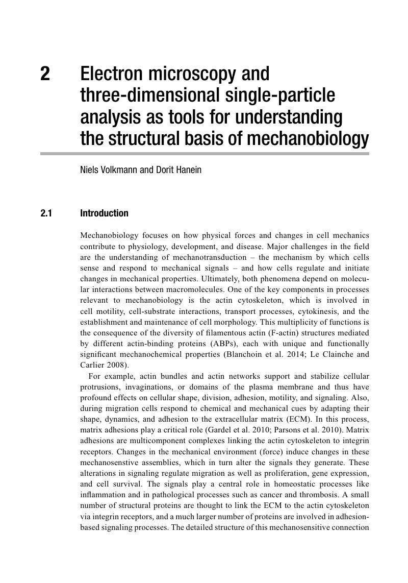
Image of the first page of this content. For PDF version, please use the ‘Save PDF’ preceeding this image.'
Type
Chapter
Information
Integrative Mechanobiology
Micro- and Nano- Techniques in Cell Mechanobiology
, pp. 15 - 31
Publisher: Cambridge University Press
Print publication year: 2015

Access options

Get access to the full version of this content by using one of the access options below. (Log in options will check for institutional or personal access. Content may require purchase if you do not have access.)

References

Anthis, N. J., Wegener, K. L., Ye, F., Kim, C., Goult, B. T., Lowe, E. D., Vakonakis, I., et al. (2009). “The structure of an integrin/talin complex reveals the basis of inside-out signal transduction.” EMBO J 28: 36233632.CrossRefGoogle ScholarPubMed
Bai, X. C., Fernandez, I. S., McMullan, G., and Scheres, S. H.. (2013). “Ribosome structures to near-atomic resolution from thirty thousand cryo-EM particles.” elife 2: e00461.CrossRefGoogle ScholarPubMed
Bakolitsa, C., Cohen, D. M., Bankston, L. A., Bobkov, A. A., Cadwell, G. W., Jennings, L., Critchley, D. R., et al. (2004). “Structural basis for vinculin activation at sites of cell adhesion.” Nature 430: 583586.CrossRefGoogle ScholarPubMed
Bakolitsa, C., de Pereda, J. M., Bagshaw, C. R., Critchley, D. R., and Liddington, R. C.. (1999). “Crystal structure of the vinculin tail suggests a pathway for activation.” Cell 99: 603613.CrossRefGoogle ScholarPubMed
Barstead, R. J., and Waterston, R. H.. (1991). “Vinculin is essential for muscle function in the nematode.” J Cell Biol 114: 715724.CrossRefGoogle ScholarPubMed
Bartesaghi, A., Lecumberry, F., Sapiro, G., and Subramaniam, S.. (2012). “Protein secondary structure determination by constrained single-particle cryo-electron tomography.” Structure 20: 20032013.CrossRefGoogle ScholarPubMed
Blanchoin, L., Boujemaa-Paterski, R., Sykes, C., and Plastino, J.. (2014). “Actin dynamics, architecture, and mechanics in cell motility.” Physiol Rev 94: 235263.CrossRefGoogle ScholarPubMed
Burridge, K., and Mangeat, P.. (1984). “An interaction between vinculin and talin.” Nature 308: 744746.CrossRefGoogle ScholarPubMed
Chafel, M. M., Shen, W., and Matsudaira, P.. (1995). “Sequential expression and differential localization of I-, L-, and T-fimbrin during differentiation of the mouse intestine and yolk sac.” Dev Dyn 203: 141151.CrossRefGoogle Scholar
Chen, H., Cohen, D. M., Choudhury, D. M., Kioka, N., and Craig, S. W.. (2005). “Spatial distribution and functional significance of activated vinculin in living cells.” J Cell Biol 169: 459470.CrossRefGoogle ScholarPubMed
Critchley, D. R. (2009). “Biochemical and structural properties of the integrin-associated cytoskeletal protein talin.” Annu Rev Biophys 38: 235254.CrossRefGoogle ScholarPubMed
Critchley, D. R., and Gingras, A. R.. (2008). “Talin at a glance.” J Cell Sci 121: 13451347.CrossRefGoogle ScholarPubMed
Debrand, E., El Jai, Y., Spence, L., Bate, N., Praekelt, U., Pritchard, C. A., Monkley, S. J., and Critchley, D. R.. (2009). “Talin 2 is a large and complex gene encoding multiple transcripts and protein isoforms.” FEBS J 276: 16101628.CrossRefGoogle ScholarPubMed
DeMali, K. A., Barlow, C. A., and Burridge, K.. (2002). “Recruitment of the Arp2/3 complex to vinculin: coupling membrane protrusion to matrix adhesion.” J Cell Biol 159: 881891.CrossRefGoogle ScholarPubMed
DeRosier, D. J., and Moore, P. B.. (1970). “Reconstruction of three-dimensional images from electron micrographs of structures with helical symmetry.” J Mol Biol 52: 355369.CrossRefGoogle ScholarPubMed
Dubochet, J., Adrian, M., Chang, J.-J., Homo, J.-C., Lepault, J., McDowall, A. W., and Schultz, P.. (1988). “Cryo-electron microscopy of vitrified specimens.” Q Rev Biophys 21: 129228.CrossRefGoogle ScholarPubMed
Egelman, E. H. (2000). “A robust algorithm for the reconstruction of helical filaments using single-particle methods.” Ultramicroscopy 85: 225–34.CrossRefGoogle ScholarPubMed
Egelman, E. H. (2007). “Single-particle reconstruction from EM images of helical filaments.” Curr Opin Struct Biol 17: 556561.CrossRefGoogle ScholarPubMed
Elliott, P. R., Goult, B. T., Kopp, P. M., Bate, N., Grossmann, J. G., Roberts, G. C., Critchley, D. R., et al. (2010). “The structure of the talin head reveals a novel extended conformation of the FERM domain.” Structure 18: 12891299.CrossRefGoogle ScholarPubMed
Frank, J. (2006a). Electron tomography: methods for three-dimensional visualization of structures in the cell. New York: Springer.CrossRefGoogle Scholar
Frank, J. (2006b). Three-dimensional electron microscopy of macromolecular assemblies: visualization of biological molecules in their native state. Oxford: Oxford University Press.CrossRefGoogle Scholar
Frank, J. (2013). “Story in a sample – the potential (and limitations) of cryo-electron microscopy applied to molecular machines.” Biopolymers 99: 832836.CrossRefGoogle Scholar
Galkin, V. E., Orlova, A., Cherepanova, O., Lebart, M. C., and Egelman, E. H.. (2008). “High-resolution cryo-EM structure of the F-actin-fimbrin/plastin ABD2 complex.” Proc Natl Acad Sci USA 105: 14941498.CrossRefGoogle ScholarPubMed
Gardel, M. L., Schneider, I. C., Aratyn-Schaus, Y., and Waterman, C. M.. (2010). “Mechanical integration of actin and adhesion dynamics in cell migration.” Annu Rev Cell Dev Biol 26: 315333.CrossRefGoogle ScholarPubMed
Giannone, G., Jiang, G., Sutton, D. H., Critchley, D. R., and Sheetz, M. P.. (2003). “Talin1 is critical for force-dependent reinforcement of initial integrin-cytoskeleton bonds but not tyrosine kinase activation.” J Cell Biol 163: 409419.CrossRefGoogle Scholar
Gingras, A. R., Bate, N., Goult, B. T., Hazelwood, L., Canestrelli, I., Grossmann, J. G., Liu, H., et al. (2007). “The structure of the C-terminal actin-binding domain of talin.” EMBO J 27: 458469.CrossRefGoogle ScholarPubMed
Gingras, A. R., Ziegler, W. H., Frank, R., Barsukov, I. L., Roberts, G. C., Critchley, D. R., and Emsley, J.. (2005). “Mapping and consensus sequence identification for multiple vinculin binding sites within the talin rod.” J Biol Chem 280: 3721737224.CrossRefGoogle ScholarPubMed
Goult, B. T., Xu, X. P., Gingras, A. R., Swift, M., Patel, B., Bate, N., Kopp, P. M., et al. (2013). “Structural studies on full-length talin1 reveal a compact auto-inhibited dimer: Implications for talin activation.” J Struc Biol 184: 2132.CrossRefGoogle ScholarPubMed
Grashoff, C., Hoffman, B. D., Brenner, M. D., Zhou, R., Parsons, M., Yang, M. T., McLean, M. A., et al. (2010). “Measuring mechanical tension across vinculin reveals regulation of focal adhesion dynamics.” Nature 466: 263266.CrossRefGoogle ScholarPubMed
Hanein, D., Matsudaira, P., and DeRosier, D. J.. (1997). “Evidence for a conformational change in actin induced by fimbrin (N375) binding.” J Cell Biol 139: 387396.CrossRefGoogle ScholarPubMed
Hanein, D., and Volkmann, N.. (2011). “Correlative light-electron microscopy.” Adv Protein Chem Struct Biol 82: 9199.CrossRefGoogle ScholarPubMed
Hanein, D., Volkmann, N., Goldsmith, S., Michon, A. M., Lehman, W., Craig, R., DeRosier, D., et al. (1998). “An atomic model of fimbrin binding to F-actin and its implications for filament crosslinking and regulation.” Nat Struct Biol 5: 787792.CrossRefGoogle ScholarPubMed
Hemmings, L., Rees, D. J., Ohanian, V., Bolton, S. J., Gilmore, A. P., Patel, B., Priddle, H., et al. (1996). “Talin contains three actin-binding sites each of which is adjacent to a vinculin-binding site.” J Cell Sci 109: 27152726.CrossRefGoogle ScholarPubMed
Higgs, H. N., and Pollard, T. D.. (1999). “Regulation of actin polymerization by Arp2/3 complex and WASp/Scar proteins.” J Biol Chem 274: 325313254.CrossRefGoogle ScholarPubMed
Holmes, K. C., Angert, I., Kull, F. J., Jahn, W., and Schroder, R. R.. (2003). “Electron cryo-microscopy shows how strong binding of myosin to actin releases nucleotide.” Nature 425: 423427.CrossRefGoogle ScholarPubMed
Janssen, M. E., Kim, E., Liu, H., Fujimoto, L. M., Bobkov, A., Volkmann, N., and Hanein, D.. (2006). “Three-dimensional structure of vinculin bound to actin filaments.” Mol Cell 21: 271281.CrossRefGoogle ScholarPubMed
Janssen, M. E., Liu, H., Volkmann, N., and Hanein, D.. (2012). “The C-terminal tail domain of metavinculin, vinculin’s splice variant, severs actin filaments.” J Cell Biol 197: 585593.CrossRefGoogle ScholarPubMed
Jiang, G., Giannone, G., Critchley, D. R., Fukumoto, E., and Sheetz, M. P.. (2003). “Two-piconewton slip bond between fibronectin and the cytoskeleton depends on talin.” Nature 424: 334337.CrossRefGoogle ScholarPubMed
Jockusch, B. M., and Isenberg, G.. (1981). “Interaction of alpha-actinin and vinculin with actin: opposite effects on filament network formation.” Proc Natl Acad Sci USA 78: 30053009.CrossRefGoogle ScholarPubMed
Jockusch, B. M., and Rudiger, M.. (1996). “Crosstalk between cell adhesion molecules: vinculin as a paradigm for regulation by conformation.” Trends Cell Biol 6: 311315.CrossRefGoogle ScholarPubMed
Johnson, R. P., and Craig, S. W.. (1995). “F-actin binding site masked by the intramolecular association of vinculin head and tail domains.” Nature 373: 261264.CrossRefGoogle ScholarPubMed
Johnson, R. P., and Craig, S. W.. (2000). “Actin activates a cryptic dimerization potential of the vinculin tail domain.” J Biol Chem 275: 95105.CrossRefGoogle ScholarPubMed
Kelleher, J. F., Atkinson, S. J., and Pollard, T. D.. (1995). “Sequences, structural models, and cellular localization of the actin– related proteins Arp2 and Arp3 from Acanthamoeba.” J Cell Biol 131: 385397.CrossRefGoogle ScholarPubMed
Klein, M. G., Shi, W., Ramagopal, U., Tseng, Y., Wirtz, D., Kovar, D. R., Staiger, C. J., et al. (2004). “Structure of the actin crosslinking core of fimbrin.” Structure 12: 9991013.CrossRefGoogle ScholarPubMed
Koster, A. J., Grimm, R., Typke, D., Hegerl, R., Stoschek, A., Walz, J., and Baumeister, W.. (1997). “Perspectives of molecular and cellular electron tomography.” J Struct Biol 120: 276308.CrossRefGoogle ScholarPubMed
Kühlbrandt, W. (2014). “Cryo-EM enters a new era.” elife 3: e03678.CrossRefGoogle ScholarPubMed
Le Clainche, C., and Carlier, M. F.. (2008). “Regulation of actin assembly associated with protrusion and adhesion in cell migration.” Physiol Rev 88: 489513.CrossRefGoogle ScholarPubMed
Lee, H.-S., Bellin, R. M., Walker, D. L., Patel, B., Powers, P., Liu, H., Garcia-Alvarez, B., et al. (2004). “Characterization of an actin-binding site within the talin FERM domain.” J Mol Biol 343: 771784.CrossRefGoogle ScholarPubMed
Luan, Q., and Nolen, B. J.. (2013). “Structural basis for regulation of Arp2/3 complex by GMF.” Nat Struct Mol Biol 20: 10621068.CrossRefGoogle ScholarPubMed
Lucic, V., Yang, T., Schweikert, G., Forster, F., and Baumeister, W.. (2005). “Morphological characterization of molecular complexes present in the synaptic cleft.” Structure 13: 423434.Google ScholarPubMed
Marchand, J. B., Kaiser, D. A., Pollard, T. D., and Higgs, H. N.. (2001). “Interaction of WASP/Scar proteins with actin and vertebrate Arp2/3 complex.” Nat Cell Biol 3: 7682.CrossRefGoogle ScholarPubMed
Martin, A. C., Xu, X. P., Rouiller, I., Kaksonen, M., Sun, Y., Belmont, L., Volkmann, N., et al. (2005). “Effects of Arp2 and Arp3 nucleotide-binding pocket mutations on Arp2/3 complex function.” J Cell Biol 168: 315328.CrossRefGoogle ScholarPubMed
Matsudaira, P. (1991). “Modular organization of actin crosslinking proteins.” Trends Biochem Sci 16: 8792.CrossRefGoogle ScholarPubMed
Nolen, B. J., and Pollard, T. D.. (2007). “Insights into the influence of nucleotides on actin family proteins from seven structures of Arp2/3 complex.” Mol Cell 26: 449457.CrossRefGoogle ScholarPubMed
Nolen, B. J., Littlefield, R. S., and Pollard, T. D.. (2004). “Crystal structures of actin-related protein 2/3 complex with bound ATP or ADP.” Proc Natl Acad Sci USA 101: 1562715632.CrossRefGoogle ScholarPubMed
Nolen, B. J., Tomasevic, N., Russell, A., Pierce, D. W., Jia, Z., McCormick, C. D., Hartman, J., et al. (2009). “Characterization of two classes of small molecule inhibitors of Arp2/3 complex.” Nature 460: 10311034.CrossRefGoogle ScholarPubMed
Parsons, J. T., Horwitz, A. R., and Schwartz, M. A.. (2010). “Cell adhesion: integrating cytoskeletal dynamics and cellular tension.” Nat Rev Mol Cell Biol 11: 633643.CrossRefGoogle ScholarPubMed
Penczek, P. A. (2010). “Image restoration in cryo-electron microscopy.” Methods Enzymol 482: 3572.CrossRefGoogle ScholarPubMed
Peng, X., Nelson, E. S., Maiers, J. L., and Demali, K. A.. (2011). “New insights into vinculin function and regulation.” Int Rev Cell Mol Biol 287: 191231.CrossRefGoogle ScholarPubMed
Pfaendtner, J., Volkmann, N., Hanein, D., Dalhaimer, P., Pollard, T. D., and Voth, G. A.. (2012). “Key structural features of the actin filament Arp2/3 complex branch junction revealed by molecular simulation.” J Mol Biol 416: 148161.CrossRefGoogle ScholarPubMed
Pollard, T. D. (2007). “Regulation of actin filament assembly by Arp2/3 complex and formins.” Annu Rev Biophys Biomol Struct 36: 451477.CrossRefGoogle ScholarPubMed
Radermacher, M. (1988). “Three-dimensional reconstruction of single particles from random and nonrandom tilt series.” J Electron Microsc Tech 9: 359–94.CrossRefGoogle ScholarPubMed
Robinson, R. C., Turbedsky, K., Kaiser, D. A., Marchand, J. B., Higgs, H. N., Choe, S., and Pollard, T. D.. (2001). “Crystal structure of Arp2/3 complex.” Science 294: 1679–184.CrossRefGoogle ScholarPubMed
Rouiller, I., Xu, X. P., Amann, K. J., Egile, C., Nickell, S., Nicastro, D., Li, R., et al. (2008). “The structural basis of actin filament branching by Arp2/3 complex.” J Cell Biol 180: 887895.CrossRefGoogle ScholarPubMed
Saunders, R. M., Holt, M. R., Jennings, L., Sutton, D. H., Barsukov, I. L., Bobkov, A., Liddington, R. C., Adamson, E. A., Dunn, G. A., and Critchley, D. R.. (2006). “Role of vinculin in regulating focal adhesion turnover.” Eur J Cell Biol 85: 487500.CrossRefGoogle ScholarPubMed
Scheres, S. H. (2012). “A Bayesian view on cryo-EM structure determination.” J Mol Biol 415: 406418.CrossRefGoogle ScholarPubMed
Schur, F. K., Hagen, W. J., de Marco, A., and Briggs, J. A.. (2013). “Determination of protein structure at 8.5 Å resolution using cryo-electron tomography and sub-tomogram averaging.” J Struc Biol 184: 394400.CrossRefGoogle ScholarPubMed
Spahn, C. M., and Penczek, P. A.. (2009). “Exploring conformational modes of macromolecular assemblies by multiparticle cryo-EM.” Curr Opin Struct Biol 19: 623631.CrossRefGoogle ScholarPubMed
Subauste, M. C., Pertz, O., Adamson, E. D., Turner, C. E., Junger, S., and Hahn, K. M.. (2004). “Vinculin modulation of paxillin-FAK interactions regulates ERK to control survival and motility.” J Cell Biol 165: 371381.CrossRefGoogle ScholarPubMed
Thievessen, I., Thompson, P. M., Berlemont, S., Plevock, K. M., Plotnikov, S. V., Zemljic-Harpf, A., et al. (2013). “Vinculin-actin interaction couples actin retrograde flow to focal adhesions, but is dispensable for focal adhesion growth.” J Cell Biol 202: 163177.CrossRefGoogle ScholarPubMed
Ti, S. C., Jurgenson, C. T., Nolen, B. J., and Pollard, T. D.. (2011). “Structural and biochemical characterization of two binding sites for nucleation-promoting factor WASp-VCA on Arp2/3 complex.” Proc Natl Acad Sci USA 108: E463E471.CrossRefGoogle ScholarPubMed
Tilney, L. G., Egelman, E. H., DeRosier, D. J., and Saunder, J. C.. (1983). “Actin filaments, stereocilia, and hair cells of the bird cochlea. II. Packing of actin filaments in the stereocilia and in the cuticular plate and what happens to the organization when the stereocilia are bent.” J Cell Biol 96: 822834.CrossRefGoogle Scholar
Tilney, L. G., Tilney, M. S., and Guild, G. M.. (1995). “F actin bundles in Drosophila bristles. I. Two filament cross-links are involved in bundling.” J Cell Biol 130: 629638.CrossRefGoogle ScholarPubMed
van Heel, M. (1987). “Angular reconstitution: a posteriori assignment of projection directions for 3D reconstruction.” Ultramicroscopy 21: 111124.CrossRefGoogle Scholar
Volkmann, N. (2009). “Confidence intervals for fitting of atomic models into low-resolution densities.” Acta Crystallogr D Biol Crystallogr 65: 679689.CrossRefGoogle ScholarPubMed
Volkmann, N. (2012). “Putting structure into context: fitting of atomic models into electron microscopic and electron tomographic reconstructions.” Curr Opin Cell Biol 24: 141147.CrossRefGoogle ScholarPubMed
Volkmann, N., and Hanein, D.. (1999). “Quantitative fitting of atomic models into observed densities derived by electron microscopy.” J Struc Biol 125: 176184.CrossRefGoogle ScholarPubMed
Volkmann, N., and Hanein, D.. (2009). “Electron microscopy in the context of systems biology.” In Structural Bioinformatics, Gu, J. and Bourne, P. E., eds. New York: Wiley-Blackwell, 143170.Google Scholar
Volkmann, N., Page, C., Li, R., and Hanein, D.. (2014). “Three-dimensional reconstructions of actin filaments capped by Arp2/3 complex.” Eur J Cell Biol 93: 179183.CrossRefGoogle ScholarPubMed
Volkmann, N., Amann, K. J., Stoilova-McPhie, S., Egile, C., Winter, D. C., Hazelwood, L., Heuser, J. E., et al. (2001). “Structure of Arp2/3 complex in its activated state and in actin filament branch junctions.” Science 293: 24562459.CrossRefGoogle ScholarPubMed
Wegener, K. L., Partridge, A. W., Han, J., Pickford, A. R., Liddington, R. C., Ginsberg, M. H., and Campbell, I.D.. (2007). “Structural basis of integrin activation by talin.” Cell 128: 171182.CrossRefGoogle ScholarPubMed
Winkler, H., and Taylor, K. A.. (1996). “Three-dimensional distortion correction applied to tomographic reconstructions of sectioned crystals.” Ultramicroscopy 63: 125132.CrossRefGoogle ScholarPubMed
Xu, W., Baribault, H., and Adamson, E. D.. (1998). “Vinculin knockout results in heart and brain defects during embryonic development.” Development 125: 327337.CrossRefGoogle ScholarPubMed
Xu, X. P., Rouiller, I., Slaughter, B. D., Egile, C., Kim, E., Unruh, J. R., Fan, X., et al. (2011). “Three-dimensional reconstructions of Arp2/3 complex with bound nucleation promoting factors.” EMBO J 31: 236247.CrossRefGoogle ScholarPubMed
Zamir, E., Katz, M., Posen, Y., Erez, N., Yamada, K. M., Katz, B. Z., Lin, S., et al. (2000). “Dynamics and segregation of cell-matrix adhesions in cultured fibroblasts.” Nat Cell Biol 2: 191196.CrossRefGoogle ScholarPubMed

Save book to Kindle

To save this book to your Kindle, first ensure coreplatform@cambridge.org is added to your Approved Personal Document E-mail List under your Personal Document Settings on the Manage Your Content and Devices page of your Amazon account. Then enter the ‘name’ part of your Kindle email address below. Find out more about saving to your Kindle.

Note you can select to save to either the @free.kindle.com or @kindle.com variations. ‘@free.kindle.com’ emails are free but can only be saved to your device when it is connected to wi-fi. ‘@kindle.com’ emails can be delivered even when you are not connected to wi-fi, but note that service fees apply.

Find out more about the Kindle Personal Document Service.

Available formats
×

Save book to Dropbox

To save content items to your account, please confirm that you agree to abide by our usage policies. If this is the first time you use this feature, you will be asked to authorise Cambridge Core to connect with your account. Find out more about saving content to Dropbox.

Available formats
×

Save book to Google Drive

To save content items to your account, please confirm that you agree to abide by our usage policies. If this is the first time you use this feature, you will be asked to authorise Cambridge Core to connect with your account. Find out more about saving content to Google Drive.

Available formats
×