Skip to main content Accessibility help
×
Hostname: page-component-848d4c4894-m9kch Total loading time: 0 Render date: 2024-05-03T18:09:12.864Z Has data issue: false hasContentIssue false

8 - The Adnexa and Other Pathology

Published online by Cambridge University Press:  20 October 2017

Emma Kirk
Affiliation:
Royal Free Hospital, London
Get access

Summary

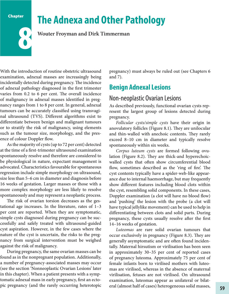
Image of the first page of this content. For PDF version, please use the ‘Save PDF’ preceeding this image.'
Type
Chapter
Information
Early Pregnancy Ultrasound
A Practical Guide
, pp. 59 - 68
Publisher: Cambridge University Press
Print publication year: 2017

Access options

Get access to the full version of this content by using one of the access options below. (Log in options will check for institutional or personal access. Content may require purchase if you do not have access.)

References

Further Reading

Hoover, K, Jenkins, TR (2011). Evaluation and management of adnexal mass in pregnancy. Am J Obstet Gynecol 205: 97102.Google Scholar
Kaijser, J, Bourne, T, Valentin, L, Sayasneh, A, Van Holsbeke, C, Vergote, I, et al. (2013). Improving strategies for diagnosing ovarian cancer: a summary of the International Ovarian Tumor Analysis (IOTA) studies. Ultrasound Obstet Gynecol 41: 920.Google Scholar
Condous, G, Khalid, A, Okaro, E, Bourne, T (2004). Should we be examining ovaries during pregnancy? Prevalence and natural history of adnexal pathology detected at first trimester sonography. Ultrasound Obstet Gynecol 24: 6266.Google Scholar
Sayasneh, A, Ekechi, C, Ferrara, L, Kaijser, J, Stalder, C, Sur, S et al. (2015). The characteristic ultrasound features of specific types of ovarian pathology (review). Int J Oncol 46(2): 445458.Google Scholar
Chiang, G, Levine, D (2004). Imaging of adnexal masses in pregnancy. J Ultrasound Med 23: 805819.CrossRefGoogle ScholarPubMed
Van Holsbeke, C, Amant, F, Veldman, J, De Boodt, A, Moerman, P, Timmerman, D (2009). Hyperreactio luteinalis in a spontaneously conceived singleton pregnancy. Ultrasound Obstet Gynecol 33: 371373.Google Scholar
Mascilini, F, Moruzzi, C, Giansiracusa, C, Guastafierro, F, Savelli, L, De Meis, L et al. (2014). Imaging of gynecological disease: clinical and ultrasound characteristics of decidualized endometriomas surgically removed during pregnancy. Ultrasound Obstet Gynecol 44: 354360.CrossRefGoogle ScholarPubMed
Paladini, D, Testa, A, Van Holsbeke, C, Mancari, R, Timmerman, D, Valentin, L (2009). Imaging in gynecological disease (5): clinical and ultrasound characteristics in fibroma and fibrothecoma of the ovary. Ultrasound Obstet Gynecol 34: 188195.CrossRefGoogle ScholarPubMed
De Haan, J, Verheecke, M, Amant, F (2015). Management of ovarian cysts and cancer in pregnancy. Facts Views Vis Obgyn 7(1): 2531.Google Scholar
Testa, A, Ferrandina, G, Timmerman, D, Savelli, L, Ludovisi, M, Van Holsbeke, C et al. (2007). Imaging in gynecological disease (1): ultrasound features of metastases in the ovaries differ depending on the origin of the primary tumor. Ultrasound Obstet Gynecol 29: 505511.Google Scholar
Abbasi, N, Patanaude, V, Abenhaim, HA (2014). Management and outcomes of acute appendicitis in pregnancy-population-based study of over 7000 cases. BJOG 121(12): 15091514.CrossRefGoogle ScholarPubMed
Haider, Z, Condous, G, Ahmed, S, Kirk, E, Bourne, T (2006). Transvaginal sonographic diagnosis of appendicitis in acute pelvic pain. J Ultrasound Med 25: 12431244.Google Scholar
Singer, A, Simmons, MZ, Maldjian, PD (2008). Spectrum of congenital renal anomalies presenting in adulthood. Clin Imaging 32(3): 183191.Google Scholar
Nowitzki, KM, Hoimes, ML, Chen, B, Zheng, LZ, Kim, YH (2015). Ultrasonography of intrauterine devices. Ultrasonography 34(3): 183194.CrossRefGoogle ScholarPubMed
Moschos, E, Twickler, DM (2011). Intrauterine devices in early pregnancy: findings on ultrasound and clinical outcomes. Am J Obstet Gynecol 204: 427.e16.Google Scholar
Brahmi, D, Steenland, MW, Renner, R-M, Gaffield, ME, Curtis, KM (2012). Pregnancy outcomes with an IUD in situ: a systematic review. Contraception 85: 131139.Google Scholar
Legendre, G, Levaillant, JM, Faivre, E, Deffieux, X, Gervaise, A, Fernandez, H (2011). 3D ultrasound to assess the position of tubal sterilization microinserts. Hum Reprod 26: 26832689.Google Scholar

Save book to Kindle

To save this book to your Kindle, first ensure coreplatform@cambridge.org is added to your Approved Personal Document E-mail List under your Personal Document Settings on the Manage Your Content and Devices page of your Amazon account. Then enter the ‘name’ part of your Kindle email address below. Find out more about saving to your Kindle.

Note you can select to save to either the @free.kindle.com or @kindle.com variations. ‘@free.kindle.com’ emails are free but can only be saved to your device when it is connected to wi-fi. ‘@kindle.com’ emails can be delivered even when you are not connected to wi-fi, but note that service fees apply.

Find out more about the Kindle Personal Document Service.

Available formats
×

Save book to Dropbox

To save content items to your account, please confirm that you agree to abide by our usage policies. If this is the first time you use this feature, you will be asked to authorise Cambridge Core to connect with your account. Find out more about saving content to Dropbox.

Available formats
×

Save book to Google Drive

To save content items to your account, please confirm that you agree to abide by our usage policies. If this is the first time you use this feature, you will be asked to authorise Cambridge Core to connect with your account. Find out more about saving content to Google Drive.

Available formats
×