Skip to main content Accessibility help
×
Hostname: page-component-848d4c4894-4rdrl Total loading time: 0 Render date: 2024-06-27T22:30:39.706Z Has data issue: false hasContentIssue false

Chapter 9 - Placental Histopathology Findings in Fetal Growth Restriction

from Section 2 - Maternal Cardiovascular Characteristics and the Placenta

Published online by Cambridge University Press:  23 July 2018

Christoph Lees
Affiliation:
Imperial College London
Gerard H. A. Visser
Affiliation:
Universiteit Utrecht, The Netherlands
Kurt Hecher
Affiliation:
University Medical Centre, Hamburg
Get access

Summary

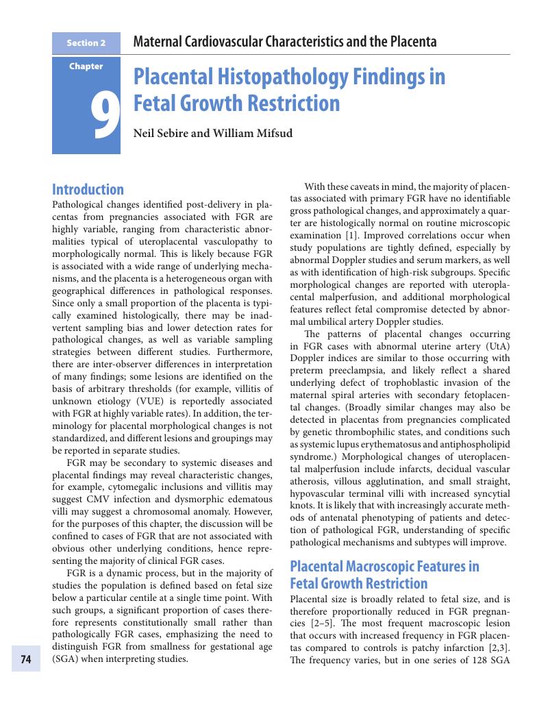
Image of the first page of this content. For PDF version, please use the ‘Save PDF’ preceeding this image.'
Type
Chapter
Information
Publisher: Cambridge University Press
Print publication year: 2018

Access options

Get access to the full version of this content by using one of the access options below. (Log in options will check for institutional or personal access. Content may require purchase if you do not have access.)

References

Fox, H, Sebire, NJ. The placenta in abnormalities and disorders of the fetus. In Fox, H, Sebire, NJ, editors. Pathology of the Placenta (Major Problems in Pathology 7). Philadelphia, PA: Saunders Elsevier.Google Scholar
Bjoro, K. Gross pathology of the placenta in intrauterine growth restriction. Ann Chir Gynaecol 1981;70(6):316–22.Google Scholar
Salafia, CM, Vintzileos, AM, Silberman, L, Bantham, KF, Vogel, CA. Placental pathology of idiopathic intrauterine growth retardation at term. Am J Perinatol. Department of Laboratory Medicine, Danbury Hospital, Connecticut; 1992;9(3):179–84.Google Scholar
Biswas, S, Ghosh, SK. Gross morphological changes of placentas associated with intrauterine growth restriction of fetuses: A case control study. Early Hum Dev 2008;84(6):357–62.Google Scholar
Tomas, SZ, Roje, D, Prusac, IK, Tadin, I, Capkun, V. Morphological characteristics of placentas associated with idiopathic intrauterine growth retardation: A clinicopathologic study. Eur J Obstet Gynecol Reprod Biol. School of Medicine University Split, Soltanska 2, 21000 Split, Croatia. sandrazt@online.hr; 2010;152(1):3943.Google Scholar
Harris, RD, Simpson, WA, Pet, LR, Marin-Padilla, M, Crow, HC. Placental hypoechoic-anechoic areas and infarction: Sonographic-pathologic correlation. Radiology 1990;176(1):7580.CrossRefGoogle ScholarPubMed
Sebire, NJ. Pathophysiological significance of abnormal umbilical cord coiling index. Ultrasound Obstet Gynecol 2007;30(6):804–6.Google Scholar
Gerretsen, GG, Huisjes, HJ, Elema, JD. Morphological changes of the spiral arteries in the placental bed in relation to pre-eclampsia and fetal growth retardation. BJOG 1981;88(9):876–81.Google Scholar
Olofsson, P, Laurini, RN, Marsál, K. A high uterine artery pulsatility index reflects a defective development of placental bed spiral arteries in pregnancies complicated by hypertension and fetal growth retardation. Eur J Obstet Gynecol Reprod Biol. Department of Obstetrics and Gynecology, University of Lund, Malmö General Hospital, Sweden; 1993;49(3):161–8.Google Scholar
Lin, S, Shimizu, I, Suehara, N, Nakayama, M, Aono, T. Uterine artery Doppler velocimetry in relation to trophoblast migration into the myometrium of the placental bed. Obstet Gynecol. Department of Obstetrics and Pathology, Osaka Medical Center, Japan; 1995;85(5 Pt 1):760–5.Google Scholar
Khong, TY, Chambers, HM. Alternative method of sampling placentas for the assessment of uteroplacental vasculature. J Clin Pathol. Department of Pathology, Adelaide Medical Centre for Women and Children, Queen Victoria Hospital, Australia: BMJ Group; 1992;45(10):925–7.Google Scholar
Wigglesworth, JS. The gross and microscopic pathology of the prematurely delivered placenta. J Obstet Gynaecol Br Emp 1962;69:934–43.Google Scholar
Wigglesworth, JS. Morphological variations in the insufficient placenta. J Obstet Gynaecol Br Commonw 1964;71:871–84.Google Scholar
Altshuler, G, Russell, P, Ermocilla, R. The placental pathology of small-for-gestational age infants. Am J Obstet Gynecol. Elsevier Inc; 1975;121(3):351–9.Google Scholar
Garcia, AG. Placental morphology of low-birth-weight infants born at term. Contrib Gynecol Obstet 1982;9:100–12.Google Scholar
Salafia, CM, Minior, VK, Pezzullo, JC, Popek, EJ, Rosenkrantz, TS, Vintzileos, AM. Intrauterine growth restriction in infants of less than thirty-two weeks’ gestation: Associated placental pathologic features. Am J Obstet Gynecol. Division of Anatomic Pathology, University of Connecticut Health Center, Farmington, USA: Elsevier Inc; 1995;173(4):1049–57.Google Scholar
Oliveira, LH, Xavier, CC, Lana, AMA. [Changes in placental morphology of small for gestational age newborns]. J Pediatr (Rio J). Universidade Federal de Juiz de Fora, MG. lucio@icb.ufjf.br; 2002;78(5):397402.Google Scholar
Labarrere, CC, Althabe, OO, Telenta, MM. Chronic villitis of unknown aetiology in placentae of idiopathic small for gestational age infants. Placenta. Elsevier Ltd.; 1982;3(3):309–17.Google Scholar
Van der Veen, F, Fox, H. The human placenta in idiopathic intrauterine growth retardation: A light and electron microscopic study. Placenta. Elsevier Ltd.; 1983;4(1):6577.Google Scholar
Boyd, PA, Scott, A. Quantitative structural studies on human placentas associated with pre-eclampsia, essential hypertension and intrauterine growth retardation. BJOG 1985;92(7):714–21.Google Scholar
Macara, L, Kingdom, JC, Kaufmann, P, Kohnen, G, Hair, J, More, IA, et al. Structural analysis of placental terminal villi from growth-restricted pregnancies with abnormal umbilical artery Doppler waveforms. Placenta. Department of Obstetrics and Gynaecology, University of Glasgow, Scotland, UK: Elsevier Ltd.; 1996;17(1):3748.Google Scholar
Krebs, C, Macara, LM, Leiser, R, Bowman, AW, Greer, IA, Kingdom, JC. Intrauterine growth restriction with absent end-diastolic flow velocity in the umbilical artery is associated with maldevelopment of the placental terminal villous tree. Am J Obstet Gynecol. Department of Veterinary Anatomy, Justus Liebig University, Giessen, Germany: Elsevier Inc; 1996;175(6):1534–42.Google Scholar
Todros, T, Sciarrone, A, Piccoli, E, Guiot, C, Kaufmann, P, Kingdom, J. Umbilical Doppler waveforms and placental villous angiogenesis in pregnancies complicated by fetal growth restriction. Obstet Gynecol. Department of Obstetrics and Gynaecology, University of Torino, Italy. todros@iol.it; 1999;93(4):499503.Google Scholar
Mayhew, TM, Ohadike, C, Baker, PN, Crocker, IP, Mitchell, C, Ong, SS. Stereological investigation of placental morphology in pregnancies complicated by pre-eclampsia with and without intrauterine growth restriction. Placenta. Elsevier Ltd.; 2003;24(2–3):219–26.Google Scholar
Sheppard, BL, Bonnar, JJ. An ultrastructural study of utero-placental spiral arteries in hypertensive and normotensive pregnancy and fetal growth retardation. BJOG 1981;88(7):695705.Google Scholar
Brosens, IA, Robertson, WB, Dixon, HG. The role of the spiral arteries in the pathogenesis of preeclampsia. Obstet Gynecol Annu 1972;1:177–91.Google Scholar
De Wolf, F, Brosens, I, Renaer, M. Fetal growth retardation and the maternal arterial supply of the human placenta in the absence of sustained hypertension. BJOG 1980;87(8):678–85.Google Scholar
Khong, TY, De Wolf, F, Robertson, WB, Brosens, I. Inadequate maternal vascular response to placentation in pregnancies complicated by pre-eclampsia and by small-for-gestational age infants. BJOG 1986;93(10):1049–59.Google Scholar
Voigt, HJ, Becker, V. Doppler flow measurements and histomorphology of the placental bed in uteroplacental insufficiency. J Perinat Med. Department of Obstetrics and Gynecology, University of Erlangen-Nuernberg, Fed. Rep. of Germany; 1992;20(2):139–47.Google Scholar
Roberts, JM, Redman, CW. Pre-eclampsia: More than pregnancy-induced hypertension. Lancet. Department of Obstetrics, Gynecology, and Reproductive Sciences, Magee Women’s Hospital, University of Pittsburgh, Pennsylvania 15213; 1993;341(8858):1447–51.Google Scholar
Salafia, CM, Pezzullo, JC, López-Zeno, JA, Simmens, S, Minior, VK, Vintzileos, AM. Placental pathologic features of preterm preeclampsia. Am J Obstet Gynecol. Division of Anatomic Pathology, University of Connecticut Health Center, Farmington, USA: Elsevier Inc.; 1995;173(4):1097–105.CrossRefGoogle ScholarPubMed
Mifsud, W, Sebire, NJ. Placental pathology in early-onset and late-onset fetal growth restriction. Fetal Diagn Ther 2014.Google Scholar
Sağol, S, Sağol, O, Ozdemir, N. Stereological quantification of placental villus vascularization and its relation to umbilical artery Doppler flow in intrauterine growth restriction. Prenat Diagn. Department of Obstetrics and Gynaecology, Medical Faculty, Ege University, Turkey. sagol@med.ege.edu.tr; 2002;22(5):398403.Google Scholar
Aardema, MW, Oosterhof, H, Timmer, A, Van Rooy, I, Aarnoudse, JG. Uterine artery Doppler flow and uteroplacental vascular pathology in normal pregnancies and pregnancies complicated by pre-eclampsia and small for gestational age fetuses. Placenta 2001;22(5):405–11.CrossRefGoogle ScholarPubMed
Madazli, R, Somunkiran, A, Calay, Z, Ilvan, S, Aksu, MF. Histomorphology of the placenta and the placental bed of growth restricted foetuses and correlation with the Doppler velocimetries of the urine and umbilical arteries. Placenta 2003;24(5):510–16.Google Scholar
McFadyen, IR, Price, a B, Geirsson, RT. The relation of birthweight to histological appearances in vessels of the placental bed. BJOG 1986;93(5):476–81.Google Scholar
Frusca, T, Morassi, L, Pecorelli, S, Grigolato, P, Gastaldi, A. Histological features of uteroplacental vessels in normal and hypertensive patients in relation to birthweight. BJOG 1989;96(7):835–9.Google Scholar
Kupferminc, MJ, Fait, G, Many, A, Gordon, D, Eldor, A, Lessing, JB. Severe preeclampsia and high frequency of genetic thrombophilic mutations. Obstet Gynecol 2000;96(1):45–9.Google Scholar
Driul, L, Damante, G, D’Elia, A, Ianni, A, Springolo, F, Marchesoni, D. Genetic thrombophilias and uterine artery Doppler velocimetry and preeclampsia. Int J Gynecol Obstet 2005;88(3):265–70.Google Scholar
Mousa, HA, Alfirevic1, Z. Do placental lesions reflect thrombophilia state in women with adverse pregnancy outcome? Hum Reprod 2000;15(8):1830–3.Google Scholar
Many, A, Schreiber, L, Rosner, S, Lessing, JB, Eldor, A, Kupferminc, MJ. Pathologic features of the placenta in women with severe pregnancy complications and thrombophilia. Obstet Gynecol 2001;98(6):1041–4.Google Scholar
Sikkema, JM, Franx, A, Bruinse, HW, Van Der Wijk, NG, De Valk, HW, Nikkels, PGJ. Placental pathology in early onset pre-eclampsia and intra-uterine growth restriction in women with and without thrombophilia. Placenta. Department of Obstetrics, University Medical Center, Utrecht, The Netherlands. J.M.Sikkema@azu.nl: Elsevier Ltd.; 2002;23(4):337–42.Google Scholar
Lockwood, CJ, Romero, R, Feinberg, RF, Clyne, LP, Coster, B, Hobbins, JC. The prevalence and biologic significance of lupus anticoagulant and anticardiolipin antibodies in a general obstetric population. Am J Obstet Gynecol 1989;161(2):369–73.Google Scholar
Branch, DW, Andres, R, Digre, KB, Rote, NS, Scott, JR. The association of antiphospholipid antibodies with severe preeclampsia. Obstet Gynecol 1989;541–5.Google Scholar
Hayslett, JP. The effect of systemic lupus erythematosus on pregnancy and pregnancy outcome. Am J Reprod Immunol 28(3–4):199204.Google Scholar
Venkat-Raman, N, Backos, M, Teoh, TG, Lo, WT, Regan, L. Uterine artery Doppler in predicting pregnancy outcome in women with antiphospholipid syndrome. Obstet Gynecol 2001;98(2):235–42.Google Scholar
Abramowsky, CR, Vegas, ME, Swinehart, G, Gyves, MT. Decidual vasculopathy of the placenta in lupus erythematosus. N Engl J Med 1980;303(12):668–72.CrossRefGoogle ScholarPubMed
Sebire, NJ, Fox, H, Backos, M, Rai, R, Paterson, C, Regan, L. Defective endovascular trophoblast invasion in primary antiphospholipid antibody syndrome-associated early pregnancy failure. Hum Reprod 2002;17(4):1067–71.Google Scholar
Stone, S, Pijnenborg, R, Vercruysse, L, Poston, R, Khamashta, MA, Hunt, BJ, et al. The placental bed in pregnancies complicated by primary antiphospholipid syndrome. Placenta 2006;27(4–5):457–67.Google Scholar
Mulla, MJ, Myrtolli, K, Brosens, JJ, Chamley, LW, Kwak-Kim, JY, Paidas, MJ, et al. Antiphospholipid antibodies limit trophoblast migration by reducing IL-6 production and STAT3 activity. Am J Reprod Immunol. Department of Obstetrics, Gynecology & Reproductive Sciences, Yale University School of Medicine, New Haven, CT 06510, USA; 2010;63(5):339–48.Google Scholar
Viall, CA, Chamley, LW. Histopathology in the placentae of women with antiphospholipid antibodies: A systematic review of the literature. Autoimmun Rev. Elsevier B.V.; 2015;14(5):120.Google Scholar
Nicolaides, KH, Bilardo, CM, Soothill, PW, Campbell, S. Absence of end diastolic frequencies in umbilical artery: A sign of fetal hypoxia and acidosis. BMJ. Harris Birthright Research Centre for Fetal Medicine, King’s College School of Medicine and Dentistry, London; 1988;297(6655):1026–7.Google Scholar
Sebire, NJ. Umbilical artery Doppler revisited: Pathophysiology of changes in intrauterine growth restriction revealed. Ultrasound Obstet Gynecol 2003;21(5):419–22.Google Scholar
Fok, RY, Pavlova, Z, Benirschke, K, Paul, RH, Platt, LD. The correlation of arterial lesions with umbilical artery Doppler velocimetry in the placentas of small-for-dates pregnancies. Obstet Gynecol. Department of Obstetrics-Gynecology, Los Angeles County/University of Southern California Medical Center; 1990;75(4):578–83.Google Scholar
Redline, RW, Boyd, T, Campbell, V, Hyde, S, Kaplan, C, Khong, TY, et al. Maternal vascular underperfusion: Nosology and reproducibility of placental reaction patterns. Pediatr Dev Pathol. Department of Pathology, University Hospitals of Cleveland and Case Western Reserve University, 1100 Euclid Avenue, 44106, USA. raymond.redline@uhhs.com; 2004;7(3):237–49.Google Scholar
Fitzgerald, B, Kingdom, J, Keating, S. Distal villous hypoplasia. Diagnostic Histopathol. Elsevier Ltd.; 2012;18(5):195200.Google Scholar
Lang, U, Baker, RS, Khoury, J, Clark, KE. Fetal umbilical vascular response to chronic reductions in uteroplacental blood flow in late-term sheep. Am J Obstet Gynecol. Department of Obstetrics and Gynecology, Justus-Liebig University Giessen, Germany: Elsevier Inc.; 2002;187(1):178–86.Google Scholar
Howard, RB, Hosokawa, T, Maguire, MH. Hypoxia-induced fetoplacental vasoconstriction in perfused human placental cotyledons. Am J Obstet Gynecol. Department of Pharmacology, Toxicology, and Therapeutics, University of Kansas Medical Center, Kansas City 66103: Elsevier Inc; 1987;157(5):1261–6.Google Scholar
Hampl, V, Bíbová, J, Stranák, Z, Wu, X, Michelakis, ED, Hashimoto, K, et al. Hypoxic fetoplacental vasoconstriction in humans is mediated by potassium channel inhibition. Am J Physiol Heart Circ Physiol. Department of Physiology, Charles University Second Medical School, 15000 Prague 5, Czech Republic. vaclav.hampl@lfmotol.cuni.cz; 2002;283(6):H2440–9.CrossRefGoogle ScholarPubMed
Jakoubek, V, Bíbová, J, Hampl, V. Voltage-gated calcium channels mediate hypoxic vasoconstriction in the human placenta. Placenta. Department of Physiology, Charles University Second Medical School, Prague, Czech Republic: Elsevier Ltd.; 2006;27(910):1030–3.Google Scholar
Wareing, M, Greenwood, SL, Baker, PN. Reactivity of human placental chorionic plate vessels is modified by level of oxygenation: Differences between arteries and veins. Placenta. Maternal and Fetal Health Research Centre, University of Manchester, St. Mary’s Hospital, Hathersage Road, Manchester M13 0JH, UK. mark.wareing@man.ac.uk: Elsevier Ltd.; 2006;27(1):42–8.Google Scholar
Sebire, NJ, Talbert, D. The role of intraplacental vascular smooth muscle in the dynamic placenta: A conceptual framework for understanding uteroplacental disease. Med Hypotheses. Department of Histopathology, Great Ormond Street Hospital, London, UK. 2002;58(4):347–51.Google Scholar
Talbert, D, Sebire, NJ. The dynamic placenta: I. Hypothetical model of a placental mechanism matching local fetal blood flow to local intervillus oxygen delivery. Med Hypotheses. Institute of Reproduction and Developmental Biology, ICSM, Hammersmith Hospital, Du Cane Road, London, UK; 2004;62(4):511–19.CrossRefGoogle ScholarPubMed
Sebire, NJ, Talbert, D. The dynamic placenta: II. Hypothetical model of a fetus driven transplacental water balance mechanism producing low apparent permeability in a highly permeable placenta. Med Hypotheses. Department of Histopathology, Camelia Botnar Laboratories, Great Ormond Street Hospital, Great Ormond Street, London WC1N 3JH, UK. SebirN@gosh.nhs.uk; 2004;62(4):520–8.Google Scholar
Cindrova-Davies, T, Herrera, EA, Niu, Y, Kingdom, J, Giussani, DA, Burton, GJ. Reduced cystathionine γ-lyase and increased miR-21 expression are associated with increased vascular resistance in growth-restricted pregnancies. Am J Pathol. American Society for Investigative Pathology; 2013;182(4):1448–58.Google Scholar
Redline, RW. Distal villous immaturity. Diagnostic Histopathol. Elsevier Ltd.; 2012;18(5):189–94.Google Scholar
Khong, TY, Staples, A, Moore, L, Byard, RW. Observer reliability in assessing villitis of unknown aetiology. J Clin Pathol. Department of Pathology, Queen Victoria Hospital, Australia: BMJ Group; 1993;46(3):208–10.Google Scholar
Altemani, AM, Gonzatti, AR. [Villitis of unknown etiology in placentas of pregnancies with hypertensive disorders and of small-for-gestational-age infants]. Rev Assoc Med Bras. Departamento de Anatomia Patológica, Faculdade de Ciências Mêdicas, UNICAMP, Campinas, SP, Brasil; 2003;49(1):6771.Google Scholar
Styer, AK, Parker, HJ, Roberts, DJ, Palmer-Toy, D, Toth, TL, Ecker, JL. Placental villitis of unclear etiology during ovum donor in vitro fertilization pregnancy. Am J Obstet Gynecol. Department of Obstetrics and Gynecology, Vincent Center for Reproductive Biology, Boston, MA, USA: Elsevier Inc.; 2003;189(4):1184–6.Google Scholar
Redline, RW, Abramowsky, CR. Clinical and pathologic aspects of recurrent placental villitis. Hum Pathol. 1985;16(7):727–31.Google Scholar
Becroft, DM, Thompson, JM, Mitchell, EA. Placental villitis of unknown origin: Epidemiologic associations. Am J Obstet Gynecol. Department of Obstetrics and Gynaecology, University of Auckland, New Zealand: Elsevier Inc.; 2005;192(1):264–71.Google Scholar
Jacques, SM, Qureshi, F. Chronic villitis of unknown etiology in twin gestations. Pediatr Pathol. Department of Pathology, Hutzel Hospital, Detroit, Michigan 48201; 1994;14(4):575–84.Google Scholar
Pathak, S, Lees, CC, Hackett, G, Jessop, F, Sebire, NJ. Frequency and clinical significance of placental histological lesions in an unselected population at or near term. Virchows Arch 2011;459(6):565–72.Google Scholar
Boyd, TK, Redline, RW. Chronic histiocytic intervillositis: A placental lesion associated with recurrent reproductive loss. Hum Pathol 2000;31(11):1389–96.Google Scholar
Gruenwald, P. Abnormalities of placental vascularity in relation to intrauterine deprivation and retardation of fetal growth. Significance of avascular chorionic villi. N Y State J Med 1961;61:1508–13.Google Scholar

Save book to Kindle

To save this book to your Kindle, first ensure coreplatform@cambridge.org is added to your Approved Personal Document E-mail List under your Personal Document Settings on the Manage Your Content and Devices page of your Amazon account. Then enter the ‘name’ part of your Kindle email address below. Find out more about saving to your Kindle.

Note you can select to save to either the @free.kindle.com or @kindle.com variations. ‘@free.kindle.com’ emails are free but can only be saved to your device when it is connected to wi-fi. ‘@kindle.com’ emails can be delivered even when you are not connected to wi-fi, but note that service fees apply.

Find out more about the Kindle Personal Document Service.

Available formats
×

Save book to Dropbox

To save content items to your account, please confirm that you agree to abide by our usage policies. If this is the first time you use this feature, you will be asked to authorise Cambridge Core to connect with your account. Find out more about saving content to Dropbox.

Available formats
×

Save book to Google Drive

To save content items to your account, please confirm that you agree to abide by our usage policies. If this is the first time you use this feature, you will be asked to authorise Cambridge Core to connect with your account. Find out more about saving content to Google Drive.

Available formats
×