Skip to main content Accessibility help
×
Hostname: page-component-848d4c4894-v5vhk Total loading time: 0 Render date: 2024-07-07T22:23:51.916Z Has data issue: false hasContentIssue false

16 - Micropost arrays as a means to assess cardiac muscle cells

from Part II - Recent progress in cell mechanobiology

Published online by Cambridge University Press:  05 November 2015

Yu Sun
Affiliation:
University of Toronto
Deok-Ho Kim
Affiliation:
University of Washington
Craig A. Simmons
Affiliation:
University of Toronto
Get access

Summary

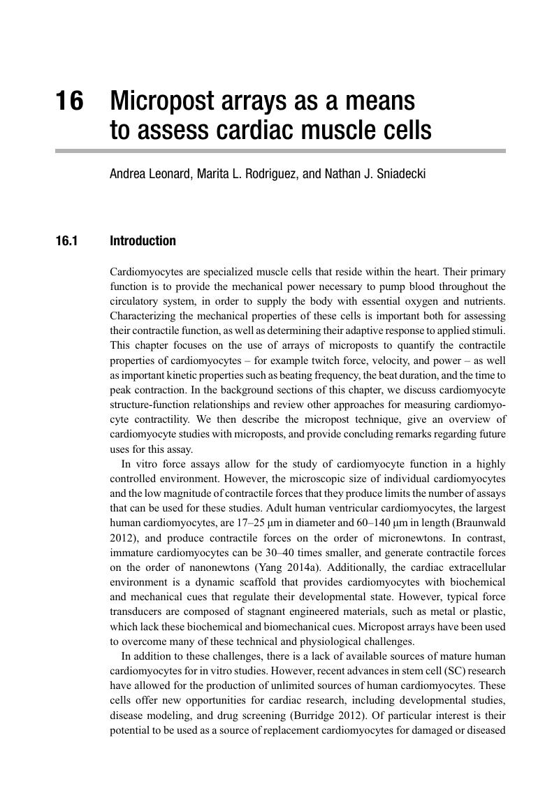
Image of the first page of this content. For PDF version, please use the ‘Save PDF’ preceeding this image.'
Type
Chapter
Information
Integrative Mechanobiology
Micro- and Nano- Techniques in Cell Mechanobiology
, pp. 295 - 315
Publisher: Cambridge University Press
Print publication year: 2015

Access options

Get access to the full version of this content by using one of the access options below. (Log in options will check for institutional or personal access. Content may require purchase if you do not have access.)

References

Araujo, A., and Walker, J. W. (1994). Kinetics of tension development in skinned cardiac myocytes measured by photorelease of Ca2+. Am J Physiol 267: H1643H1653.Google ScholarPubMed
Azeloglu, E. U., and Costa, K. D. (2010). Cross-bridge cycling gives rise to spatiotemporal heterogeneity of dynamic subcellular mechanics in cardiac myocytes probed with atomic force microscopy. Am J Physiol Heart Circ Physiol 298: H853H860.CrossRefGoogle ScholarPubMed
Balaban, N. Q., Schwarz, U. S., Riveline, D., et al. (2001). Force and focal adhesion assembly: a close relationship studied using elastic micropatterned substrates. Nature Cell Biology 3: 466472.CrossRefGoogle ScholarPubMed
Bluhm, W. F., Mcculloch, A. D., and Lew, W. Y. (1995). Active force in rabbit ventricular myocytes. J Biomech 28: 11191122.CrossRefGoogle ScholarPubMed
Borg, T. K., Rubin, K., Lundgren, E., Borg, K., and Obrink, B. (1984). Recognition of extracellular matrix components by neonatal and adult cardiac myocytes. Dev Biol 104: 8696.CrossRefGoogle ScholarPubMed
Boudou, T., Legant, W. R., Mu, A. B., et al. (2012). A microfabricated platform to measure and manipulate the mechanics of engineered cardiac microtissues. Tissue Engineering Part A 18: 910919.CrossRefGoogle ScholarPubMed
Brady, A. J. (1991). Mechanical properties of isolated cardiac myocytes. Physiol Rev 71: 413428.CrossRefGoogle ScholarPubMed
Brady, A. J., Tan, S. T., and Ricchiuti, N. V. (1979). Contractile force measured in unskinned isolated adult rat heart fibres. Nature 282: 728729.CrossRefGoogle ScholarPubMed
Braunwald, E., and Bonow, R. O. (2012). Braunwald’s heart disease: a textbook of cardiovascular medicine. Philadelphia: Saunders.Google Scholar
Bray, M. A., Sheehy, S. P., and Parker, K. K. (2008). Sarcomere alignment is regulated by myocyte shape. Cell Motility and the Cytoskeleton 65: 641651.CrossRefGoogle ScholarPubMed
Burridge, P. W., Keller, G., Gold, J. D., and Wu, J. C. (2012). Production of de novo cardiomyocytes: human pluripotent stem cell differentiation and direct reprogramming. Cell Stem Cell 10: 1628.CrossRefGoogle ScholarPubMed
Chang, W. T., Yu, D., Lai, Y. C., Lin, K. Y., and Liau, I. (2013). Characterization of the mechanodynamic response of cardiomyocytes with atomic force microscopy. Anal Chem 85: 13951400.CrossRefGoogle ScholarPubMed
Chattergoon, N. N., Giraud, G. D., Louey, S., et al. (2012). Thyroid hormone drives fetal cardiomyocyte maturation. FASEB J 26: 397408.CrossRefGoogle ScholarPubMed
Cheng, Q., Sun, Z., Meininger, G. A., and Almasri, M. (2010). Note: Mechanical study of micromachined polydimethylsiloxane elastic microposts. Rev Sci Instrum 81: 106104.CrossRefGoogle ScholarPubMed
Copelas, L., Briggs, M., Grossman, W., and Morgan, J. P. (1987). A method for recording isometric tension development by isolated cardiac myocytes: transducer attachment with fibrin glue. Pflugers Arch 408: 315317.CrossRefGoogle ScholarPubMed
Danowski, B. A., Imanaka-Yoshida, K., Sanger, J. M., and Sanger, J. W. (1992). Costameres are sites of force transmission to the substratum in adult rat cardiomyocytes. J Cell Biol 118: 14111420.CrossRefGoogle Scholar
Dembo, M., Oliver, T., Ishihara, A., and Jacobson, K. (1996). Imaging the traction stresses exerted by locomoting cells with the elastic substratum method. Biophys J 70: 20082022.CrossRefGoogle ScholarPubMed
Dillmann, W. H. (2002). Cellular action of thyroid hormone on the heart. Thyroid 12: 447452.CrossRefGoogle ScholarPubMed
Domke, J., Parak, W. J., George, M., Gaub, H. E., and Radmacher, M. (1999). Mapping the mechanical pulse of single cardiomyocytes with the atomic force microscope. Eur Biophys J 28: 179186.CrossRefGoogle ScholarPubMed
Engler, A. J., Carag-Krieger, C., Johnson, C. P., et al. (2008). Embryonic cardiomyocytes beat best on a matrix with heart-like elasticity: scar-like rigidity inhibits beating. J Cell Sci 121: 37943802.CrossRefGoogle Scholar
Eschenhagen, T., Fink, C., Remmers, U., et al. (1997). Three-dimensional reconstitution of embryonic cardiomyocytes in a collagen matrix: a new heart muscle model system. FASEB J 11: 683694.CrossRefGoogle Scholar
Fabiato, A., and Fabiato, F. (1975). Contractions induced by a calcium-triggered release of calcium from the sarcoplasmic reticulum of single skinned cardiac cells. J Physiol 249: 469495.CrossRefGoogle ScholarPubMed
Forte, G., Pagliari, S., Ebara, M., et al. (2012). Substrate stiffness modulates gene expression and phenotype in neonatal cardiomyocytes in vitro. Tissue Eng Part A 18: 18371848.CrossRefGoogle ScholarPubMed
Garcia-Webb, M. G., Taberner, A. J., Hogan, N. C., and Hunter, I. W. (2007). A modular instrument for exploring the mechanics of cardiac myocytes. Am J Physiol Heart Circ Physiol 293: H866H874.CrossRefGoogle ScholarPubMed
Harris, A. K., Wild, P., and Stopak, D. (1980). Silicone rubber substrata: a new wrinkle in the study of cell locomotion. Science 208: 177179.CrossRefGoogle Scholar
Hasenfuss, G., Mulieri, L. A., Blanchard, E. M., et al. (1991). Energetics of isometric force development in control and volume-overload human myocardium. Comparison with animal species. Circulation Research 68: 836846.CrossRefGoogle ScholarPubMed
Hazeltine, L. B., Simmons, C. S., Salick, M. R., et al. (2012). Effects of substrate mechanics on contractility of cardiomyocytes generated from human pluripotent stem cells. Int J Cell Biol 2012: 508294.CrossRefGoogle ScholarPubMed
Hersch, N., Wolters, B., Dreissen, G., et al. (2013). The constant beat: cardiomyocytes adapt their forces by equal contraction upon environmental stiffening. Biol Open 2: 351361.CrossRefGoogle ScholarPubMed
Iribe, G., Helmes, M., and Kohl, P. (2007). Force-length relations in isolated intact cardiomyocytes subjected to dynamic changes in mechanical load. Am J Physiol Heart Circ Physiol 292: H1487H1497.CrossRefGoogle ScholarPubMed
Ivashchenko, C. Y., Pipes, G. C., Lozinskaya, I. M., et al. (2013). Human-induced pluripotent stem cell-derived cardiomyocytes exhibit temporal changes in phenotype. Am J Physiol Heart Circ Physiol 305: H913H922.CrossRefGoogle ScholarPubMed
Jacot, J. G., Martin, J. C., and Hunt, D. L. (2010). Mechanobiology of cardiomyocyte development. J Biomech 43: 9398.CrossRefGoogle ScholarPubMed
Jacot, J. G., Mcculloch, A. D., and Omens, J. H. (2008). Substrate stiffness affects the functional maturation of neonatal rat ventricular myocytes. Biophys J 95: 34793487.CrossRefGoogle ScholarPubMed
Kajzar, A., Cesa, C. M., Kirchgessner, N., Hoffmann, B., and Merkel, R. (2008). Toward physiological conditions for cell analyses: forces of heart muscle cells suspended between elastic micropillars. Biophys J 94: 18541866.CrossRefGoogle ScholarPubMed
Kim, J., Park, J., Na, K., et al. (2008). Quantitative evaluation of cardiomyocyte contractility in a 3D microenvironment. J Biomech 41: 23962401.CrossRefGoogle Scholar
Kim, J., Park, J., Ryu, S. K., et al. (2006). Realistic computational modeling for hybrid biopolymer microcantilevers. Conf Proc IEEE Eng Med Biol Soc 1: 21022105.CrossRefGoogle Scholar
Kim, K., Taylor, R., Sim, J. Y., et al. (2011). Calibrated micropost arrays for biomechanical characterisation of cardiomyocytes. Micro and Nano Letters 6: 317.CrossRefGoogle Scholar
Klein, I., Daood, M., and Whiteside, T. (1985). Development of heart cells in culture: studies using an affinity purified antibody to a myosin light chain. J Cell Physiol 124: 4953.CrossRefGoogle ScholarPubMed
Klein, I., and Ojamaa, K. (2001). Thyroid hormone and the cardiovascular system. N Engl J Med 344: 501509.CrossRefGoogle ScholarPubMed
Korte, F. S., Dai, J., Buckley, K., et al. (2011). Upregulation of cardiomyocyte ribonucleotide reductase increases intracellular 2 deoxy-ATP, contractility, and relaxation. J Mol Cell Cardiol 51: 894901.CrossRefGoogle ScholarPubMed
Kraft, T., and Brenner, B. (1997). Force enhancement without changes in cross-bridge turnover kinetics: the effect of EMD 57033. Biophys J 72: 272281.CrossRefGoogle ScholarPubMed
Kresh, J. Y., and Chopra, A. (2011). Intercellular and extracellular mechanotransduction in cardiac myocytes. Pflugers Arch 462: 7587.CrossRefGoogle ScholarPubMed
Kruger, M., Sachse, C., Zimmermann, W. H., et al. (2008). Thyroid hormone regulates developmental titin isoform transitions via the phosphatidylinositol-3-kinase/ AKT pathway. Circ Res 102: 439447.CrossRefGoogle ScholarPubMed
Le Guennec, J. Y., Peineau, N., Argibay, J. A., Mongo, K. G., and Garnier, D. (1990). A new method of attachment of isolated mammalian ventricular myocytes for tension recording: length dependence of passive and active tension. J Mol Cell Cardiol 22: 10831093.CrossRefGoogle ScholarPubMed
Lee, J., Leonard, M., Oliver, T., Ishihara, A., and Jacobson, K. (1994). Traction forces generated by locomoting keratocytes. J Cell Biol 127: 19571964.CrossRefGoogle ScholarPubMed
Lee, Y. K., Ng, K. M., Chan, Y. C., et al. (2010). Triiodothyronine promotes cardiac differentiation and maturation of embryonic stem cells via the classical genomic pathway. Mol Endocrinol 24: 17281736.CrossRefGoogle ScholarPubMed
Lin, G., Palmer, R. E., Pister, K. S., and Roos, K. P. (2001). Miniature heart cell force transducer system implemented in MEMS technology. IEEE Trans Biomed Eng 48: 9961006.Google ScholarPubMed
Lin, G., Pister, K. S. J., and Roos, K. P. (1995). Microscale force-transducer system to quantify isolated heart cell contractile characteristics. Sensors and Actuators A: Physical 46: 233236.CrossRefGoogle Scholar
Lin, G., Pister, K. S. J., and Roos, K. P. (2000). Surface micromachined polysilicon heart cell force transducer. Journal of Microelectromechanical Systems 9: 917.CrossRefGoogle Scholar
Lipscomb-Allhouse, S., Mulligan, I. P., and Ashley, C. C. (2001). The effects of the inotropic agent EMD 57033 on activation and relaxation kinetics in frog skinned skeletal muscle. Pflugers Arch 442: 171177.Google ScholarPubMed
Liu, J., Sun, N., Bruce, M. A., Wu, J. C., and Butte, M. J. (2012a). Atomic force mechanobiology of pluripotent stem cell-derived cardiomyocytes. PLoS One 7: e37559.CrossRefGoogle ScholarPubMed
Liu, Y., Feng, J., Shi, L., et al. (2012b). In situ mechanical analysis of cardiomyocytes at nano scales. Nanoscale 4: 99102.CrossRefGoogle ScholarPubMed
Lundgren, E., Terracio, L., and Borg, T. K. (1985a). Adhesion of cardiac myocytes to extracellular matrix components. Basic Res Cardiol 80(Suppl 1): 6974.Google ScholarPubMed
Lundgren, E., Terracio, L., Mardh, S. and Borg, T. K. (1985b). Extracellular matrix components influence the survival of adult cardiac myocytes in vitro. Exp Cell Res 158: 371381.CrossRefGoogle ScholarPubMed
Luo, C. H., and Tung, L. (1991). Null-balance transducer for isometric force measurements and length control of single heart cells. IEEE Trans Biomed Eng 38: 11651174.CrossRefGoogle ScholarPubMed
Majkut, S. F., and Discher, D. E. (2012). Cardiomyocytes from late embryos and neonates do optimal work and striate best on substrates with tissue-level elasticity: metrics and mathematics. Biomech Model Mechanobiol 11: 12191225.CrossRefGoogle Scholar
Mann, J. M., Lam, R. H., Weng, S., Sun, Y., and Fu, J. (2012). A silicone-based stretchable micropost array membrane for monitoring live-cell subcellular cytoskeletal response. Lab Chip 12: 731740.CrossRefGoogle ScholarPubMed
McDermott, P. J., and Morgan, H. E. (1989). Contraction modulates the capacity for protein synthesis during growth of neonatal heart cells in culture. Circ Res 64: 542553.CrossRefGoogle ScholarPubMed
McDonald, K. S., Leiden, J. M., Field, L. J., et al. (1993). Length-dependence of Ca-2+ sensitivity of tension in transgenic mouse myocytes expressing skeletal troponin-C. Circulation 88: 8686.Google Scholar
Metzger, J. M. (1995). Myosin binding-induced cooperative activation of the thin filament in cardiac myocytes and skeletal muscle fibers. Biophys J 68: 14301442.CrossRefGoogle ScholarPubMed
Nishimura, S., Yasuda, S., Katoh, M., et al. (2004). Single cell mechanics of rat cardiomyocytes under isometric, unloaded, and physiologically loaded conditions. Am J Physiol Heart Circ Physiol 287: H196H202.CrossRefGoogle ScholarPubMed
Nowakowski, S. G., Kolwicz, S. C., Korte, F. S., et al. (2013). Transgenic overexpression of ribonucleotide reductase improves cardiac performance. Proc Natl Acad Sci USA 110: 61876192.CrossRefGoogle ScholarPubMed
Oliver, T., Lee, J., and Jacobson, K. 1994. Forces exerted by locomoting cells. Semin Cell Biol 5: 139147.CrossRefGoogle ScholarPubMed
Park, J., Kim, I. C., Cha, J., et al. (2007). Mechanotransduction of cardiomyocytes interacting with a thin membrane transducer. Journal of Micromechanics and Microengineering 17: 11621167.CrossRefGoogle Scholar
Park, J., Kim, J., Roh, D., et al. (2006). Fabrication of complex 3D polymer structures for cell-polymer hybrid systems. Journal of Micromechanics and Microengineering 16: 16141619.CrossRefGoogle Scholar
Park, J., Ryu, J., Choi, S. K., et al. (2005). Real-time measurement of the contractile forces of self-organized cardiomyocytes on hybrid biopolymer microcantilevers. Anal Chem 77: 65716580.CrossRefGoogle ScholarPubMed
Parker, K. K., and Ingber, D. E. (2007). Extracellular matrix, mechanotransduction and structural hierarchies in heart tissue engineering. Philos Trans R Soc Lond B Biol Sci 362: 12671279.CrossRefGoogle ScholarPubMed
Pillekamp, F., Halbach, M., Reppel, M., et al. (2007a). Neonatal murine heart slices. A robust model to study ventricular isometric contractions. Cell Physiol Biochem 20: 837846.CrossRefGoogle ScholarPubMed
Pillekamp, F., Reppel, M., Rubenchyk, O., et al. (2007b). Force measurements of human embryonic stem cell-derived cardiomyocytes in an in vitro transplantation model. Stem Cells 25: 174180.CrossRefGoogle Scholar
Puceat, M., Clement, O., Lechene, P., et al. (1990). Neurohormonal control of calcium sensitivity of myofilaments in rat single heart cells. Circ Res 67: 517524.CrossRefGoogle ScholarPubMed
Qin, L., Huang, J. Y., Xiong, C. Y., Zhang, Y. Y., and Fang, J. (2007). Dynamical stress characterization and energy evaluation of single cardiac myocyte actuating on flexible substrate. Biochem Biophys Res Commun 360: 352356.CrossRefGoogle ScholarPubMed
Regnier, M., and Homsher, E. (1998a). The effect of ATP analogs on posthydrolytic and force development steps in skinned skeletal muscle fibers. Biophys J 74: 30593071.CrossRefGoogle ScholarPubMed
Regnier, M., Lee, D. M. and Homsher, E. (1998b). ATP analogs and muscle contraction: mechanics and kinetics of nucleoside triphosphate binding and hydrolysis. Biophys J 74: 30443058.CrossRefGoogle ScholarPubMed
Regnier, M., Martyn, D. A. and Chase, P. B. (1998c). Calcium regulation of tension redevelopment kinetics with 2-deoxy-ATP or low [ATP] in rabbit skeletal muscle. Biophys J 74: 20052015.CrossRefGoogle ScholarPubMed
Regnier, M., Rivera, A. J., Chen, Y., and Chase, P. B. (2000). 2-deoxy-ATP enhances contractility of rat cardiac muscle. Circ Res 86: 12111217.CrossRefGoogle ScholarPubMed
Rodriguez, A. G., Han, S. J., Regnier, M., and Sniadecki, N. J. (2011). Substrate stiffness increases twitch power of neonatal cardiomyocytes in correlation with changes in myofibril structure and intracellular calcium. Biophys J 101: 24552464.CrossRefGoogle ScholarPubMed
Rodriguez, A. G., Rodriguez, M. L., Han, S. J., Sniadecki, N. J., and Regnier, M. (2013). Enhanced contractility with 2-deoxy-ATP and EMD 57033 is correlated with reduced myofibril structure and twitch power in neonatal cardiomyocytes. Integr Biol (Camb) 5: 13661373.CrossRefGoogle ScholarPubMed
Rodriguez, M., Graham, B. T., Pabon, L. M., et al. (2014). Measuring the contractile forces of human induced pluripotent stem cell-derived cardiomyocytes with arrays of microposts. J Biomech Eng 136: 051005.CrossRefGoogle ScholarPubMed
Schaaf, S., Shibamiya, A., Mewe, M., et al. (2011). Human engineered heart tissue as a versatile tool in basic research and preclinical toxicology. Plos One 6.CrossRefGoogle ScholarPubMed
Schoen, I., Hu, W., Klotzsch, E., and Vogel, V. (2010). Probing cellular traction forces by micropillar arrays: contribution of substrate warping to pillar deflection. Nano Lett 10 182318182330.CrossRefGoogle ScholarPubMed
Shepherd, N., Vornanen, M., and Isenberg, G. (1990). Force measurements from voltage-clamped guinea pig ventricular myocytes. Am J Physiol 258: H452H459.Google ScholarPubMed
Shroff, S. G., Saner, D. R., and Lal, R. (1995). Dynamic micromechanical properties of cultured rat atrial myocytes measured by atomic force microscopy. Am J Physiol 269: C286C292.CrossRefGoogle ScholarPubMed
Simpson, D. G., Majeski, M., Borg, T. K., and Terracio, L. (1999). Regulation of cardiac myocyte protein turnover and myofibrillar structure in vitro by specific directions of stretch. Circ Res 85: e59e69.CrossRefGoogle ScholarPubMed
Simpson, D. G., Sharp, W. W., Borg, T. K., et al. (1996). Mechanical regulation of cardiac myocyte protein turnover and myofibrillar structure. Am J Physiol 270: C1075C1087.CrossRefGoogle ScholarPubMed
Sniadecki, N. J., et al. (2007a). Magnetic microposts as an approach to apply forces to living cells. Proc Natl Acad Sci USA, 104: 1455314558.CrossRefGoogle ScholarPubMed
Sniadecki, N. J. et al. (2007b). Microfabricated silicone elastomeric post arrays for measuring traction forces of adherent cells. Methods in Cell Biology 83: 313328.CrossRefGoogle ScholarPubMed
Solaro, R. J., Gambassi, G., Warshaw, D. M., et al. (1993). Stereoselective actions of thiadiazinones on canine cardiac myocytes and myofilaments. Circ Res 73: 981990.CrossRefGoogle ScholarPubMed
Sonnenblick, E. H. (1962). Implications of muscle mechanics in the heart. Fed Proc 21: 975990.Google ScholarPubMed
Soufivand, A. A., Soleimani, M., and Navidbakhsh, M. (2014). Is it appropriate to apply Hertz model to describe cardiac myocytes’ mechanical properties by atomic force microscopy nanoindentation? Micro & Nano Letters 9: 153156.CrossRefGoogle Scholar
Strang, K. T., Sweitzer, N. K., Greaser, M. L., and Moss, R. L. (1994). Beta-adrenergic receptor stimulation increases unloaded shortening velocity of skinned single ventricular myocytes from rats. Circ Res 74: 542549.CrossRefGoogle ScholarPubMed
Strauss, J. D., Bletz, C., and Ruegg, J. C. (1994). The calcium sensitizer EMD 53998 antagonizes phosphate-induced increases in energy cost of isometric tension in cardiac skinned fibres. Eur J Pharmacol 252: 219224.CrossRefGoogle ScholarPubMed
Tan, J. L., Tien, J., Pirone, D. M., et al. (2003). Cells lying on a bed of microneedles: an approach to isolate mechanical force. Proc Natl Acad Sci USA 100: 14841489.CrossRefGoogle Scholar
Tanaka, Y., Morishima, K., Shimizu, T., et al. (2006). Demonstration of a PDMS-based bio-microactuator using cultured cardiomyocytes to drive polymer micropillars. Lab Chip 6: 230235.CrossRefGoogle ScholarPubMed
Tarr, M., Trank, J. W., Leiffer, P., and Shepherd, N. (1981). Evidence that the velocity of sarcomere shortening in single frog atrial cardiac cells is load dependent. Circ Res 48: 200206.CrossRefGoogle ScholarPubMed
Tasche, C., Meyhofer, E., and Brenner, B. (1999). A force transducer for measuring mechanical properties of single cardiac myocytes. Am J Physiol 277: H2400H4008.Google ScholarPubMed
Taylor, R. E., Kim, K., Sun, N., et al. (2013). Sacrificial layer technique for axial force post assay of immature cardiomyocytes. Biomed Microdevices 15: 171181.CrossRefGoogle ScholarPubMed
Terracio, L., Rubin, K., Gullberg, D., et al. (1991). Expression of collagen binding integrins during cardiac development and hypertrophy. Circ Res 68: 734744.CrossRefGoogle ScholarPubMed
Tulloch, N. L., Muskheli, V., Razumova, M. V., et al. (2011). Growth of engineered human myocardium with mechanical loading and vascular coculture. Circ Res 109: 4759.CrossRefGoogle ScholarPubMed
Tung, L. (1986). An ultrasensitive transducer for measurement of isometric contractile force from single heart cells. Pflugers Arch 407: 109115.CrossRefGoogle ScholarPubMed
van der Velden, J., Klein, L. J., Van Der Bijl, M., et al. (1998). Force production in mechanically isolated cardiac myocytes from human ventricular muscle tissue. Cardiovasc Res 38: 414423.CrossRefGoogle ScholarPubMed
Vannier, C., Chevassus, H., and Vassort, G. (1996). Ca-dependence of isometric force kinetics in single skinned ventricular cardiomyocytes from rats. Cardiovasc Res 32: 580586.CrossRefGoogle ScholarPubMed
Wang, I. N., Wang, X., Ge, X., et al. (2012). Apelin enhances directed cardiac differentiation of mouse and human embryonic stem cells. PLoS One 7: e38328.CrossRefGoogle ScholarPubMed
Xi, J., Khalil, M., Shishechian, N., et al. (2010). Comparison of contractile behavior of native murine ventricular tissue and cardiomyocytes derived from embryonic or induced pluripotent stem cells. FASEB J 24: 27392751.CrossRefGoogle ScholarPubMed
Xing, R., Li, S., Liu, K., et al. (2014). HIP-55 negatively regulates myocardial contractility at the single-cell level. J Biomech 47: 27152720.CrossRefGoogle ScholarPubMed
Yang, X., Pabon, L. and Murry, C. E. (2014a). Engineering adolescence: maturation of human pluripotent stem cell-derived cardiomyocytes. Circ Res 114: 511523.CrossRefGoogle ScholarPubMed
Yang, X., Rodriguez, M., Pabon, L., et al. (2014b). Tri-iodo-l-thyronine promotes the maturation of human cardiomyocytes-derived from induced pluripotent stem cells. J Mol Cell Cardiol 72: 296304.CrossRefGoogle ScholarPubMed
Yasuda, S. I., Sugiura, S., Kobayakawa, N., et al. (2001). A novel method to study contraction characteristics of a single cardiac myocyte using carbon fibers. Am J Physiol Heart Circ Physiol 281: H1442H1446.CrossRefGoogle ScholarPubMed
Yin, S., Zhang, X., Zhan, C., et al. (2005). Measuring single cardiac myocyte contractile force via moving a magnetic bead. Biophys J 88: 14891495.CrossRefGoogle ScholarPubMed
Zhao, Y., and Zhang, X. (2005). Contraction force measurements in cardiac myocytes using PDMS pillar arrays. 18th IEEE International Conference on Micro Electro Mechanical Systems: 834837.Google Scholar
Zhao, Y., and Zhang, X. (2006). Cellular mechanics study in cardiac myocytes using PDMS pillars array. Sensors and Actuators A: Physical 125: 398404.CrossRefGoogle Scholar

Save book to Kindle

To save this book to your Kindle, first ensure coreplatform@cambridge.org is added to your Approved Personal Document E-mail List under your Personal Document Settings on the Manage Your Content and Devices page of your Amazon account. Then enter the ‘name’ part of your Kindle email address below. Find out more about saving to your Kindle.

Note you can select to save to either the @free.kindle.com or @kindle.com variations. ‘@free.kindle.com’ emails are free but can only be saved to your device when it is connected to wi-fi. ‘@kindle.com’ emails can be delivered even when you are not connected to wi-fi, but note that service fees apply.

Find out more about the Kindle Personal Document Service.

Available formats
×

Save book to Dropbox

To save content items to your account, please confirm that you agree to abide by our usage policies. If this is the first time you use this feature, you will be asked to authorise Cambridge Core to connect with your account. Find out more about saving content to Dropbox.

Available formats
×

Save book to Google Drive

To save content items to your account, please confirm that you agree to abide by our usage policies. If this is the first time you use this feature, you will be asked to authorise Cambridge Core to connect with your account. Find out more about saving content to Google Drive.

Available formats
×