


- AP-1
activating protein-1
- CRP
C-reactive protein
- LPS
lipopolysaccharide
- MAPK
mitogen-activated protein kinase
It is widely recognized that lifestyle factors, particularly diet, play a paramount role in the development or prevention of degenerative diseases(Reference Hamer and Steptoe1, Reference Serafini, Villano and Spera2). Thus, there is growing interest worldwide in the prospect that overall diet as well as particular foods can promote and help maintain a good health status. Evidence has been provided that suggests that dietary patterns rich in foods of plant origin, such as fruits and vegetables, play a key role in disease prevention through a multi-factorial action involving a modulation of the immune system and the inflammatory response(Reference Raskin, Ribnicky and Komamytsky3, Reference Rates4). If the inflammatory response is not properly controlled, excess inflammatory stress may be induced, becoming a key modulator of endothelial damage playing a role in the pathogenesis of risk factors for CVD including obesity, hyperglycaemia and dyslipidaemia(Reference Hamer and Steptoe1). Inflammatory response mediated by acute-phase proteins such as C-reactive protein (CRP) and cytokines such as IL-6 may directly influence plaque vulnerability and rupture(Reference Blake and Ridker5). TNFα promotes the inflammatory cascade within the arterial wall, by inducing endothelial cell injury, as well as regulating leucocyte activation, maturation, cytokine and chemokine release, and production of reactive oxygen and nitrogen intermediates(Reference McKellar, McCarey and Sattar6). Soluble forms of cellular adhesion molecules, such as intercellular adhesion molecule-1 and vascular cell adhesion molecule-1, are considered to be markers of endothelial activation(Reference Frijns, Kappelle and van Gijn7) elevated in patients suffering from CVD(Reference Zeitler, Ko and Zimmermann8).
Polyphenols are secondary metabolites of plants involved in pigmentation, reproduction and protection against pathogens(Reference Manach, Scalbert and Morand9). Currently, there are more than 8000 known polyphenols sharing a common chemical structure (hydroxyl group on aromatic ring) with different constituents. Flavonoids are the most abundant polyphenols present in the human diet and represent a class of molecules characterized by a C6–C3–C6 backbone structure(Reference Manach, Scalbert and Morand9). Flavonoids can be divided into several subclasses according to different constituents such as flavanones, flavone, flavanols and flavonols. They can be found in almost all foods of vegetable origin and are present in high amounts in apples, onions, red wine, grapes, citrus fruits, tea, berries and olive oil. Among the different flavonols, myricetin, kaempferol and quercetin are the most representative. Catechins are the most abundant flavanols and are mainly contained in tea leaves. Flavanones are mainly represented by taxifolin, naringinin and hesperitin. The main sources of flavanones are citrus fruits. Flavones, luteolin, wogonin and apigenin are less common and identified in sweet red pepper and celery. In addition to these flavonoids, other subclasses are present such as proanthocyanidins and their oligomers present in cocoa products.
A growing number of observational epidemiological studies have examined the association between the intake of foods rich in polyphenols (onions, apples, tea, cocoa and red wine) as well as of individual dietary flavonoids (mainly flavonols, flavones and catechins) and chronic diseases(Reference Arts and Hollman10). Overall, the epidemiological evidence generally shows that a higher flavonoid intake is associated with lower CVD(Reference Zern and Fernandez11) and cancer risk(Reference Butt and Sultan12, Reference Ramos13). However, intervention trials in human subjects using pure compounds are scarce and studies with flavonoid-rich foods provided contrasting findings, failing to identify the flavonoids as the molecules responsible for the observed effect(Reference Taubert, Roesen and Lehmann14–Reference Serafini, Testa and Villaño16). However, it must be considered that the biological activities of flavonoid-rich foods are critically determined by their bioavailability. The most abundant flavonoids in the diet are not always those able to reach the highest levels in human circulation. We showed(Reference Serafini, Bugianesi and Salucci17) that, in healthy subjects, plasma concentrations of caffeic acid (34 μg/l basal, 63 μg/l at 3 h), p-coumaric acid (46 μg/l basal, 85 μg/l at 3 h) and quercetin (46 μg/l basal, 66 μg/l at 3 h) after ingestion of 250 g lettuce do not reflect their content in food (31·7 mg caffeic acid, 7·3 mg p-coumaric acid and 12·7 mg quercetin). Flavonoids can be absorbed in the stomach and at small intestine level by passive diffusion or active transport(Reference Passamonti, Vrbovsek and Vazo18, Reference Manach and Donovan19). Once absorbed, metabolism of flavonoids in humans involves a biotransformation through enzymic conjugation with sulphate, methyl or glucuronide groups both in the small intestine epithelial cells and liver(Reference Manach and Donovan19). Variable amounts of flavonoids, not absorbed in the upper gastrointestinal tract, reach the colon where they are subject to the action of the colon microflora, resulting in cleavage of glycosidic linkages and the breakdown of the flavonoid heterocycle into phenolic acids and aldehydes(Reference Aura, Martin-Lopez and O'Leary20–Reference Vitaglione, Donnarumma and Napolitano24). These microbial catabolites are absorbed into the circulatory system from the large intestine(Reference Manach, Williamson and Morand25). Upon absorption, polyphenols are readily metabolized in intestinal cells to form glucuronide and sulphate conjugates that appear in the portal blood.
The present paper will review the more recent evidence regarding the role of flavonoids as dietary modulators of the cascade of events associated with inflammatory responses.
Anti-inflammatory properties of flavonoids: evidence from in vitro and cellular models
The molecular mechanisms involved in the anti-inflammatory activities of flavonoids have been suggested to include: inhibition of pro-inflammatory enzymes, such as cyclooxygenase-2, lipoxygenase and inducible NO synthase, inhibition of NF-κB and activating protein-1 (AP-1) and activation of phase II antioxidant detoxifying enzymes, mitogen-activated protein kinase (MAPK), protein kinase C and nuclear factor-erythroid 2-related factor 2(Reference Santangelo, Varì and Scazzocchio26–Reference Yoon and Baek28). Cyclooxygenase-2 is inhibited by quercetin and kaempferol in rat peritoneal macrophages(Reference Welton, Tobias, Fiedler-Nagy, Cody, Middleton and Harborne29). Catechin weakly inhibits cyclooxygenase-2 but at a very high concentration (100 μm) with respect to the serum concentrations found following the ingestion of flavonoid-rich foods(Reference Noreen, Serrano and Perera30). Flavonols such as kaemferol, quercetin, morin and myricetin were found to be better lipoxygenase inhibitors than flavones. Flavanones such as naringenin were ineffective. In lipopolysaccharide (LPS)/cytokine-treated macrophages or macrophage cell lines, quercetin, apigenin and luteolin were found to inhibit NO production(Reference Kim, Murakami and Nakamura31). Using LPS-treated RAW cell lines, it was found that catechins and flavanones were not active in reducing NO production below a concentration of 100 μM(Reference Liang, Huang and Tsai32). Further evidence(Reference Kim, Cheon and Kim33) showed that the inhibitory effect of apigenin, genistein and kaempferol on NO production is mediated by an effect on inducible NO synthase down-regulation.
Inflammatory cytokines produced and regulated at the transcriptional level can either enhance or inhibit the inflammatory process. It has been observed that several flavonoids are able to decrease the expression of different pro-inflammatory cytokines/chemokines, including TNFα, IL-1β, IL-6, IL-8 and monocyte-chemoattractant protein-1, in different cell types such as RAW macrophages, Jurkat T-cells and peripheral blood mononuclear cells(Reference Santangelo, Varì and Scazzocchio26). Quercetin and catechins coupled their inhibitory action on TNFα and IL-1β to an enhanced release of the anti-inflammatory cytokine IL-10(Reference Santangelo, Varì and Scazzocchio26). Molecular mechanisms involved in their cytokine-modulating activity, including polyphenol-mediated inhibition of transcription factors NF-κB and AP-1 and reduction of MAPK activity, have been suggested as relevant anti-inflammatory pathways(Reference Santangelo, Varì and Scazzocchio26, Reference Bode and Dong34). Genistein has been reported to inhibit IL-1β, IL-6 and TNFα production in LPS-induced human blood monocytes(Reference Geng, Zhang and Lotz35). Genistein and silybin were shown to inhibit TNFα production from LPS-treated RAW cells(Reference Cho, Kim and Park36). Quercetin has been shown to affect inducible NO synthase and TNFα expression from LPS-induced RAW cells by inhibiting MAPK and AP-1 DNA binding(Reference Wadsworth, McDonald and Koop37, Reference Wadsworth and Koop38). Quercetin was shown to also affect NF-κB activation by extracellular signal-regulated kinase and p38 kinase inhibition(Reference Cho, Park and Kwon39). The effect on NF-κB was shown also for genistein, apigenin, kaempferol and epigallocatechin 3-gallate in LPS-stimulated macrophages. Evidence suggests that flavonoids are able to inhibit the expression of inflammation-related enzymes/proteins, partly by suppressing the activation of NF-κB, AP-1 and MAPK(Reference Santangelo, Varì and Scazzocchio26). Specifically, quercetin showed an inhibitory effect on extracellular signal-regulated kinase and c-Jun N-terminal kinase, while catechin inhibited p38 and c-Jun N-terminal kinase(Reference Santangelo, Varì and Scazzocchio26). The AP-1 transcription factors and AP-1 factor-associated signal transduction, implicated in inflammatory response, are important targets of flavonoid action(Reference Bode and Dong34, Reference Balasubramanian, Efimova and Eckert40). In addition, induction of nuclear factor-erythroid 2-related factor, the transcription factor responsible for both constitutive and inducible expressions of the antioxidant responsive element-regulated genes(Reference Gopalakrishnan and Tony Kong41), suppressed MCP-1 and vascular cell adhesion molecule-1 expression, monocyte adhesion to endothelial cells and transmigration, activation of p38 MAPK and inhibited atherosclerotic lesion formation in mice and rabbit(Reference Chen, Dodd and Thomas42). The body of evidence suggests that dietary flavonoids can modulate inflammatory responses also through an activation of pathways inducing antioxidant transcription and detoxification defence systems, such as glutathione peroxidase, haem oxygenases, γ-glutamylcysteine synthetase, superoxide dismutase and glutathione reductase, through anti-oxidant responsive element(Reference Masella, Di Benedetto and Varì43–Reference Mann, Rowlands and Li46).
Anti-inflammatory properties of flavonoids: human studies
Dietary intervention trials investigating the effect of flavonoids on markers of inflammation in human subjects are scarce and are focused on a limited number of foods of plant origin such as black and green teas, fruit juices, grape extract and red wine, as described and summarized in Table 1. Four-week administration of black tea, green tea and green tea extracts had no effect on the inflammatory markers IL-6, IL-1β, TNFα, CRP and fibrinogen(Reference de Maat, Pijl and Kluft47). However, the same period of time was enough to show a reduction of P-selectin levels concomitantly with an increase of urinary 4-O-methyl gallic acid, following black tea supplementation(Reference Hodgson, Puddey and Mori48). In agreement with this evidence, Murphy et al.(Reference Murphy, Chronopoulos and Singh15) showed an effect on P-selectin plasma levels together with an increase of plasma catechins after 4 weeks of cocoa tablet administration(Reference Murphy, Chronopoulos and Singh15). Widlansky et al.(Reference Widlansky, Duffy and Hamburg49) observed, in patients with coronary artery disease, an increase in plasma catechins concentration after 4 weeks of daily ingestion of 900 ml black tea without any effect on CRP. In diabetic subjects, CRP and IL-6, were unaffected by green tea (900 ml) administration(Reference Ryu, Lee and Lee50) as were fibrinogen, TNFα, intercellular adhesion molecule and vascular cell adhesion molecule after 6 months of daily consumption of black tea in diabetic subjects(Reference Mukamal, MacDermott and Vinson51). Steptoe et al.(Reference Steptoe, Gibson and Vuononvirta52) showed a reduction in CRP levels after 6 weeks of black tea consumption; however, no evidence was provided regarding the extent of flavonoid absorption.
Table 1. Overview of human intervention studies on the anti-inflammatory effects of plant foods and flavonoids
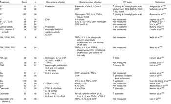
PP, polyphenol; n, number of total subjects; ↓, decrease; ↑, increase; BT, black tea; GT, green tea; GTE, green tea extract; RGJ, red grape juice; RW, red wine; DRW, de-alcoholized red wine; GE, grape extract; PRJ, polyphenol-rich juices; ICAM-1, intercellular adhesion molecule-1; VCAM-1, vascular cell adhesion molecule-1; CRP, C-reactive protein; NK, natural killer; TGF-β, transforming growth factor-β; IFN-γ, interferon-γ; COX-2, cyclooxygenase 2; ECG, epicatechin gallate; EGCG, epigallocatechin gallate; EGC, epigallocatechin; EC, epicatechin.
Studies with alternative sources of dietary flavonoids such as grape juice and red wine were also contradictory. Watzl et al. showed that both acute(Reference Watzl, Bub and Briviba53) and chronic(Reference Watzl, Bub and Pretzer54) administration of red wine, de-alcoholized red wine and red grape juice had no effect on cytokine production, phagocytic activity of neutrophils and monocytes, lymphocyte proliferation and lytic activity of natural killer cells. However, Estruch et al.(Reference Estruch, Sacanella and Badia55) found reduced plasma levels of fibrinogen, IL-1α, CRP, vascular cell adhesion molecule-1 and intercellular adhesion molecule-1 and an increase in plasma levels of epigallocatechin, after 4 weeks of red wine consumption. Chronic supplementation with red grape juice and grape extract reduced neutrophil NADPH oxidase activity(Reference Castilla, Dávalos and Teruel56) and TNFα in postmenopausal women(Reference Zern, Wood and Greene57). Polyphenol-rich juices enhanced immune function as showed by increases in lymphocyte proliferation, IL-2 secretion and lytic activity of natural killer cells(Reference Bub, Watzl and Blockhaus58). In all these studies, neither plasma nor urinary flavonoid levels were measured.
Long-term intervention studies conducted using soya as a source of bioactive molecules showed reduction in levels of vascular cell adhesion molecule-1 and CRP(Reference Fanti, Asmis and Stephenson59, Reference Nasca, Zhou and Welty60), but also a lack of effect on CRP(Reference Jenkins, Kendall and Connelly61–Reference Ryan-Borchers, Park and Chew63), TNFα(Reference Jenkins, Kendall and Connelly61, Reference Ryan-Borchers, Park and Chew63) and IL-6(Reference Nasca, Zhou and Welty60, Reference Maskarinec, Oum and Chaptman62). Increases in total isoflavones, genistein and daidzein(Reference Fanti, Asmis and Stephenson59) and genistein levels(Reference Ryan-Borchers, Park and Chew63) induced by long-term soya consumption were not associated with CRP reductions(Reference Fanti, Asmis and Stephenson59, Reference Ryan-Borchers, Park and Chew63).
When pure molecules were utilized, CRP and IL-8 were lowered by quercetin supplementation in runners(Reference Nieman, Henson and Davis64) and bikers(Reference Nieman, Henson and Davis65), while quercetin plasma levels increased. Other cytokines, such as IL-6(Reference Nieman, Henson and Davis64–Reference Bae, Jung and Lee66) and TNFα(Reference Nieman, Henson and Davis65, Reference Bae, Jung and Lee66), were unaffected by quercetin supplementation, also when consumed in combination with vitamin C(Reference Bae, Jung and Lee66).
Conclusions
Although existing evidence indicates that flavonoids potentially display a multitargeting anti-inflammatory action, a clear conclusion on their effects in human subjects cannot be drawn. The large body of evidence in vitro and on cellular models might be somehow biased by the non-physiological concentrations (in the range of 5–100 μM) utilized. Human intervention studies have shown that absorption of flavonoids in the gastrointestinal tract is typically between 1 and 5% of the ingested dose, with the overall result of reaching plasma concentrations not higher than 1 μM following ingestion of flavonoid-rich food(Reference Manach, Scalbert and Morand9). Moreover, in vivo, flavonoids are extensively metabolized and transformed in molecules with different chemical structures and activities compared with the ones originally present in the food. The large majority of in vitro and cellular experiments have not been performed with the metabolites present in body fluids, thereby increasing the chance of misinterpretation of the results.
The experimental evidence in human subjects suggests a direct role for plant foods in modulating the inflammatory response in vivo. However, the mechanism and the molecules responsible for this effect have not been identified. The assumption that flavonoids might be responsible for the anti-inflammatory effect of plant food is not fully justified on the basis of the current in vivo evidence. Studies investigating the effect of flavonoids on markers of inflammation have been mainly focused on flavonoid-rich foods and not on pure molecules. Moreover, most of the studies lack assessment of flavonoid absorption or failed to associate the anti-inflammatory effect following ingestion of plant foods with changes in circulating levels of flavonoids. Plant foods are rich in flavonoids as well as other components such as antioxidants, vitamins, fibre and nutrients that may be involved in their biological activity. Specifically, planned human trials with proper placebo and pure molecules are needed to clarify whether flavonoids represent ancillary ingredients or key molecules involved in the anti-inflammatory properties of plant foods.
Acknowledgements
The authors declare that there is no conflict of interest. This research received no specific grant from any funding agency in the public, commercial or not-for-profit sectors.

