Non-alcoholic fatty liver disease (NAFLD) is the hepatic consequence of the metabolic syndrome. It is highly prevalent in Western populations and puts a huge economic burden on health services(Reference Younossi and Blissett1). NAFLD begins with the hepatic accumulation of triacylglycerol in lipid droplets within the hepatocytes (steatosis). A greater understanding of the impact of specific nutrients on NAFLD development and progression is needed to help identify better dietary recommendations, treatments and prevention strategies. This study aimed to determine whether HepG2 cells, a commonly used human hepatoma cell line, respond to nutritional stimuli in a similar manner to primary human hepatocytes (PHH) in regards to lipid accumulation.
PHH were obtained from 3 patients undergoing elective liver surgery and were obtained with full ethical approval and informed consent. HepG2 cells or PHH seeded in collagen coated 96-well plates were treated with glucose (5mM or 11mM), fructose (0mM, 2mM or 8mM), fatty acids (0μM or 200μM of a mixture of palmitic, oleic and linoleic acids in the ratio 2:2:1) or vehicle controls for 48 hours before determining relative intracellular lipid content using a Nile Red(Reference Greenspan and Mayer2) staining assay. Cells were then lysed by freezing and the DNA content measured to normalise for cell number. Data was analysed using three-way ANOVA.
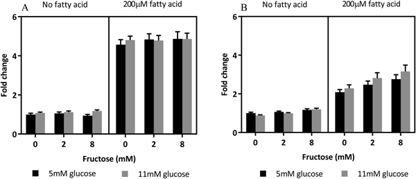
HepG2 cells only accumulated lipid in response to treatment with 200μM fatty acids (P < 0.0001) whereas PHH accumulated lipid in response to fatty acids (P < 0.0001) and fructose (P < 0.0001) (Figure 1). Fatty acid×glucose and fructose×fatty acid interactions were suggested in PHH (P = 0.057 and P = 0.076 respectively) and were significant in some individual livers.

Fig. 1. Nutrient effects on intracellular lipid content (A) HepG2 cells and (B) primary human hepatocytes. Lipid was measured by Nile Red staining after 48 hours of treatment and normalised to DNA content. Fold change relative to 5mM gluc + 0mM fruc + 0μM fatty acid. Values are expressed as mean + SEM. A: n = 20 (4 plates each with n = 5), B: n = 30 (3 livers each with 2 plates with n = 5 replicates)
In conclusion, HepG2 cells and primary human hepatocytes respond differently to glucose and fructose treatment in terms of lipid accumulation. The only consistent effect was increased intracellular lipid with fatty acid treatment, with a greater magnitude in HepG2 cells. Further investigation into genetic factors, gene regulation and substrate utilisation will help to elucidate the mechanisms behind these differences. HepG2 cells may not be a physiologically relevant model for studying NAFLD.
This work was funded by The University of Nottingham and The Rank Prize Funds.