

Methods, Techniques, and Biological Applications
Morphological Properties of the Two Types of Caudate Interneurons: Kohonen Self-Organizing Maps and Correlation-Comparison Analysis by I Grbatinić, B Krstonošić, D Marić, and N Milošević, Microsc Microanal | doi: 10.1017/S1431927618015337
The caudate cluster consists of two subclusters of morphologically different interneurons. Binary images of human caudate interneurons (Figure) were analyzed by using 46 computational neuromorphological parameters to quantify the size of a neuron, neuronal shape, compartmental length, dendritic branching, neuromorphological organization, and dendritic tree complexity. Each individual interneuron was assigned to a cluster according to morphological criteria by using the trained Kohonen self-organizing map. After assignment to a cluster, analysis was continued by feature-wise determination of morphological differences found between clusters and then finished by defining correlation-based, mutual, inter-parametric relations for each of the clusters. The first was performed by using single-factor analysis and the second by correlation-comparison analysis. Single-factor analysis showed 34 significant parameters that distinguish between the clusters. Correlation-comparison analysis extended the results of single-factor analysis by demonstrating significance for 198 inter-parametric correlation pairs, representing 19.13% of mismatched correlations (significant inter-cluster separation zone). The two clusters of caudate interneurons were found to be significantly morphologically different, which may be important for their different neurofunctional behavior.
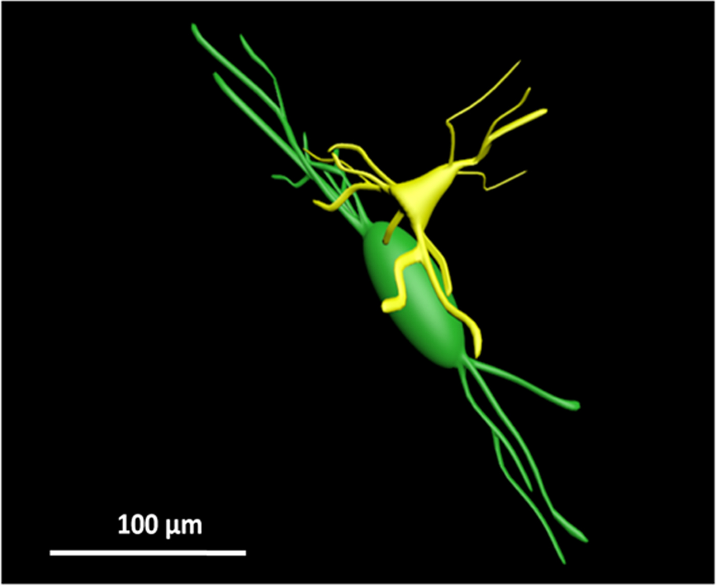
3D reconstruction of cluster 1 (green) and cluster II (yellow) caudate interneurons. Cluster I interneurons are larger with a more elongated cell body, longer dendrites, and a larger area of spreading. Cluster II interneurons have a more complex dendritic arborization and thicker dendrites relative to the soma size. These structural variations may imply functional specificity.
Techniques and Material Applications
Compressed Sensing of Scanning Transmission Electron Microscopy (STEM) with Nonrectangular Scans by Xin Li, Ondrej Dyck, Sergei V. Kalinin, and Stephen Jesse, Microsc Microanal | doi: 10.1017/S143192761801543X
Scanning transmission electron microscopy (STEM) has become the mainstay for materials characterization on the atomic level, with applications ranging from visualization of defects to mapping order parameter fields. In recent years, attention has been focused on the potential of STEM to explore beam-induced chemical processes, especially manipulating atomic motion and enabling atom-by-atom fabrication. These applications together with traditional imaging of beam-sensitive materials, necessitate an increase in the dynamic range of STEM in imaging and manipulation modes, increasing the absolute scanning speed that can be achieved by combining sparse sensing methods with nonrectangular scanning trajectories. This work presents a general method for real-time reconstruction of sparsely sampled images from high-speed, noninvasive, and diverse scanning pathways, such as spiral and Lissajous scans. This approach is demonstrated on both the synthetic data and experimental STEM data on the beam-sensitive material graphene. This work opens the door for comprehensive investigation and optimal design of dose efficient scanning strategies and real-time adaptive inference and control of e-beam induced atomic fabrication.
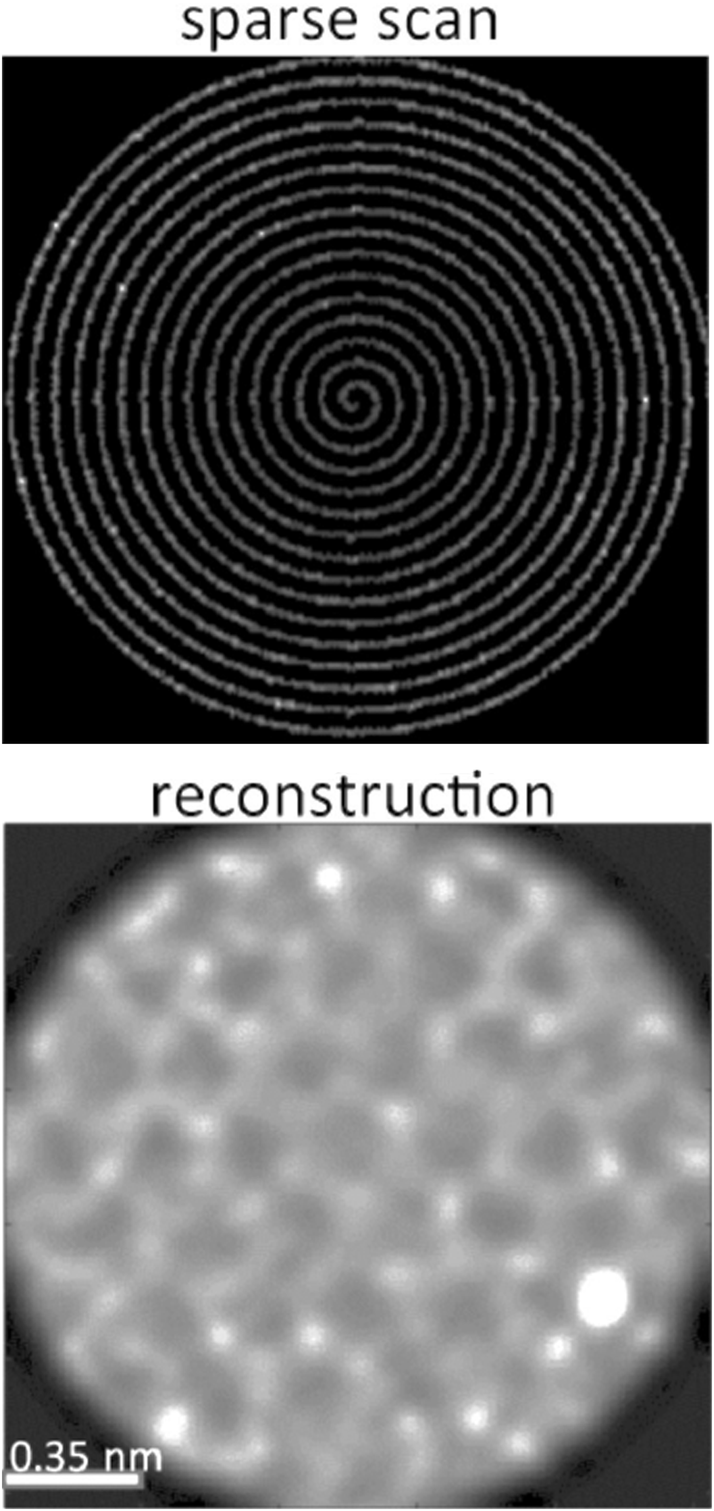
Schematic diagram showing a real-time sparse spiral scan (top); reconstruction of image of graphene with a silicon dopant (bottom). This result was finished in milliseconds.
Techniques and Material Applications
Multi-Angle Plasma FIB Curtaining Artefact Correction Using a Fourier-Based Linear Optimization Model by CW Schankula, CK Anand, and ND Bassim, Microsc Microanal | doi: 10.1017/S1431927618015234
We present a flexible linear optimization model for correcting multi-angle curtaining effects in plasma focused ion beam scanning electron microscopy (PFIB-SEM) images produced by rocking-polishing schemes. When PFIB-SEM is employed in a serial sectioning tomography workflow, it is capable of imaging large 3D volumes quickly, providing rich information in the critical 10–100 nm feature length scale. During tomogram acquisition, a “rocking polish” is often used to reduce straight-line “curtaining” gradations in the milled sample surface. While this mitigation scheme is effective for deep curtains, it leaves shallower line artefacts at two discretized angles. Segmentation and other automated processing of the image set requires that these artefacts be corrected for accurate microstructural quantification. Our work details a new Fourier-based linear optimization model for correcting curtaining artefacts by targeting curtains at two discrete angles. We demonstrate its capabilities by processing images from a tomogram from a multiphase, heterogeneous concrete sample. Our model provides effective multi-angle curtain correction without introducing artefacts.

Two examples of curtaining artefacts in an ultra-high performance concrete sample, with the original image on the left and the corrected image on the right. Note the preservation of contrast, including that of voids.
