


Introduction
Microscopy and imaging in the life sciences are advancing from traditional lamps to solid-state light-emitting diode (LED) technology. Moving to LEDs allows users to benefit from long lifetimes with increased stability while negating the need to replace or dispose of toxic bulb waste. Microscopy and fluorescence excitation have traditionally relied on the spectral properties of the mercury arc lamps, which has defined the chemistry of fluorophores, as well as the excitation and emission filters used in fluorescence imaging. Mercury arc lamps have discrete peaks around which the most common fluorophores, namely DAPI, FITC, and TRITC, have been developed and used for decades. With technology moving to LEDs, users must be aware of the differences in the peak optical power between lamps and LEDs and ensure optimization of their filters to achieve maximum excitation efficiency of their fluorophores.
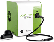
The X-Cite NOVEM (Figure 1) was awarded the 2021 Microscopy Today Innovation Award, which recognizes innovations to make microscopy more powerful, productive, and easier to accomplish [Reference Lyman1]. The X-Cite NOVEM is a nine-channel LED illumination system for fluorescence imaging, with configurations spanning from near-UV wavelengths of the spectrum to the near-IR. The unit features a patented LaserLED Hybrid Drive® technology, which provides users with high-power excitation in the previously challenging 500–600 nm Green Gap. Each of the nine channels can be controlled individually or in combination with other channels. Rapid channel switching allows for high-throughput imaging.

Figure 1: The X-Cite NOVEM, winner of a 2021 Microscopy Today Innovation Award.
The unit has five independent LEDs, outfitted with excitation filters, allowing for imaging of popular fluorophores such as DAPI, CFP, GFP/FITC, and Cy5. Four additional channels (500–600 nm) are powered by the LaserLED Hybrid Drive® technology with a 4-position motorized filter changer to select specific wavelengths within this range for popular green excited fluorophores such as YFP, TRITC, and mCherry. The preinstalled filters and motorized filter changer make it easy to image with multiband filter cubes without bleed-through (Table 1). The X-Cite NOVEM couples to most standard research microscopes with a liquid light guide and an X-Cite microscope adaptor (Figure 1). Each of the nine channels can be individually controlled or combined with others.
Table 1: Fluorophores excited by the X-Cite NOVEM with suggested filters for each wavelength.

Intensity control of 1% per channel can be achieved using the intuitive speedDIAL hand controller, or via USB, TTL, or third-party software control. Even operating at full power, the acoustic noise generated by the X-Cite NOVEM is exceptionally low when compared to the X-Cite Xylis and 120Q systems (Figure 2).

Figure 2: Acoustic noise comparison of the X-Cite 120Q, Xylis, and NOVEM lamp systems. The new NOVEM, even when operated at 100%, has lower noise levels.
Imaging with Fura-2 to IR800/Indocyanine Green (ICG)
Intracellular calcium signals drive many cellular processes including muscle contraction, cell proliferation, synaptic transmission, and gene transcription. Detecting changes in intracellular calcium is of interest to researchers because it provides insight to these dynamic processes in living cells. Techniques for detecting intracellular calcium signals using Fura-2 imaging are well established [Reference Barreto-Chang and Dolmetsch2,Reference Wier3]. The excitation maxima of Fura-2 shifts from 380 nm to 340 nm upon binding of calcium. This shift can be used to quantify intracellular calcium fluxes. Xenon lamps have been the gold standard for Fura-2 imaging because they provide a uniform intensity of light over a wide range of wavelengths. However, these arc lamps have their own drawbacks. Since arc lamps are broad-spectrum, they must be used in combination with filter wheels to isolate and constantly change between 340 nm and 380 nm excitation wavelengths. This results in a mechanical limit on imaging speed. In addition, xenon lamps require frequent replacement, making them a nuisance and expensive to maintain. In addition, since arc lamps are broad-spectrum, they must be used in combination with filter wheels to isolate and constantly flip between the 340 nm and 380 nm excitation wavelengths, putting a mechanical limit on imaging speed.
Hence, there has been a general move toward dyes like Fluo-4, which are single-excitation. Single-wavelength dyes can detect changes in calcium but cannot measure calcium concentrations in the cytoplasm. Fura-2 imaging requires alternately exciting the fluorophore at 340 nm and 380 nm. Image pairs are recorded, and the signal excited by 340 nm is divided by the signal excited by 380 nm to produce a ratiometric image. A change in the ratio over time corresponds to a change in intracellular calcium [Reference Barreto-Chang and Dolmetsch2]. With the development of LEDs at lower wavelengths, it is now possible to use solid-state technology for this application.
In an LED system, each wavelength can be controlled separately, allowing simple adjustment of the 340 nm versus 380 nm LEDs to capture the images at the two wavelengths at similar excitation intensities. This alleviates the need for using cumbersome xenon lamp technology for this application, which requires the user to align and replace bulbs every few hundred hours. LED technology can be used reliably for tens of thousands of hours without replacement. Having a powerful, fast-switching light source providing filtered LED excitation negates the need for reliance on a filter wheel, thereby speeding up data acquisition. Here we demonstrate how the X-Cite NOVEM allows researchers to exploit the benefits of Fura-2 imaging (quantitation and ability to correct for bleaching) without the need for arc lamps while providing the speed and economy of use of single-wavelength probes.
The near-infrared (NIR) version of NOVEM allows imaging in the NIR fluorescence region. Research interest in this this region (using indocyanine green (ICG) and IR800) is growing because of its potential use in image-guided surgery [Reference Ma4]. IR800 has high water solubility and stability, low toxicity, and high signal in human tumor cells due to low background autofluorescence in the NIR region. NIR wavelengths also allow for greater depth penetration in thicker tissues and living animals.
Examples of Imaging with the NOVEM X-Cite LED Technology
Below are some case studies describing the use of the NOVEM in calcium imaging using Fura-2 ratiometric dye. The laboratories have traditionally used xenon lamps in their past research and have generously given their time to test the X-Cite NOVEM for similar work.
Case study 1:
Fura-2 Calcium Imaging of HeLa cells Using Ionomycin. Callen T. Wallace, Senior Research Specialist, Center for Biologic Imaging, University of Pittsburgh (unpublished data).
Images were acquired on a Nikon Ti inverted microscope with a 40 × 1.30 N.A. objective and a Photometrics Prime BSI sCMOS camera using the X-Cite NOVEM light source (Figure 3). HeLa cells were loaded with Fura-2 AM at 5 μM concentration for 30 minutes before imaging. Time-lapse imaging was performed, triggering between 340 nm and 385 nm excitation at 100 ms intervals, with a continuous acquisition to capture baseline fluorescence in each channel (Figure 3A) and the ratiometric change due to agonist (Figure 3B). To induce calcium flux, 1 mM of ionomycin was added to 2 mL of media in real-time during imaging.

Figure 3: Images were acquired using triggered acquisition with the light source at 100 ms exposure for each channel. The ratiometric shift occurs instantaneously on the addition of Ionomycin. (A) Pre-stimulation and (B) post-stimulation.
Case study 2:
Capsaicin Elicits Calcium Responses in Rat DRG Sensory Neurons. Dong Liu, Matt Reissman, Mahsa Sadeghi, Adina Hazan, and Robert Petroski, Neuroservice USA (unpublished data).
Sensory neuron cultures were prepared from adult rat dorsal root ganglia and plated on a monolayer of astrocytes growing on PDL-coated glass imaging dishes (MatTek). Calcium imaging was performed on Fura-2 AM-loaded cells at five days in vitro. Images were acquired with an Olympus IX73 inverted microscope with the X-Cite NOVEM and Hamamatsu ORCA Fusion BT camera. MetaFluor imaging software was used for data acquisition and analysis. Adult rat DRG neuron cultured cells were loaded with 3 μM Fura-2 AM in 0.025% Pluronic F-127. Capsaicin was used to evoke calcium signals by activating native TRPV1 receptors (Figures 4 and 5).

Figure 4: (A) Phase-contrast and (B–D) ratiometric pseudo-color images of Fura-2-loaded DRG sensory neurons. (B, C) Low capsaicin (30 nM, TRPV1 agonist) elicited calcium responses in sensory neurons, while (D) high capsaicin (1 μM) elicited larger calcium responses in sensory neurons.

Figure 5: Time course of calcium responses in individual Fura-2AM-loaded DRG sensory neurons indicated by colored lines. Following a 1-minute baseline period, calcium responses of sensory neurons were detected in a low (30 nM) then high (1 μM) concentration of capsaicin for 2 minutes each. All cells responded to depolarization by 60 mM KCl, indicating healthy, electrically excitable neurons.
Case study 3:
Glutamate Elicits Calcium Fluxes in Rat Pyramidal Neurons. Dong Liu, Matt Reissman, Mahsa Sadeghi, Adina Hazan, and Robert Petroski, Neuroservice USA (unpublished data).
Neuron cultures were prepared from E18 rat cortex and plated on a monolayer of astrocytes growing on PDL-coated glass imaging dishes (MatTek). Calcium imaging was performed in Fura-2AM-loaded cells at 9 days in vitro (Figures 6 and 7).

Figure 6: (A) Low resting baseline calcium level (blue) in rat cortical pyramidal neurons and the response to (B) 100 μM glutamate that triggers a rapid calcium flux (green).

Figure 7: Calcium responses to glutamate in E18 cortical neurons were concentration-dependent. Depolarization by 60 mM KCl also elicited a comparable calcium signal.
Case study 4:
NMDA Elicits Calcium Fluxes in Rat Cortical Neurons. Dong Liu, Matt Reissman, Mahsa Sadeghi, Adina Hazan, and Robert Petroski, Neuroservice USA (unpublished data).
Neuron cultures were prepared from embryonic day 18 (E18) rat cortex and plated on a monolayer of astrocytes growing on PDL-coated glass imaging dishes (MatTek). Calcium imaging was performed in Fura-2AM-loaded cells at 13 days in vitro (Figures 8 and 9). The cells were loaded with 3 μM Fura-2AM for 30 minutes at 37° and cells rinsed for 30–90 minutes before imaging. MetaFluor imaging software was used to rapidly switch between 340 nm and 380 nm excitation wavelengths, and image pairs were acquired at 515 nm emission every 6 seconds at 390 nm. Test solutions were applied to the recording chamber by manual pipette.

Figure 8: (A) Phase contrast and (B, C) ratiometric pseudo-colored images of Fura-2-loaded rat cortical pyramidal neurons. (B) Cells display low resting calcium at baseline. (C) 100 μM NMDA triggers a rapid calcium flux.

Figure 9: (A) Course of calcium responses in individual Fura-2AM-loaded E18 cortical neurons indicated by colored lines. (B) 2-minute application of NMDA rapidly produced a calcium response. Calcium responses to NMDA in E18 cortical neurons were concentration-dependent with an EC50 of 12.9 μM.
The Green Gap
Manufacturers are challenged by designing fluorescence illumination systems that cover the same spectrum as the traditionally used mercury arc lamp to adequately excite common fluorophores used in fluorescence studies. The challenging wavelength band is between 540–590 nm, known as the “Green Gap.” LEDs in this region of the spectrum are fundamentally limited by the lack of semiconductor materials to efficiently emit light at this wavelength. Some manufacturers have generated innovative solutions to bridge the gap, including LED arrays, wavelength conversion phosphor technology, and laser solutions.
The Solution Wavelength conversion using phosphor materials has been used in the lighting industry for a long time. Shorter wavelength light (blue, violet, UV) is absorbed by a material that re-emits light of a longer wavelength via phosphor conversion. The X-Cite NOVEM uses patented (US #9,239,133 B2) laser phosphor conversion technology to generate high-power light in the 540–590 nm region. The term LaserLED Hybrid Drive refers to the combination of laser and LED technology within the unit to generate high-power excitation light. To successfully take advantage of the LaserLED Hybrid Drive, the X-Cite systems take into account all thermal, electrical, and optical parameters to maximize light conversion and delivery of light to the sample. The LaserLED Hybrid Drive is more efficient than other phosphor conversion techniques and was recognized with a 2016 Microscopy Today Innovation Award [Reference Lyman5]. Combining LED, laser, and phosphor technologies can create an effective solution to fill the Green Gap and produce an optical spectrum that meets the needs of users interested in exciting common fluorescent proteins and fluorophores. The X-Cite NOVEM uses the LaserLED Hybrid Drive to provide superior illumination uniformity and maximum light delivery at all wavelengths, enabling users to excite common fluorophores including mCherry (formerly in the Green Gap) used in fluorescence detection and imaging applications.
Summary
The X-Cite NOVEM provides full spectral resolution and is available in four configurations, each covering a wide range of wavelengths. The system allows ratiometric imaging of Fura-2 at the low end of the spectrum, and coverage extends all the way to the near-IR for deeper imaging with Cy7, ICG, or IR800. High power, rich spectrum, and convenient design features make the X-Cite NOVEM more than just an arc lamp replacement. The X-Cite NOVEM can be used in any laboratory or imaging instrument that requires fluorescence excitation. The nine-channel switching capability allows for fast imaging for high-throughput imaging but also allows users in a multi-imaging facility to benefit from all the wavelengths. Whether the users are interested in Fura-2, or any of the popular fluorescence proteins (BFP, CFP, GFP, YFP, RFP), one system can serve all these applications of interest. The NIR version of NOVEM will be for users who want to image in the near-infrared fluorescence region. Interest for this region (using ICG and IR800) is growing because of its potential use in image-guided surgery.


