

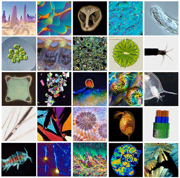
Competition finalists shown on the cover are identified below. The identities and affiliations of the submitters were not known to the judges. The larger number of Open category finalists reflects the larger number of entries in that category. Prize winners will be named in the Microscopy Today section of the awards presentation at the virtual M&M meeting in August. Finalist micrographs may be viewed at the 2021 gallery (see website at the bottom of this page). The microscopy community is encouraged to vote for the People's Choice Award at the same website.

Column 1 (Published category)
C1-1Exfoliated WSe2 flake on silicon nanopillars (atomic force microscopy) by Radek Dao
C1-2Tetraselmis suecica alga (differential interference contrast light microscopy) by Gerd Günther
C1-3Biddulphia antediluviana cushion diatom (phase contrast light microscopy) by Elaine Humphrey
C1-4Quail feather (light microscopy) by Soho Soliman
C1-5Harpacticoid copepod under ultraviolet light (fluorescent microscopy) by Håkan Kvarnström
Columns 2, 3, and 4 (Open category of unpublished micrographs)
C2-1Natural diamond crystal (differential interference contrast) by Nathan Renfro
C2-2Cross section through a stalk of the plant Senecio (polarized light microscopy) by Marek Mis
C2-3Hydrothermal synthesis of BaFeSi4O10 (scanning electron microscopy) by Eric Formo
C2-4Graphite intergrown with feldspars (polarized light microscopy) by Bernardo Cesare
C2-5Campo del Cielo meteorite (reflected light microscopy) by Gaby Ketzer-Raichle
C3-1Sea urchin three-jawed appendage (secondary electron SEM at 15 kV) by Kenneth Bart
C3-2Coated grains of aragonite in sedimentary rock (light microscopy) by Bernardo Cesare
C3-3Symbiosis between fungi and algae in lichen (dark-field light microscopy) by Patrick Jung
C3-4Red fossil corals within agate from the Miocene epoch (light microscopy) by Norm Barker
C3-5Mycelium matrix of fungi in a wood-wide web (polarized light microscopy) by Karl Gaff
C4-1Sildenafil citrate crystallized from solution (polarized light microscopy) by Bernardo Cesare
C4-2Fresh water desmid of the genus Micrasterias (light microscopy focus stack) by Michael Much
C4-3Tracheal bladders of midge larva of genus Chaoborus (light microscopy) by Andrei Savitsky
C4-4Water flea (Daphnia sp.) with eggs (dark-field and polarized light microscopy) by Marek Mis
C4-5Planktonic foraminifera (Mg distribution map by EPMA-WDS) by Anette von Der Handt
Column 5 (Video category)
C5-1Cell division in Tradescantia virginiana (differential interference contrast) by Gerd Günther
C5-2Tube-dwelling amphipod (likely Cerapus sp.) in its tube (light microscopy) by Julia Van Etten
C5-3Daphnia pulex water flea with emerging young (dark-field light microscopy) by Andrei Savitsky
C5-4SiC-SiC ceramic composite in 3D (synchrotron X-ray computed tomography) by Aly Badran
C5-5Crystallization of taurine (dark-field polarized light microscopy) by José Martinez-Lòpez
Vote for your favorite micrograph
at the 2021 image gallery:
www.microscopy.org/awards/micrograph_gallery.cfm
[Please put “at the 2021 image gallery:” on line 2 (different than July 2020 issue).]
