


Crossref Citations
This article has been cited by the following publications. This list is generated based on data provided by Crossref.
Sladky, Ronald
Höflich, Anna
Atanelov, Jacqueline
Kraus, Christoph
Baldinger, Pia
Moser, Ewald
Lanzenberger, Rupert
Windischberger, Christian
and
Zhang, Nanyin
2012.
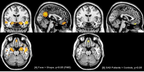
Increased Neural Habituation in the Amygdala and Orbitofrontal Cortex in Social Anxiety Disorder Revealed by fMRI.
PLoS ONE,
Vol. 7,
Issue. 11,
p.
e50050.
Comments
No Comments have been published for this article.