


A 73-year-old male with a history of chronic ataxia presented with transient facial droop to the Emergency Department. A CT angiogram and MRI with diffusion weighted imaging (DWI) were negative for stroke. However, incidental note was made of numerous giant arachnoid granulation pits in the posterior fossa predominantly involving the left occipital bone (Figure 1). These arachnoid pits demonstrated multiple foci of herniation of the adjacent cerebellar parenchyma into the pits with gliosis of the herniated parenchyma and focal encephalomalacia of the subjacent cerebellar parenchyma. Review of bone windows on a remote CT brain performed almost 13 years earlier confirmed this to be a longstanding abnormality (Figure 2). The patient’s physical exam was suggestive of cerebellar ataxia with left-sided dysmetria on finger to nose testing and a wide-based unsteady gait.
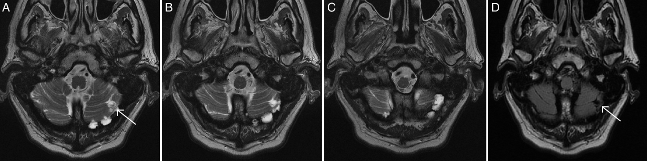
Figure 1: (A–D) MRI images of extensive arachnoid granulation pits in the posterior fossa. T2 images (A–C) from an MRI brain demonstrating extensive arachnoid granulation pits in the posterior fossa predominantly involving the left occipital bone. A FLAIR image (D) illustrates parenchymal gliosis associated with the arachnoid granulation herniation pits.
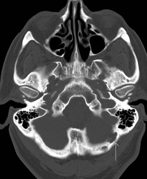
Figure 2: CT bone window image of arachnoid granulation pits in the posterior fossa.
Arachnoid granulations (also called Pacchionian granulations after the Italian physician Antonio Pacchioni) are normal anatomical structures that facilitate the return of cerebrospinal fluid to the venous system through the dural venous sinuses.Reference Ogul and Kantarci1 Giant arachnoid granulation pits are considered to be of large size (>1 cm)Reference Trimble, Harnsberger, Castillo, Brant-Zawadzki and Osborn2 or causing filling defects in the dural venous sinus.Reference Kan, Stevens and Couldwell3 They are commonly located near the cerebral venous sinuses but have been reported to be found in other locations.Reference Ogul and Kantarci1,Reference Lee, Kim, Lee, Kim, Jeon and Kim4 At times, they are noted to extend into the inner table of the skull and can create osteolytic lesions, as in this case.Reference Ogul and Kantarci1 The occipital bone is a rare location to find granulation pits.Reference Ogul and Kantarci1,Reference Lee, Kim, Lee, Kim, Jeon and Kim4 Although typically asymptomatic, their role in cerebrospinal fluid leaks has been describedReference Lee, Kim, Lee, Kim, Jeon and Kim4 and they may account for neurological symptoms when large or when they contain herniated brain parenchyma.Reference Liebo, Lane, Van Gompel, Eckel, Schwartz and Lehman5
Acknowledgements
None.
Disclosures
The authors have no conflicts of interest to declare.
Statement of Authorship
MNK, BHS, CJW, and LM were all involved with the drafting and editing of this manuscript.


