


A 71-year-old man was admitted to our hospital with a 3-month history of progressive gait disturbance. His past medical history included hypertension, diabetes mellitus, renal cell carcinoma, chronic kidney disease (on hemodialysis), and sleep apnea syndrome (on continuous positive airway pressure).
On admission, neurological examination demonstrated left hemiparesis. Diffusion-weighted MRI (DWI) showed numerous small hyperintense lesions mainly in the bilateral cerebral white matter (Figure 1). The apparent diffusion coefficient values of the lesions were almost equal to those of the surrounding parenchyma and were slightly decreased in some of the lesions (Figure 1).
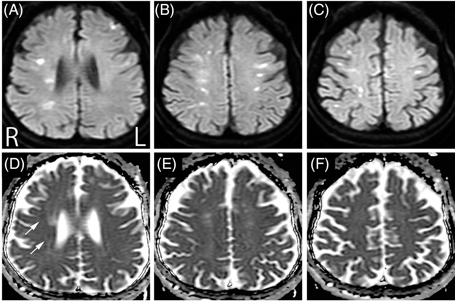
Figure 1: Diffusion-weighted MRI (DWI) (A-C) demonstrated numerous hyperintense lesions mainly in the bilateral cerebral white matter. In apparent diffusion coefficient (ADC) maps (D-F), the ADC values of the lesions were almost equal to those of the surrounding parenchyma and were slightly decreased in some of the lesions (D, arrows).
Blood tests 10 days after admission demonstrated a soluble interleukin 2 receptor (sIL-2R) level of 1929 U/mL (reference range: 145–519 U/mL). A lumbar puncture yielded watery clear cerebrospinal fluid (CSF). CSF examination showed a cell count of 7/mm3, total protein of 75 mg/dL (reference range: 8–43 mg/dL), glucose of 83 mg/dL, and sIL-2R of 2070 U/mL (reference range: <85). Random skin biopsy 12 days after admission established a diagnosis of large B-cell intravascular lymphoma (IVL).
Numerous hyperintense cerebral white matter lesions on DWI are rare in a clinical setting, but have been reported in patients with IVL. Reference Kageyama, Yamanaka, Nakamura and Suenaga1,Reference Kobayashi, Ishihara and Tomimitsu2 Compared with the previous patients, Reference Kageyama, Yamanaka, Nakamura and Suenaga1,Reference Kobayashi, Ishihara and Tomimitsu2 we could suspect IVL at very early stages based on this characteristic finding. Clinicians should be aware of IVL in patients showing progressive neurological deficits and numerous hyperintense cerebral white matter lesions on DWI.
Conflict of interest
The authors declare no conflicts of interest associated with this manuscript.
Statement of Authorship
MS analyzed the data. ZK designed and conceptualized study; major role in the acquisition of data; interpreted the data; drafted the manuscript for intellectual content. SI revised the manuscript for intellectual content. HT revised the manuscript for intellectual content.

