


A 72-year-old man was admitted with complains of recurrent falls associated with severe right leg pain and weakness over the course of 2 months. His history was significant for 15-pound weight loss over the same period. He denied having fever, abdominal pain, chest pain, cough, shortness of breath, or back pain. He had sustained injury to his right knee as the result of his last fall. He had a remote history of right middle cerebral artery ischemic stroke, history of seizure disorder since childhood well-controlled on phenytoin, dyslipidemia, and chronic obstructive pulmonary disease. He had a 60 pack-year cigarette smoker.
At presentation to the emergency department, his general medical examination was unremarkable, except for the right leg weakness, pain, and right knee effusion; the latter was thought to be secondary to a medial collateral ligament tear from his fall, as per orthopedic consultation. The laboratory investigations including cell counts, liver function tests, and electrolytes were within normal limits. Given the complexity of his medical history, a neurological consultation was made to investigate the etiology of the patient’s falls. Neurological examination revealed intact cranial nerves. On the right lower extremity, he demonstrated some degree of wasting associated with occasional fasciculation in the quadriceps muscle, and weakness in his knee extension, hip flexion, and hip adduction: 4−/5, 4/5, and 4−/5, respectively. The sensory examination showed hyperalgesia of the anterior thigh in the L2 and L3 dermatomes. There was no sensory level. The knee jerk was absent and thigh adductor reflex was 1+/4+. The ankle jerk was normal; plantar reflex was downgoing. The right upper extremity was unremarkable. On the left side, he had mild spasticity of the upper and lower extremities and an upgoing plantar reflex, which could be attributed to his old right middle cerebral artery infarct. He had an antalgic gait and his right knee buckled at midstance of his gait.
The right tibial motor, peroneal motor, peroneal sensory, and sural nerve studies were normal. A needle electromyograph (EMG) showed evidence of 4+ active denervation of the right quadriceps muscle. The right tibialis anterior and gastrocnemius muscles also showed 1+ denervation. The femoral sensory conduction and needle EMG studies of thigh adductors were not performed because of the patient’s significant discomfort and hyperalgesia at the anterior thigh. A radiculopathy versus plexopathy was suspected. A noncontrast computed tomography (CT) scan of the lumbar spine was performed, which failed to show a significant spinal stenosis, but revealed a right-sided mass within the iliopsoas muscle. An attempt to perform CT-guided biopsy of the mass was found to be technically challenging and unsuccessful. A CT scan of the chest, abdomen, and pelvis with contrast showed multiple nodules, ground glass opacities, and a large mass in the right paratracheal space with mediastinal infiltration. A 4.3 cm×5.1 cm peripherally enhancing lesion within the psoas and involving the iliacus muscle was also visualized (Figure 1). A bronchoscopy and biopsy of the lung mass revealed an invasive squamous cell carcinoma. A bone scan was normal. The patient was diagnosed with stage 4, metastatic, poorly differentiated squamous cell carcinoma of the lung. He opted not to receive chemotherapy, but agreed to have palliative radiation treatment to his lung and psoas lesions.
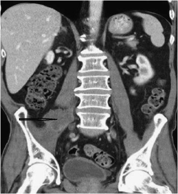
Figure 1 Computed tomography scan of the abdomen with contrast showing a large peripherally enhancing metastatic mass in the right psoas muscle.
Primary presentation of a skeletal muscle metastasis is an unusual phenomenon.Reference Razak, Chhabra, Hughes, England, Dildey and McMenemin 1 The psoas muscle is one of the targets of retroperitoneal metastasis of primary cancers. In a retrospective study of 1754 patients with biopsy-proven lung cancer, using multidetector computed tomography and a positron emission tomography scan, it was demonstrated that the psoas and buttock muscles were the most frequently (33.3%) involved skeletal metastases.Reference Bocchino, Valente, Somma, de Rosa, Bifulco and Rea 2 Psoas metastasis is rarely clinically symptomatic.Reference Strauss, Shah, Chen, Gielda and Kim 4 The initial presentation of an occult cancer with metastasis to the psoas muscle, if recognized on time, can have an impact on patient’s survival because muscle metastasis is a rare phenomenon and has a poor prognosis. We reviewed the literature for various initial presentations of psoas muscle metastasis with unknown primaries at presentation. Those cases found during the staging for a known primary cancer were excluded. We found only a few reported casesReference Strauss, Shah, Chen, Gielda and Kim 4 - Reference Tonelli, Prallet, Cartry, Fournel and Alexandre 10 ; a summary is shown in Table 1.
Table 1 Initial symptomatology of psoas muscle metastasis with unknown primaries at presentation

NSCLC = non-small cell lung cancer; SCC = squamous cell carcinoma; TCC = transitional cell carcinoma.
Among these cases, only the reports by Tonelli et alReference Tonelli, Prallet, Cartry, Fournel and Alexandre 10 and Gharaibeh et alReference Gharaibeh, Lopez-Ruiz and Yousuf 5 indicated “crural neuralgia” and “groin and anterior thigh pain,” respectively, as the initial symptoms of psoas muscle metastasis, disclosing a hidden primary cancer. However, these reports lacked an electrodiagnostic verification of femoral neuropathy. Our case report highlights the importance of maintaining a high level of suspicion in a clinical quest where the patient’s symptoms are not reasonably and collectively explained by routine workup. Our patient presented with signs and symptoms that initially suggested a possible lumbar spine radiculopathy involving the L2, L3, and L4 roots versus a plexopathy. The EMG demonstrated severe denervation of the quadriceps muscle, but some degree of denervation was also evident in the tibialis anterior and gastrocnemius muscles. The patient’s lumbar spine CT scan did not show evidence of radiculopathy. The possibility of a lateral lesion involving the lumbar plexus was highly suspected. The common differential diagnoses for the visualized psoas mass on the patient’s reconstructed lumbar spine CT scan were psoas primary sarcoma and psoas metastasis. Given the history of smoking and recent weight loss, we were encouraged to pursue further investigation by performing CT scans of the chest, abdomen, and pelvis that visualized the lung and psoas masses described previously.
When the clinical picture, imaging, and diagnostic findings do not strongly correlate, a high level of suspicion should be maintained, and further investigations should be performed to rule out a more sinister etiology for a common presentation such as leg pain and falls in an elderly man.
Acknowledgments
The authors thank Dr. Diane Colbert, Assistant Professor of Radiology, for her valuable contribution in interpreting the radiological imaging and editing the radiological discussions of this case.
Disclosures
The authors do not have anything to disclose.


