Skip to main content Accessibility help
×
Hostname: page-component-76fb5796d-22dnz Total loading time: 0 Render date: 2024-04-28T02:51:30.924Z Has data issue: false hasContentIssue false

Chapter 5 - Shaken Baby Syndrome

Abusive Head Trauma or Just a Type of Hydrocephalus?

from Section 2 - Medicine

Published online by Cambridge University Press:  07 June 2023

Keith A. Findley
Affiliation:
University of Wisconsin, Madison
Cyrille Rossant
Affiliation:
University College London
Kana Sasakura
Affiliation:
Konan University, Japan
Leila Schneps
Affiliation:
Sorbonne Université, Paris
Waney Squier
Affiliation:
John Radcliffe Hospital, Oxford
Knut Wester
Affiliation:
Universitetet i Bergen, Norway
Get access

Summary

This chapter reviews epidemiological, clinical, and pathological aspects of benign external hydrocephalus, a medical condition that is a risk factor for development of subdural haematoma, and that frequently is mistaken for abusive head trauma (AHT). For infants, there are striking epidemiological similarities regarding gender and age between external hydrocephalus, subdural haematoma (SDH), and AHT/SBS. There is a marked male preponderance, in most infants the symptom debut occurs during the first 6 months, and prematurity appears to be more frequent. External hydrocephalus is known to predispose for development of SDH. Most infants with external hydrocephalus are born with a close-to-normal head circumference (HC) that starts to grow abnormally fast during the first postnatal months; most of these infants reach HC values compatible with hydrocephalus at the age of 2 to 3 months, the peak age at which AHT/SBS most often is diagnosed. Both in infantile SDH and AHT/SBS, the subdural fluid collections appear to be chronic, not acute as one would expect after a traumatic event. There are reasons to assume that external hydrocephalus often has been and will be misdiagnosed as AHT/SBS.

Type
Chapter
Information
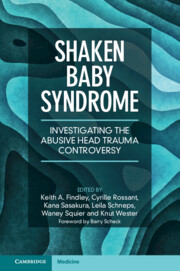
Shaken Baby Syndrome
Investigating the Abusive Head Trauma Controversy
, pp. 85 - 104
Publisher: Cambridge University Press
Print publication year: 2023

Access options

Get access to the full version of this content by using one of the access options below. (Log in options will check for institutional or personal access. Content may require purchase if you do not have access.)

References

Wester, K. Two infant boys misdiagnosed as ‘shaken baby’ and their twin sisters: A cautionary tale. Pediatric Neurology. 2019;97:311.Google Scholar
Thiblin, I, Andersson, J, Wester, K et al. Medical findings and symptoms in infants exposed to witnessed or admitted abusive shaking: A nationwide registry study. PLoS One. 2020;15(10):e0240182.Google Scholar
Wester, K, Stridbeck, U, Syse, A, Wikstrom, J. Re-evaluation of medical findings in alleged shaken baby syndrome and abusive head trauma in Norwegian courts fails to support abuse diagnoses. Acta Paediatrica. 2022;111(4):779–92.Google Scholar
Wester, K, Wikström, J, Lynøe, N, Eriksson, A. Unsubstantiated belief in the diagnostic accuracy of the triad of abusive head trauma may lead to incorrect diagnoses of alleged abuse cases. Acta Paediatrica. 2022;111(4):809–11.CrossRefGoogle ScholarPubMed
Andersson, J, Wikstrom, J, Hogberg, U, Wester, K, Thiblin, I. External hydrocephalus as a cause of infant subdural hematoma: Epidemiological and radiological investigations of infants suspected of being abused. Pediatric Neurology. 2021;126:2634.Google Scholar
Wiig, US, Zahl, SM, Egge, A, Helseth, E, Wester, K. Epidemiology of benign external hydrocephalus in Norway: A population-based study. Pediatric Neurology. 2017;73:3641.Google Scholar
Yew, AY, Maher, CO, Muraszko, KM, Garton, HJ. Long-term health status in benign external hydrocephalus. Pediatric Neurosurgery. 2011;47(1):16.Google Scholar
Lee, HC, Chong, S, Lee, JY et al. Benign extracerebral fluid collection complicated by subdural hematoma and fluid collection: Clinical characteristics and management. Child’s Nervous System. 2018;34(2):235–45.Google Scholar
McNeely, PD, Atkinson, JD, Saigal, G, O’Gorman, AM, Farmer, JP. Subdural hematomas in infants with benign enlargement of the subarachnoid spaces are not pathognomonic for child abuse. American Journal of Neuroradiology. 2006;27(8):1725–8.Google Scholar
Zahl, SM, Egge, A, Helseth, E, Wester, K. Clinical, radiological, and demographic details of benign external hydrocephalus: A population-based study. Pediatric Neurology. 2019;96:53–7.Google Scholar
Alvarez, LA, Maytal, J, Shinnar, S. Idiopathic external hydrocephalus: Natural history and relationship to benign familial macrocephaly. Pediatrics. 1986;77(6):901–7.CrossRefGoogle ScholarPubMed
Fessell, DP, Frankel, DA, Wolfson, WP. Sonography of extraaxial fluid in neurologically normal infants with head circumference greater than or equal to the 95th percentile for age. Journal of Ultrasound Medicine. 2000;19(7):443–7.Google Scholar
Fukuyama, Y, Miyao, M, Ishizu, T, Maruyama, H. Developmental changes in normal cranial measurements by computed-tomography. Developmental Medicine and Child Neurology. 1979;21(4):425–32.Google Scholar
Libicher, M, Troger, J. Us measurement of the subarachnoid space in infants: Normal values. Radiology. 1992;184(3):749–51.Google Scholar
Marino, MA, Morabito, R, Vinci, S et al. Benign external hydrocephalus in infants. A single centre experience and literature review. Neuroradiology Journal. 2014;27(2):245–50.CrossRefGoogle ScholarPubMed
Ingraham, F, Matson, D. Subdural hematoma in infancy. Journal of Pediatrics. 1944;24(1):137.Google Scholar
Hogberg, U, Andersson, J, Squier, et al. Epidemiology of subdural haemorrhage during infancy: A population-based register study. PLoS One. 2018;13(10):117.Google Scholar
Rooks, VJ, Eaton, JP, Ruess, L et al. Prevalence and evolution of intracranial hemorrhage in asymptomatic term infants. American Journal of Neuroradiology. 2008;29(6):1082–9.CrossRefGoogle ScholarPubMed
Ito, H, Yamamoto, S, Komai, T, Mizukoshi, H. Role of local hyperfibrinolysis in etiology of chronic subdural hematoma. Journal of Neurosurgery. 1976;45(1):2631.Google Scholar
Kawakami, Y, Chikama, M, Tamiya, T, Shimamura, Y. Coagulation and fibrinolysis in chronic subdural hematoma. Neurosurgery. 1989;25(1):25–9.Google Scholar
Murakami, H, Hirose, Y, Sagoh, M et al. Why do chronic subdural hematomas continue to grow slowly and not coagulate? Role of thrombomodulin in the mechanism. Journal of Neurosurgery. 2002;96(5):877–84.Google Scholar
Azais, M, Echenne, B. Idiopathic subarachnoid space enlargement (benign external hydrocephalus) in infants. Annales de pediatrie. 1992;39(9):550–8.Google Scholar
Ghosh, PS, Ghosh, D. Subdural hematoma in infants without accidental or nonaccidental injury: Benign external hydrocephalus, a risk factor. Clinical Pediatrics. 2011;50(10):897903.Google Scholar
Gout, A, Gautier, I, Bellaiche, M et al. Idiopathic subarachnoid space enlargement in infancy: Simple anatomic variant or hemorrhagic risk factor? Archives de pediatrie. 1997;4(10):983–7.Google Scholar
Hellbusch, LC. Benign extracerebral fluid collections in infancy: Clinical presentation and long-term follow-up. Journal of Neurosurgery. 2007;107(2 Suppl):119–25.Google Scholar
Kapila, A, Trice, J, Spies, WG, Siegel, BA, Gado, MH. Enlarged cerebrospinal fluid spaces in infants with subdural hematomas. Radiology. 1982;142(3):669–72.Google Scholar
Laubscher, B, Deonna, T, Uske, A, Van, MG. Primitive megalocephaly in children: Natural history, medium term prognosis with special reference to external hydrocephalus. European Journal of Pediatrics. 1990;149(7):502–7.CrossRefGoogle ScholarPubMed
McKeag, H, Christian, CW, Rubin, D et al. Subdural hemorrhage in pediatric patients with enlargement of the subarachnoid spaces Clinical article. Journal of Neurosurgery: Pediatrics. 2013;11(4):438–44.Google Scholar
Miller, D, Barnes, P, Miller, M. The significance of macrocephaly or enlarging head circumference in infants with the triad further evidence of mimics of shaken baby syndrome. American Journal of Forensic Medical Pathology. 2015;36(2):111–20.Google Scholar
Mori, K, Sakamoto, T, Nishimura, K, Fujiwara, K. Subarachnoid fluid collection in infants complicated by subdural-hematoma. Child’s Nervous System. 1993;9(5):282–4.Google Scholar
Papasian, NC, Frim, DM. A theoretical model of benign external hydrocephalus that predicts a predisposition towards extra-axial hemorrhage after minor head trauma. Pediatric Neurosurgery. 2000;33(4):188–93.CrossRefGoogle ScholarPubMed
Piatt, JH, Jr. A pitfall in the diagnosis of child abuse: External hydrocephalus, subdural hematoma, and retinal hemorrhages. Neurosurgery Focus. 1999;7(4):e4.Google Scholar
Ravid, S, Maytal, J. External hydrocephalus: A probable cause for subdural hematoma in infancy. Pediatric Neurology. 2003;28(2):139–41.Google Scholar
Tucker, J, Choudhary, AK, Piatt, J. Macrocephaly in infancy: Benign enlargement of the subarachnoid spaces and subdural collections. Journal of Neurosurgery: Pediatrics. 2016;18(1):1620.Google Scholar
Vinchon, M, Delestret, I, DeFoort-Dhellemmes, S, Desurmont, M, Noule, N. Subdural hematoma in infants: can it occur spontaneously? Data from a prospective series and critical review of the literature. Child’s Nervous System. 2010;26(9):11951205.CrossRefGoogle ScholarPubMed
Zahl, SM, Wester, K, Gabaeff, S. Examining perinatal subdural haematoma as an aetiology of extra-axial hygroma and chronic subdural haematoma. Acta Paediatrica. 2020;109(4):659–66.Google Scholar
Ingraham, FD, Matson, D. D. Subdural hematoma in infancy. Journal of Pediatrics. 1944;24(1):137.Google Scholar
Adamsbaum, C, Grabar, S, Mejean, N, Rey-Salmon, C. Abusive head trauma: Judicial admissions highlight violent and repetitive shaking. Pediatrics. 2010;126(3):546–55.Google Scholar
Hobbs, C, Childs, AM, Wynne, J, Livingston, J, Seal, A. Subdural haematoma and effusion in infancy: An epidemiological study. Archives of Disease in Childhood. 2005;90(9):952–5.Google Scholar
Vinchon, M, de Foort-Dhellemmes, S, Desurmont, M, Delestret, I. Confessed abuse versus witnessed accidents in infants: Comparison of clinical, radiological, and ophthalmological data in corroborated cases. Child’s Nervous System. 2010;26(5):637–45.Google Scholar
Alper, G, Ekinci, G, Yilmaz, Y et al. Magnetic resonance imaging characteristics of benign macrocephaly in children. Journal of Child Neurology. 1999;14(10):678–82.CrossRefGoogle ScholarPubMed
Govaert, P, Oostra, A, Matthys, D, Vanhaesebrouck, P, Leroy, J. How idiopathic is idiopathic external hydrocephalus? Developmental Medicine and Child Neurology. 1991;33(3):274–6.Google Scholar
Miller, M, Ward, T, Stolfi, A, Ayoub, D. Overrepresentation of multiple birth pregnancies in young infants with four metabolic bone disorders: Further evidence that fetal bone loading is a critical determinant of fetal and young infant bone strength. Osteoporosis International. 2014;25(7):1861–73.Google Scholar
Nogueira, GJ, Zaglul, HF. Hypodense extracerebral images on computed-tomography in children: External hydrocephalus. A misnomer. Child’s Nervous System. 1991;7(6):336–41.Google Scholar
Roshan, K, Elizabeth, C, Chacko, A et al. External hydrocephalus: A report of 16 cases from Oman. Journal of Tropical Pediatrics. 1998;44(3):153–6.Google Scholar
Sahar, A. Pseudohydrocephalus: Megalocephaly, increased intracranial pressure and widened subarachnoid space. Neuropadiatrie. 1978;9(2):131–9.Google Scholar
Binenbaum, G, Mirza-George, N, Christian, CW, Forbes, BJ. Odds of abuse associated with retinal hemorrhages in children suspected of child abuse. Journal of the American Association for Pediatric Ophthalmology. 2009;13(3):268–72.Google Scholar
Binenbaum, G, Forbes, BJ. The eye in child abuse: Key points on retinal hemorrhages and abusive head trauma. Pediatric Radiology. 2014;44 Suppl 4:S571S577.Google Scholar
Levin, AV. Retinal hemorrhage in abusive head trauma. Pediatrics. 2010;126(5):961–70.Google Scholar
Maguire, S, Pickerd, N, Farewell, D et al. Which clinical features distinguish inflicted from non-inflicted brain injury? A systematic review. Archives of Disease in Childhood. 2009;94(11):860–7.CrossRefGoogle ScholarPubMed
Maguire, SA, Lumb, RC, Kemp, AM et al. A systematic review of the differential diagnosis of retinal haemorrhages in children with clinical features associated with child abuse. Child Abuse Review. 2013;22(1):2943.CrossRefGoogle Scholar
Maguire, SA, Watts, PO, Shaw, AD et al. Retinal haemorrhages and related findings in abusive and non-abusive head trauma: A systematic review. Eye. 2013;27(1):2836.Google Scholar
Morad, Y, Wygnansky-Jaffe, T, Levin, AV. Retinal haemorrhage in abusive head trauma. Clinical and Experimental Ophthalmology. 2010;38(5):514–20.Google Scholar
Parulekar, MV, Elston, JS. The evidence base for retinal haemorrhages in shaken baby syndrome. Developmental Medicine and Child Neurology. 2008;50(10):793–4.Google Scholar
Pierre-Kahn, V, Roche, O, Dureau, P et al. Ophthalmologic findings in suspected child abuse victims with subdural hematomas. Ophthalmology. 2003;110(9):1718–23.Google Scholar
de Terson, A. L’ hemorrhagie dans le corps vitre au cours de l’hemorrhagie cerebrale. Clinical Ophthalmology. 1900;6:309–12.Google Scholar
Li, LH, Li, N, Zhao, JY et al. Findings of perinatal ocular examination performed on 3573, healthy full-term newborns. British Journal of Ophthalmology. 2013;97(5):588–91.Google Scholar
Nemivant, K, Bhalerao, A. Retinal haemorrhages in the newborn. Journal of Evolution of Medical and Dental Sciences. 2015;4(46):8028–40.Google Scholar
Vinekar, A, Govindaraj, I, Jayadev, C et al. Universal ocular screening of 1021 term infants using wide-field digital imaging in a single public hospital in India: A pilot study. Acta Ophthalmology. 2015;93(5):E372–E6.Google Scholar
Scheller, J. Infantile retinal haemorrhages in the absence of brain and bodily injury. Acta Paediatrica. 2017;106(12):1902–4.Google Scholar
Pu, QL, Li, P, Jiang, HQ et al. Factors related to retinal haemorrhage in infants born at high risk. Acta Ophthalmology. 2017;95(6):E477–E80.Google Scholar
Barnes, PD, Galaznik, J, Gardner, H, Shuman, M. Infant acute life-threatening event: Dysphagic choking versus nonaccidental injury. Seminars in Pediatric Neurology. 2010;17(1):711.Google Scholar
Kramer, K, Goldstein, B. Retinal hemorrhages following cardiopulmonary resuscitation. Clinical Pediatrics. 1993;32(6):366–8.Google Scholar
Pham, H, Enzenauer, RW, Elder, JE, Levin, AV. Retinal hemorrhage after cardiopulmonary resuscitation with chest compressions. American Journal of Forensic and Medical Pathology. 2013;34(2):122–4.Google Scholar
Polito, A, Eong, KGA, Repka, MX, Pieramici, DJ. Bilateral retinal hemorrhages in a preterm infant with retinopathy at prematurity immediately following cardiopulmonary resuscitation. Archives of Ophthalmology (Chicago). 2001;119(6):913–14.Google Scholar
Fledelius, HC. Retinal haemorrhages in premature infants: A pathogenetic alternative diagnosis to child abuse. Acta Ophthalmologica Scandinavica. 2005;83(4):424–7.Google Scholar
Binenbaum, G, Rogers, DL, Forbes, BJ et al. Patterns of retinal hemorrhage associated with increased intracranial pressure in children. Pediatrics. 2013;132(2):e430e434.Google Scholar
Thiblin, I, Andersson, J, Wester, K, Hogberg, G, Hogberg, U. Retinal haemorrhage in infants investigated for suspected maltreatment is strongly correlated with intracranial pathology. Acta Paediatrica. 2022;111(4):808–8.Google Scholar
Davies, F, Gupta, R. Apparent life threatening events in infants presenting to an emergency department. Emergency Medicine Journal. 2002;19(1):1116.Google Scholar
Meert, K, Telford, R, Holubkov, R et al. Exploring the safety and efficacy of targeted temperature management amongst infants with out-of-hospital cardiac arrest due to apparent life threatening events. Resuscitation. 2016;109:40–8.Google Scholar
Gray, C, Davies, F, Molyneux, E. Apparent life-threatening events presenting to a pediatric emergency department. Pediatric Emergency Care. 1999;15(3):195–9.Google Scholar
Maytal, J, Alvarez, LA, Elkin, CM, Shinnar, S. External hydrocephalus: Radiologic spectrum and differentiation from cerebral atrophy. American Journal of Roentgenology. 1987;148(6):1223–30.Google Scholar
Fernando, S, Obaldo, RE, Walsh, IR, Lowe, LH. Neuroimaging of nonaccidental head trauma: Pitfalls and controversies. Pediatric Radiology. 2008;38(8):827–38.Google Scholar
Ingraham, FD, Matson, DD. Subdural hematoma in infancy. Journal of Pediatrics. 1944;24(1):137.Google Scholar
Flom, L, Fromkin, J, Panigrahy, A, Tyler-Kabara, E, Berger, RP. Development of a screening MRI for infants at risk for abusive head trauma. Pediatric Radiology. 2016;46(4):519–26.Google Scholar
Raybaud, C, Greenberg, G. Imaging (normal and abnormal). In Cerebrospinal fluid disorders. Malluci, C, Sgouros, S, eds., 2010, CRC Press, Taylor & Francis, chapter 3.Google Scholar

Save book to Kindle

To save this book to your Kindle, first ensure coreplatform@cambridge.org is added to your Approved Personal Document E-mail List under your Personal Document Settings on the Manage Your Content and Devices page of your Amazon account. Then enter the ‘name’ part of your Kindle email address below. Find out more about saving to your Kindle.

Note you can select to save to either the @free.kindle.com or @kindle.com variations. ‘@free.kindle.com’ emails are free but can only be saved to your device when it is connected to wi-fi. ‘@kindle.com’ emails can be delivered even when you are not connected to wi-fi, but note that service fees apply.

Find out more about the Kindle Personal Document Service.

Available formats
×

Save book to Dropbox

To save content items to your account, please confirm that you agree to abide by our usage policies. If this is the first time you use this feature, you will be asked to authorise Cambridge Core to connect with your account. Find out more about saving content to Dropbox.

Available formats
×

Save book to Google Drive

To save content items to your account, please confirm that you agree to abide by our usage policies. If this is the first time you use this feature, you will be asked to authorise Cambridge Core to connect with your account. Find out more about saving content to Google Drive.

Available formats
×