Skip to main content Accessibility help
×
Hostname: page-component-76fb5796d-9pm4c Total loading time: 0 Render date: 2024-04-26T22:16:36.435Z Has data issue: false hasContentIssue false

16 - Neuroimaging and intellectual disabilities

from Part II - Psychopathology and special topics

Published online by Cambridge University Press:  15 December 2009

Max Pickard
Affiliation:
Consultant Psychiatrist in Mental Health of Learning Disabilities, York Clinic, Guy's Hospital, London
Dene Robertson
Affiliation:
Consultant Psychiatrist, King's College London
Nick Bouras
Affiliation:
King's College London
Geraldine Holt
Affiliation:
King's College London
Get access

Summary

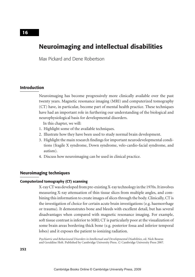
Image of the first page of this content. For PDF version, please use the ‘Save PDF’ preceeding this image.'
Type
Chapter
Information
Publisher: Cambridge University Press
Print publication year: 2007

Access options

Get access to the full version of this content by using one of the access options below. (Log in options will check for institutional or personal access. Content may require purchase if you do not have access.)

References

Barkowich, A. J., Kjos, B. O., Jackson, D. E., & Norman, D. (1988). Normal maturation of the neonatal and infant brain: MR imaging at 1.5T. Neuroradiology, 166, 173–180.Google Scholar
Bassarath, L. (2001). Neuroimaging studies of antisocial behaviour. Canadian Journal of Psychiatry, 46. 728–32.Google Scholar
Breiter, S. N., Arroyo, S., Mathews, V. P.et al. (1994). Proton, M. R. spectroscopy in patients with seizure disorders. American Journal of Neuroradiology, 15, 373–84.Google Scholar
Bruhn, H., Frahm, J., Geyngell, M. L.et al. (1989). Non-invasive differentiation of tumours with use of localised H-1 MR spectroscopy in vivo: initial experience in patients with cerebral tumours. Radiology, 172, 541–8.Google Scholar
Christophe, C., Muller, M. F., Baleriaux, D.et al. (1990). Mapping of normal brain maturation in infants on phase-sensitive inversion-recovery images. Neuroradiology, 32, 173–8.Google Scholar
Courchesne, E., Yeung-Courchesne, R., Press, G., Hussein, J. R. & Jernigan, T. L. (1988). Hypoplasia of cerebellar vermal lobules V1 and V11 in autism. New England Journal of Medicine, 318, 1349–54.Google Scholar
Deb, S. (1997). Structural neuroimaging in learning disability. British Journal of Psychiatry, 171, 417–19.Google Scholar
Debakan, A. S. & Sadowsky, D. (1978). Changes in brain weights during the span of human life: relation of brain weights to body heights and body weights. Annals of Neurology, 4, 345–356.Google Scholar
DeCarli, C. D., Murphy, D. G. M., Gillette, J. A.et al. (1994). Lack of age-related differences in temporal lobe volume of very healthy adults. American Journal of Neuroradiology, 15, 689–96.Google Scholar
Egaas, B., Courchesne, E. (1995). Reduced size of corpus callosum in autism. Archives of Neurology, 52, 794–801.Google Scholar
Eliez, S., Blassey, C. M., Freund, L. S., Hastie, T. & Reiss, A. L. (2001). Brain anatomy, gender and IQ in children and adolescents with fragile X syndrome. Brain, 214. 1610–18.Google Scholar
Eliez, S., White, C., Schmitt, E., Menon, V. & Reiss, A. (2000). Children and adolescents with velocardiofacial syndrome: a volumetric study. American Journal of Psychiatry, 157, 409–15.Google Scholar
Freund, L. S. & Reiss, A. L. (1991). Cognitive profiles associated with Fra (X) syndrome in males and females. American Journal of Medical Genetics, 38, 542–7.Google Scholar
Gafney, G. R., Tsai, L. Y., Kuperman, S. & Minchin, S. (1987). Cerebellar structure in autism. American Journal of Diseases in Children, 141, 1330–2.Google Scholar
Harden, A. Y., Minshew, N.J., Mallikarjuhn, M. & Keveshan, M. S. (2001a). Brain volume in autism. Journal of Child Neurology, 16, 421–4.Google Scholar
Harden, A. Y., Minshew, N.J., Mallikarjuhn, M. & Keveshan, M. S. (2001b). Posterior fossa magnetic resonance imaging in autism. Journal of American Child and Adolescent Psychiatry, 40, 666–72.Google Scholar
Hashimoto, T., Tayama, M., Murakawa, K.et al. (1995). Development of the brainstem and cerebellum in autistic patients. Journal of Autism and Developmental Disorders, 25, 1–18.Google Scholar
Haxby, J. V. (1989). Neuropsychological evaluation of adults with Down syndrome: patterns of selective impairment in non-demented old adults. Journal of Mental Deficiency Research, 33, 193–210.Google Scholar
Herbert, M. R., Ziegler, D. A., Deutch, C. K.et al. (2003). Dissociations of cerebral cortex, subcortical and cerebral white matter volumes in autistic boys. Brain, 126, 1182–92.Google Scholar
Holland, B. A., Haas, D. K., Norman, D., Brant-Zawadski, M. & Newton, T. H. (1986). MRI of normal brain maturation. American Journal of Neuroradiology, 7, 201–8.Google Scholar
Jernigan, T. L., Archibald, S. L., Berhow, M. T.et al. (1991a). Cerebral structure on MRI, Part 1: Localization of age-related changes. Biological Psychiatry, 29, 55–67.Google Scholar
Jernigan, T. L., Press, G. A. & Hesselink, J. R. (1990). Methods for measuring brain morphologic features on magnetic resonance images: Validation and normal aging. Archives of Neurology, 47, 27–32.Google Scholar
Jernigan, T. L., Salmon, D., Butters, N.et al. (1991b). Cerebral structure on MRI, Part II: Specific changes in Alzheimer's and Huntington's diseases. Biological Psychiatry, 29, 68–81.Google Scholar
Kates, W. R., Burnette, C. P., Jabs., E. W.et al. (2001). Regional cortical white matter reductions in velocardiofacial syndrome: A volumetric MRI analysis. Society of Biological Psychiatry, 49, 677–84.Google Scholar
Kaye, J. A., DeCarli, C. D., Luxenberg, J. S. & Rapoport, S. I. (1992). The significance of age-related enlargement of the cerebral ventricles in healthy men and women measured by quantitative computed ⅹ-ray tomography. Journal of the American Geriatric Society, 40, 225–31.Google Scholar
Kemper, M. B., Hagerman, R. J., Ahmad, R. S., Ahmad, R. S. & Mariner, R. (1986). Cognitive profiles and the spectrum of clinical manifestations in heterozygous fragile-X females. American Journal of Medical Genetics, 23, 139–56.Google Scholar
Kemper, M. B., Hagerman, R. J. & Altshul-Stark, D. (1988). Cognitive profiles of boys with the fragile-X syndrome. American Journal of Human Genetics, 30, 191–200.Google Scholar
Kesslak, J. P., Nagata, B. S., Lott, M. D. & Nalcoiglu, O. (1994). Magnetic resonance imaging analysis of brain age-related changes in the brains of individuals with Down's syndrome. Neurology, 44, 1039–45.Google Scholar
Kwon, H., Menon, V., Eliez, S.et al. (2001). Functional neuroanatomy of visuo-spatial working memory in fragile X syndrome: relation to behavioural and molecular measures. American Journal of Psychiatry, 158, 1040–51.Google Scholar
Murakami, J., Courchesne, E., Press, G., Yeung-Courchesne, R. & Hesselink, J. (1989). Reduced cerebellar hemisphere size and its relationship to vermal hypoplasia in autism. Archives of Neurology, 46, 689–94.Google Scholar
Murphy, D. G. M., DeCarli, C. D., Schapiro, M. B., Rapoport, S. I. & Horwitz, B. (1992). Age-related differences in volumes of subcortical nuclei, brain matter, and cerebrospinal fluid in healthy men as measured with MRI. Archives of Neurology, 49, 839–49.Google Scholar
Palmen, C. M. J.S, and Engeland, H. (2004). Review on structural neuroimaging findings in autism. Journal of Neural Transmission, 111, 903–29.Google Scholar
Palmini, A., Andermann, F., Olivier, A. et al. (1991). Focal neurnal migration disorders and intractable partial epilepsy: a study of 30 patients. Annals of Neurology, 30, 741–9.Google Scholar
Pfefferbaum, A., Mathalon, D. H., Sullivan, E. V.et al. (1994). A quantitative magnetic resonance imaging study of changes in brain morphology from infancy to late adulthood. Archives of Neurology, 51, 874–87.Google Scholar
Piven, J., Arndt, S., Bailey, J. & Andreasen, N. (1996). Regional brain enlargement in autism: a magnetic resonance imaging study. Journal of American Child and Adolescent Psychiatry, 35, 530–6.Google Scholar
Piven, J., Arndt, S. et al. (1995). An MRI study of brain size in autism. American Journal of Psychiatry, 152, 1145–9.Google Scholar
Piven, J., Saliba, K., Bailey, J. & Arndt, S. (1997a). An MRI study of autism: the cerebellum revisited. Neurology, 49, 546–51.Google Scholar
Pulver, A. E., Nestadt, G., Goldberg, R.et al. (1994). Psychotic illness in patients diagnosed with velo-cardio-facial syndrome and their relatives. Journal of Nervous and Mental Disease, 182, 476–8.Google Scholar
Reiss, A. L., Abrams, M. T., Greenlaw, R., Freund, L. & Denckla, M. (1995). Neurodevelopmental effects of the FMR-1 full mutation in humans. Nature Medicine, 1, 159–67.Google Scholar
Resnick, S. M., Goldszal, A. F., Davatszikos, C.et al. (2000). One year age changes in MRI brain volumes in older adults. Cerebral Cortex, 10(5), 464–72.Google Scholar
Resnick, S. M., Pham, D. L., Kraut, M. A., Zonderman, A. B. & Davastzikos, C. (2003). Longitudinal magnetic resonance imaging studies of older adults: a shrinking brain. Neuropsychologica, 23(8), 3259–301.Google Scholar
Rivera, S. M., Menon, V., White, C. D.et al. (2002). Functional brain activation during arithmetic processing in females with fragile X syndrome is related to FMR1 protein expression. Human Brain Mapping, 16, 206–18.Google Scholar
Roth, R. M. & Saykin, A. J. (2004). Executive dysfunction in attention-deficit / hyperactivity disorder: cognitive and neuroimaging findings. Psychiatric Clinics of North America, 27, 83–96.Google Scholar
Saitoh, O., Courchesne, E., Egaas, B., Lincoln, A. J. & Screibman, L. (1995). Cross-sectional area of the posterior hippocampus in autistic patients with cerebellar and corpus callosum abnormalities. Neurology, 45, 317–24.Google Scholar
Sanderson, T. L., Best, J. J. K., Doody., G. A., Cunningham Owens, D. G. & Johnstone, E. C. (1999). Neuroanatomy of comorbid schizophrenia and learning disability: a controlled study. The Lancet, 354, 1867–71.Google Scholar
Shapiro, M. B., Luxenberg, J., Kaye, J.et al. (1989). Serial quantitative CT analysis of brain morphometrics in adult Down Syndrome at different ages. Neurology, 39, 1349–53.Google Scholar
Sparks, B. F., Friedman, F. D., Shaw, D. W.et al. (2002). Brain structural abnormalities in young children with autism spectrum disorder. Neurology, 59, 184–92.Google Scholar
Sudhalter, V., Maranion, M. & Brooks, P. (1992). Expressive semantic deficit in the production language of males with fragile X syndrome. American Journal of Medical Genetics, 43 (1–2), 65–71.Google Scholar
Sudhalter, V., Scarborough, H. S. & Cohen, I. L. (1991). Syntactic delay and pragmatic deviance in the language of fragile X males. American Journal of Medical Genetics, 38, 493–7.Google Scholar
Suvossa, J., Seidl, Z.Faber, J. (1990). Hemiparesis of cerebral palsy in relation to epilepsy and mental retardation. Developmental and Child Neurology, 32, 792–4.Google Scholar
Takeda, S. & Matsuzawa, T. (1985). Age-related brain atrophy: A study with computed tomography. Journal of Gerentology, 40, 159–63.Google Scholar
Tiepel, S. J., Alexander, G. E., Schapiro, M. B.et al. (2004). Age-related cortical grey matter reductions in non-demented Down's syndrome adults determined by MRI with voxel-based morphometry. Brain, 127(Pt 4), 811–24.Google Scholar
Amelsvoort, E., Daly, E., Robertson, D.et al. (2001). Structural brain abnormalities associated with deletion at chromosome 22q11: Quantative neuroimaging study of adults with velo-cardio-facial syndrome. British Journal of Psychiatry, 178, 412–19.Google Scholar
Karnebeek, C. D., Jansweijer, M. C., Leenders, A. G., Offringa, M. & Hennekam, R. C. (2005). Diagnostic investigations in individuals with mental retardation: a systemic literature review of their usefulness. European Journal of Human Genetics, 13(1), 6–25.Google Scholar
Wolinsky, J. S., Narayana, P. A. & Fenstermacher, M. J. (1990). Proton magnetic resonance spectroscopy in multiple sclerosis. Neurology, 40, 1764–9.Google Scholar
Zilbovicius, M., Garrau, B., Samson, Y.et al. (1995). Delayed maturation of the frontal cortex in childhood autism. American Journal of Psychiatry, 152, 248–52.Google Scholar

Save book to Kindle

To save this book to your Kindle, first ensure coreplatform@cambridge.org is added to your Approved Personal Document E-mail List under your Personal Document Settings on the Manage Your Content and Devices page of your Amazon account. Then enter the ‘name’ part of your Kindle email address below. Find out more about saving to your Kindle.

Note you can select to save to either the @free.kindle.com or @kindle.com variations. ‘@free.kindle.com’ emails are free but can only be saved to your device when it is connected to wi-fi. ‘@kindle.com’ emails can be delivered even when you are not connected to wi-fi, but note that service fees apply.

Find out more about the Kindle Personal Document Service.

  • Neuroimaging and intellectual disabilities
    • By Max Pickard, Consultant Psychiatrist in Mental Health of Learning Disabilities, York Clinic, Guy's Hospital, London, Dene Robertson, Consultant Psychiatrist, King's College London
  • Edited by Nick Bouras, King's College London, Geraldine Holt, King's College London
  • Book: Psychiatric and Behavioural Disorders in Intellectual and Developmental Disabilities
  • Online publication: 15 December 2009
  • Chapter DOI: https://doi.org/10.1017/CBO9780511543616.017
Available formats
×

Save book to Dropbox

To save content items to your account, please confirm that you agree to abide by our usage policies. If this is the first time you use this feature, you will be asked to authorise Cambridge Core to connect with your account. Find out more about saving content to Dropbox.

  • Neuroimaging and intellectual disabilities
    • By Max Pickard, Consultant Psychiatrist in Mental Health of Learning Disabilities, York Clinic, Guy's Hospital, London, Dene Robertson, Consultant Psychiatrist, King's College London
  • Edited by Nick Bouras, King's College London, Geraldine Holt, King's College London
  • Book: Psychiatric and Behavioural Disorders in Intellectual and Developmental Disabilities
  • Online publication: 15 December 2009
  • Chapter DOI: https://doi.org/10.1017/CBO9780511543616.017
Available formats
×

Save book to Google Drive

To save content items to your account, please confirm that you agree to abide by our usage policies. If this is the first time you use this feature, you will be asked to authorise Cambridge Core to connect with your account. Find out more about saving content to Google Drive.

  • Neuroimaging and intellectual disabilities
    • By Max Pickard, Consultant Psychiatrist in Mental Health of Learning Disabilities, York Clinic, Guy's Hospital, London, Dene Robertson, Consultant Psychiatrist, King's College London
  • Edited by Nick Bouras, King's College London, Geraldine Holt, King's College London
  • Book: Psychiatric and Behavioural Disorders in Intellectual and Developmental Disabilities
  • Online publication: 15 December 2009
  • Chapter DOI: https://doi.org/10.1017/CBO9780511543616.017
Available formats
×