Skip to main content Accessibility help
×
Hostname: page-component-848d4c4894-r5zm4 Total loading time: 0 Render date: 2024-06-26T20:41:18.024Z Has data issue: false hasContentIssue false

Section 2 - Regional Paediatric Orthopaedics

Published online by Cambridge University Press:  30 January 2024

Sattar Alshryda
Affiliation:
Al Jalila Children’s Specialty Hospital, Dubai Academic Health Corporation, Dubai UAE
Stan Jones
Affiliation:
Al Ahli Hospital, Qatar
Paul A. Banaszkiewicz
Affiliation:
Queen Elizabeth Hospital, Gateshead
Get access
Type
Chapter
Information
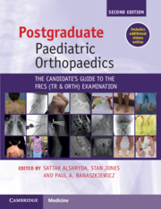
Postgraduate Paediatric Orthopaedics
The Candidate's Guide to the FRCS(Tr&Orth) Examination
, pp. 37 - 296
Publisher: Cambridge University Press
Print publication year: 2024

Access options

Get access to the full version of this content by using one of the access options below. (Log in options will check for institutional or personal access. Content may require purchase if you do not have access.)

References

References

Lode, RT, et al. Valgus slipped capital femoral epiphysis. J Pediatr Orthop. 2006;26(5):594600.Google Scholar
Kelsey, JL, Keggi, KJ, Southwick, WO. The incidence and distribution of slipped capital femoral epiphysis in Connecticut and Southwestern United States. J Bone Joint Surg Am. 1970;52(6):1203–16.Google Scholar
Loder, RT. The demographics of slipped capital femoral epiphysis. An international multicenter study. Clin Orthop Relat Res. 1996;322:827.Google Scholar
Ninomiya, S, Nagasaka, Y, Tagawa, H. Slipped capital femoral epiphysis. A study of 68 cases in the eastern half area of Japan. Clin Orthop Relat Res. 1976;119:172–6.Google Scholar
Loder, RT, et al. Slipped capital femoral epiphysis. Instr Course Lect. 2001;50:555–70.Google Scholar
Loder, RT, Greenfield, ML. Clinical characteristics of children with atypical and idiopathic slipped capital femoral epiphysis: description of the age–weight test and implications for further diagnostic investigation. J Pediatr Orthop. 2001;21(4):481–7.Google Scholar
Lehmann, TG, et al. Radiological findings that may indicate a prior silent slipped capital femoral epiphysis in a cohort of 2072 young adults. Bone Joint J. 2013;95-B(4):452–8.Google Scholar
Kallio, PE, et al. Slipped capital femoral epiphysis. Incidence and clinical assessment of physeal instability. J Bone Joint Surg Br. 1995;77(5):752–5.Google ScholarPubMed
Kallio, PE, et al. Classification in slipped capital femoral epiphysis. Sonographic assessment of stability and remodeling. Clin Orthop Relat Res. 1993;294:196203.Google Scholar
Loder, RT, Richards, BS, Shapiro, PS, Reznick, LR, Aronson, DD. Acute slipped capital femoral epiphysis: the importance of physeal stability. J Bone Joint Surg Am. 1993;75(8):1134–40.Google Scholar
Naseem, H, et al. Treatment of stable slipped capital femoral epiphysis: systematic review and exploratory patient level analysis. J Orthop Traumatol. 2017;18(4):379–94.Google Scholar
Alshryda, S, et al. Severe slipped upper femoral epiphysis; fish osteotomy versus pinning-in-situ: an eleven year perspective. Surgeon. 2014;12(5):244–8.Google Scholar
Ziebarth, K, et al. Clinical stability of slipped capital femoral epiphysis does not correlate with intraoperative stability. Clin Orthop Relat Res. 2012;470(8):2274–9.Google Scholar
Wilson, PD, Jacobs, B, Schecter, L. Slipped capital femoral epiphysis: an end-result study. J Bone Joint Surg Am. 1965;47:1128–45.Google Scholar
Southwick, WO. Osteotomy through the lesser trochanter for slipped capital femoral epiphysis. J Bone Joint Surg Am. 1967;49(5):807–35.CrossRefGoogle ScholarPubMed
Montgomery, R. Slipped upper femoral epiphysis. Orthop Trauma. 2009;23(3 June):169–83.Google Scholar
Thawrani, DP, Feldman, DS, Sala, DA. Current practice in the management of slipped capital femoral epiphysis. J Pediatr Orthop. 2016;36(3):e2737.Google Scholar
Lehman, WB, et al. A method of evaluating possible pin penetration in slipped capital femoral epiphysis using a cannulated internal fixation device. Clin Orthop Relat Res. 1984;186:6570.Google Scholar
Fish, JB. Cuneiform osteotomy of the femoral neck in the treatment of slipped capital femoral epiphysis. A follow-up note. J Bone Joint Surg Am. 1994;76(1):4659.CrossRefGoogle ScholarPubMed
Dunn, DM, Angel, JC. Replacement of the femoral head by open operation in severe adolescent slipping of the upper femoral epiphysis. J Bone Joint Surg Br. 1978;60-B(3):394403.Google Scholar
Ganz, R, et al. Surgical dislocation of the adult hip a technique with full access to the femoral head and acetabulum without the risk of avascular necrosis. J Bone Joint Surg Br. 2001;83(8):1119–24.Google Scholar
Fish, JB. Cuneiform osteotomy in treatment of slipped capital femoral epiphysis. N Y State J Med. 1972;72(21):2633–40.Google Scholar
Kramer, WG, Craig, WA, Noel, S. Compensating osteotomy at the base of the femoral neck for slipped capital femoral epiphysis. J Bone Joint Surg Am. 1976;58(6):796800.Google Scholar
Barmada, R, et al. Base of the neck extracapsular osteotomy for correction of deformity in slipped capital femoral epiphysis. Clin Orthop Relat Res. 1978;132:98101.Google Scholar
Imhauser, G. [Late results of Imhauser’s osteotomy for slipped capital femoral epiphysis (author’s transl)]. Z Orthop Ihre Grenzgeb. 1977;115(5):716–25.Google Scholar
Parsch, K, Weller, S, Parsch, D. Open reduction and smooth Kirschner wire fixation for unstable slipped capital femoral epiphysis. J Pediatr Orthop. 2009;29(1):18.Google Scholar
Crepeau, A, et al. Intracapsular pressures after stable slipped capital femoral epiphysis. J Pediatr Orthop. 2015;35(8):e90–2.Google Scholar
Ibrahim, T, et al. Hip decompression of unstable slipped capital femoral epiphysis: a systematic review and meta-analysis. J Child Orthop. 2015;9(2):113–20.Google Scholar
Ziebarth, K, et al. Capital realignment for moderate and severe SCFE using a modified Dunn procedure. Clin Orthop Relat Res. 2009;467(3):704–16.Google Scholar
Alshryda, S, et al. Evidence based treatment for unstable slipped upper femoral epiphysis: systematic review and exploratory patient level analysis. Surgeon. 2018;16(1):4654.Google Scholar
Lowndes, S, Khanna, A, Emery, D, Sim, J, Maffulli, N. Management of unstable slipped upper femoral epiphysis: a meta-analysis. Br Med Bull. 2009;90:133–46.Google Scholar
Peterson, MD, et al. Acute slipped capital femoral epiphysis: the value and safety of urgent manipulative reduction. J Pediatr Orthop. 1997;17(5):648–54.Google Scholar
Kalogrianitis, S, et al. Does unstable slipped capital femoral epiphysis require urgent stabilization? J Pediatr Orthop B. 2007;16(1):69.Google Scholar
Baghdadi, YM, et al. The fate of hips that are not prophylactically pinned after unilateral slipped capital femoral epiphysis. Clin Orthop Relat Res. 2013;471(7):2124–31.Google Scholar
Sankar, WN, et al. What are the risks of prophylactic pinning to prevent contralateral slipped capital femoral epiphysis? Clin Orthop Relat Res. 2012;471(7):2118–23.Google Scholar
Kroin, E, et al. Two cases of avascular necrosis after prophylactic pinning of the asymptomatic, contralateral femoral head for slipped capital femoral epiphysis: case report and review of the literature. J Pediatr Orthop. 2015;35(4):363–6.Google Scholar
Jerre, R, et al. The contralateral hip in patients primarily treated for unilateral slipped upper femoral epiphysis. Long-term follow-up of 61 hips. J Bone Joint Surg Br. 1994;76(4):563–7.Google Scholar
Stasikelis, PJ, et al. Slipped capital femoral epiphysis. Prediction of contralateral involvement. J Bone Joint Surg Am. 1996;78(8):1149–55.Google Scholar
Phillips, PM, et al. Posterior sloping angle as a predictor of contralateral slip in slipped capital femoral epiphysis. J Bone Joint Surg Am. 2013;95(2):146–50.Google Scholar
Herring, JA. Tachdjians’ Pediatric Orthopaedics, 4th ed, Vol. 1. Philadelphia, PA: Saunders Elsevier; 2008.Google Scholar
Murphey, MD, et al. ACR appropriateness criteria osteonecrosis of the hip. J Am Coll Radiol. 2016;13(2):147–55.Google Scholar
Ilchmann, T, Parsch, K. Complications at screw removal in slipped capital femoral epiphysis treated by cannulated titanium screws. Arch Orthop Trauma Surg. 2006;126(6):359–63.Google Scholar
Bellemans, J, et al. Pin removal after in-situ pinning for slipped capital femoral epiphysis. Acta Orthop Belg. 1994;60(2):170–2.Google ScholarPubMed
Holm, AG, Reikeras, O, Terjesen, T. Long-term results of a modified Spitzy shelf operation for residual hip dysplasia and subluxation. A fifty year follow-up study of fifty six children and young adults. Int Orthop. 2017;41(2):415–21.Google Scholar

References

Heikkinen, E, et al. Venous drainage of the femoral neck in various stages of activity in Perthes’ disease. Rontgenblatter. 1979;32(1):46–9.Google Scholar
Balasa, VV, et al. Legg–Calvé–Perthes disease and thrombophilia. J Bone Joint Surg Am. 2004;86-A(12):2642–7.Google Scholar
Thomas, DP, Morgan, G, Tayton, K. Perthes’ disease and the relevance of thrombophilia. J Bone Joint Surg Br. 1999;81(4):691–5.Google Scholar
Hayek, S, et al. Does thrombophilia play an aetiological role in Legg–Calvé–Perthes disease? J Bone Joint Surg Br. 1999;81(4):686–90.Google Scholar
Hresko, MT, et al. Prospective reevaluation of the association between thrombotic diathesis and legg–perthes disease. J Bone Joint Surg Am. 2002;84-A(9):1613–18.Google Scholar
Gallistl, S, et al. The role of inherited thrombotic disorders in the etiology of Legg–Calvé–Perthes disease. J Pediatr Orthop. 1999;19(1):82–3.Google Scholar
Landin, LA, Danielsson, LG, Wattsgård, C. Transient synovitis of the hip. Its incidence, epidemiology and relation to Perthes’ disease. J Bone Joint Surg Br. 1987;69(2):238–42.Google Scholar
Pinheiro, M, et al. New insights into the biomechanics of Legg–Calvé–Perthes’ disease: the role of epiphyseal skeletal immaturity in vascular obstruction. Bone Joint Res. 2018;7(2):148–56.Google Scholar
Kim, HK, Herring, JA. Pathophysiology, classifications, and natural history of Perthes disease. Orthop Clin North Am. 2011;42(3):285–95, v.Google Scholar
Neal, DC, et al. Quantitative assessment of synovitis in Legg–Calvé–Perthes disease using gadolinium-enhanced MRI. J Pediatr Orthop B. 2015;24(2):8994.Google Scholar
Joseph, B, Pydisetty, RK. Chondrolysis and the stiff hip in Perthes’ disease: an immunological study. J Pediatr Orthop. 1996;16(1):1519.Google Scholar
Kamegaya, M, et al. Arthrography of early Perthes’ disease. Swelling of the ligamentum teres as a cause of subluxation. J Bone Joint Surg Br. 1989;71(3):413–17.Google Scholar
Joseph, B. Morphological changes in the acetabulum in Perthes’ disease. J Bone Joint Surg Br. 1989;71(5):756–63.Google Scholar
Green, NE, Beauchamp, RD, Griffin, PP. Epiphyseal extrusion as a prognostic index in Legg–Calvé–Perthes disease. J Bone Joint Surg Am. 1981;63(6):900–5.Google Scholar
Joseph, B, et al. Natural evolution of Perthes disease: a study of 610 children under 12 years of age at disease onset. J Pediatr Orthop. 2003;23(5):590600.Google Scholar
Theissen, P, et al. The early diagnosis of Perthes disease: the value of bone scintigraphy and magnetic resonance imaging in comparison with x-ray findings. Nuklearmedizin. 1991;30(6):265–71.Google Scholar
Catterall, A. The natural history of Perthes’ disease. J Bone Joint Surg Br. 1971;53(1):3753.Google Scholar
Stulberg, SD, Cooperman, DR, Wallensten, R. The natural history of Legg–Calvé–Perthes disease. J Bone Joint Surg Am. 1981;63(7):1095–108.Google Scholar
Wiig, O, Terjesen, T, Svenningsen, S. Inter-observer reliability of the Stulberg classification in the assessment of Perthes disease. J Child Orthop. 2007;1(2):101–5.Google Scholar
Rich, MM, Schoenecker, PL. Management of Legg–Calvé–Perthes disease using an A-frame orthosis and hip range of motion: a 25-year experience. J Pediatr Orthop. 2013;33(2):112–19.Google Scholar
Axer, A. Subtrochanteric osteotomy in the treatment of Perthes’ disease: a preliminary report. J Bone Joint Surg Br. 1965;47:489–99.Google Scholar
Axer, A, et al. Indications for femoral osteotomy in Legg–Calvé–Perthes disease. Clin Orthop Relat Res. 1980;150:7887.Google Scholar
Hoikka, V, Lindholm, TS, Poussa, M. Intertrochanteric varus osteotomy in Legg–Calvé–Perthes disease: a report of 112 hips. J Pediatr Orthop. 1986;6(5):600–4.Google Scholar
Kitakoji, T, Hattori, T, Iwata, H. Femoral varus osteotomy in Legg–Calvé–Perthes disease: points at operation to prevent residual problems. J Pediatr Orthop. 1999;19(1):7681.Google Scholar
Copeliovitch, L. Femoral varus osteotomy in Legg–Calvé–Perthes disease. J Pediatr Orthop. 2011;31(2 Suppl):S189–91.Google Scholar
Joseph, B, Srinivas, G, Thomas, R. Management of Perthes disease of late onset in southern India. The evaluation of a surgical method. J Bone Joint Surg Br. 1996;78(4):625–30.Google Scholar
Salter, RB. Legg–Perthes disease: the scientific basis for the methods of treatment and their indications. Clin Orthop Relat Res. 1980;150:811.Google Scholar
Kumar, D, Bache, CE, O’Hara, JN. Interlocking triple pelvic osteotomy in severe Legg–Calvé–Perthes disease. J Pediatr Orthop. 2002;22(4):464–70.Google Scholar
Lim, KS, Shim, JS. Outcomes of combined shelf acetabuloplasty with femoral varus osteotomy in severe Legg–Calvé–Perthes (LCP) disease: advanced containment method for severe LCP disease. Clin Orthop Surg. 2015;7(4):497504.Google Scholar
Olney, BW, Asher, MA. Combined innominate and femoral osteotomy for the treatment of severe Legg–Calvé–Perthes disease. J Pediatr Orthop. 1985;5(6):645–51.Google Scholar
Javid, M, Wedge, JH. Radiographic results of combined Salter innominate and femoral osteotomy in Legg–Calvé–Perthes disease in older children. J Child Orthop. 2009;3(3):229–34.Google Scholar
Kamegaya, M, et al. Single versus combined procedures for severely involved Legg–Calvé–Perthes disease. J Pediatr Orthop. 2018;38(6):312–19.Google Scholar
Mosow, N, et al. Outcome after combined pelvic and femoral osteotomies in patients with Legg–Calvé–Perthes disease. J Bone Joint Surg Am. 2017;99(3):207–13.Google Scholar
Kruse, RW, Guille, JT, Bowen, JR. Shelf arthroplasty in patients who have Legg–Calvé–Perthes disease. A study of long-term results. J Bone Joint Surg Am. 1991;73(9):1338–47.Google Scholar
Willett, K, Hudson, I, Catterall, A. Lateral shelf acetabuloplasty: an operation for older children with Perthes’ disease. J Pediatr Orthop. 1992;12(5):563–8.Google Scholar
Daly, K, Bruce, C, Catterall, A. Lateral shelf acetabuloplasty in Perthes’ disease. A review of the end of growth. J Bone Joint Surg Br. 1999;81(3):380–4.Google Scholar
Carsi, B, Judd, J, Clarke, NM. Shelf acetabuloplasty for containment in the early stages of Legg–Calvé–Perthes disease. J Pediatr Orthop. 2015;35(2):151–6.Google Scholar
Laklouk, MA, Hosny, GA. Hinged distraction of the hip joint in the treatment of Perthes disease: evaluation at skeletal maturity. J Pediatr Orthop B. 2012;21(5):386–93.CrossRefGoogle ScholarPubMed
Aly, TA, Amin, OA. Arthrodiatasis for the treatment of Perthes’ disease. Orthopedics. 2009;32(11):817.Google Scholar
Segev, E, et al. Treatment of severe late-onset Perthes’ disease with soft tissue release and articulated hip distraction: revisited at skeletal maturity. J Child Orthop. 2007;1(4):229–35.Google Scholar
Joseph, B. Management of Perthes’ disease. Indian J Orthop. 2015;49(1):1016.Google Scholar
Nguyen, NA, et al. Operative versus nonoperative treatments for Legg–Calvé–Perthes disease: a meta-analysis. J Pediatr Orthop. 2012;32(7):697705.Google Scholar
Saran, N, Varghese, R, Mulpuri, K. Do femoral or Salter innominate osteotomies improve femoral head sphericity in Legg–Calvé–Perthes disease? A meta-analysis. Clin Orthop Relat Res. 2012;470(9):2383–93.CrossRefGoogle ScholarPubMed
Herring, JA, Kim, HT, Browne, R. Legg–Calvé–Perthes disease. Part II: Prospective multicenter study of the effect of treatment on outcome. J Bone Joint Surg Am. 2004;86-A(10):2121–34.Google Scholar
Wiig, O, Terjesen, T, Svenningsen, S. Prognostic factors and outcome of treatment in Perthes’ disease: a prospective study of 368 patients with five-year follow-up. J Bone Joint Surg Br. 2008;90(10):1364–71.Google Scholar
Bankes, MJ, Catterall, A, Hashemi-Nejad, A. Valgus extension osteotomy for ‘hinge abduction’ in Perthes’ disease. Results at maturity and factors influencing the radiological outcome. J Bone Joint Surg Br. 2000;82(4):548–54.Google Scholar
Yoo, WJ, et al. Valgus femoral osteotomy for hinge abduction in Perthes’ disease. Decision-making and outcomes. J Bone Joint Surg Br. 2004;86(5):726–30.Google Scholar
Choi, IH, Yoo, WJ, Cho, T-J, Moon, HJ. Principles of treatment in late stages of Perthes disease. Orthop Clin North Am. 2011;42(3):341–8, vi.Google Scholar
Young, ML, Little, DG, Kim, HK. Evidence for using bisphosphonate to treat Legg–Calvé–Perthes disease. Clin Orthop Relat Res. 2012;470(9):2462–75.Google Scholar
Kumar, V, et al. Do bisphosphonates alter the clinico-radiological profile of children with Perthes disease? A systematic review and meta-analysis. Eur Rev Med Pharmacol Sci. 2021;25(15):4875–94.Google Scholar

References

de Hundt, M, et al. Risk factors for developmental dysplasia of the hip: a meta-analysis. Eur J Obstet Gynecol Reprod Biol. 2012;165(1):817.Google Scholar
Ortiz-Neira, CL, Paolucci, EO, Donnon, T. A meta-analysis of common risk factors associated with the diagnosis of developmental dysplasia of the hip in newborns. Eur J Radiol. 2011;81(3):e344–51.Google Scholar
Herring, JA. Tachdjians’ Pediatric Orthopaedics, 4th ed, Vol. 1. Philadelphia, PA: Saunders Elsevier; 2008.Google Scholar
Scheuer, L, Black, SM. Developmental Juvenile Osteology. Oxford: Elsevier Academic Press; 2000.Google Scholar
Jari, S, Paton, RW, Srinivasan, MS. Unilateral limitation of abduction of the hip. A valuable clinical sign for DDH? J Bone Joint Surg Br. 2002;84(1):104–7.Google Scholar
Jones, D. Neonatal detection of developmental dysplasia of the hip (DDH). J Bone Joint Surg Br. 1998;80(6):943–5.Google Scholar
Czubak, J, et al. Ultrasound measurements of the newborn hip. Comparison of two methods in 657 newborns. Acta Orthop Scand. 1998;69(1):21–4.Google Scholar
Diaz, A, Cuervo, M, Epeldegui, T. Simultaneous ultrasound studies of developmental dysplasia of the hip using the Graf, Harcke, and Suzuki approaches. J Pediatr Orthop B. 1994;3(2):185–9.Google Scholar
Sakkers, R, Pollet, V. The natural history of abnormal ultrasound findings in hips of infants under six months of age. J Child Orthop. 2018;12(4):302–7.Google Scholar
Bialik, V, Bialik, GM, Wiener, F. Prevention of overtreatment of neonatal hip dysplasia by the use of ultrasonography. J Pediatr Orthop B. 1998;7(1):3942.Google Scholar
Ashoor, M, et al. Evidence based treatment for developmental dysplasia of the hip in children under 6 months of age. Systematic review and exploratory analysis. Surgeon. 2021;19(2):7786.Google Scholar
O’Beirne, JG, et al. International Interdisciplinary Consensus Meeting on the evaluation of developmental dysplasia of the hip. Ultraschall Med. 2019;40(4):454–64.Google Scholar
Thallinger, C, et al. Long-term results of a nationwide general ultrasound screening system for developmental disorders of the hip: the Austrian hip screening program. J Child Orthop. 2014;8(1):310.Google Scholar
Ulziibat, M, et al. Implementation of a nationwide universal ultrasound screening programme for developmental dysplasia of the neonatal hip in Mongolia. J Child Orthop. 2020;14(4):273–80.Google Scholar
Biedermann, R, et al. Results of universal ultrasound screening for developmental dysplasia of the hip: a prospective follow-up of 28 092 consecutive infants. Bone Joint J. 2018;100-b(10):1399–404.Google Scholar
Thaler, M, et al. Cost-effectiveness of universal ultrasound screening compared with clinical examination alone in the diagnosis and treatment of neonatal hip dysplasia in Austria. J Bone Joint Surg Br. 2011;93(8):1126–30.Google Scholar
Mahan, ST, Katz, JN, Kim, YJ. To screen or not to screen? A decision analysis of the utility of screening for developmental dysplasia of the hip. J Bone Joint Surg Am. 2009;91(7):1705–19.Google Scholar
Shorter, D, Hong, T, Osborn, DA. Cochrane review: screening programmes for developmental dysplasia of the hip in newborn infants. Evid Based Child Health. 2013;8(1):1154.Google Scholar
Shipman, SA, et al. Screening for developmental dysplasia of the hip: a systematic literature review for the US Preventive Services Task Force. Pediatrics. 2006;117(3):e557–76.Google Scholar
Patel, H. Preventive health care, 2001 update: screening and management of developmental dysplasia of the hip in newborns. CMAJ. 2001;164(12):1669–77.Google Scholar
Murnaghan, ML, et al. Femoral nerve palsy in Pavlik harness treatment for developmental dysplasia of the hip. J Bone Joint Surg Am. 2011;93(5):493–9.Google Scholar
Ramsey, PL, Lasser, S, MacEwen, GD. Congenital dislocation of the hip: use of the Pavlik harness in the child during the first six months of life. 1976. J Bone Joint Surg Am. 2002;84-A(8):1478; discussion 1478.Google Scholar
Gardner, RO, et al. The incidence of avascular necrosis and the radiographic outcome following medial open reduction in children with developmental dysplasia of the hip: a systematic review. Bone Joint J. 2014;96-B(2):279–86.Google Scholar
Gardner, ROE, et al. Evidence-based management of developmental dysplasia of the hip. In: Alshryda, S, Huntley, JS, Banaszkiewicz, PA, eds. Paediatric Orthopaedics: An Evidence-Based Approach to Clinical Questions. Cham: Springer; 2016. pp. 2742.Google Scholar
Salter, RB, Dubos, JP. The first fifteen year’s personal experience with innominate osteotomy in the treatment of congenital dislocation and subluxation of the hip. Clin Orthop Relat Res. 1974;98:72103.Google Scholar
Jospeh, B, et al. Paediatric Orthopaedics. A System of Decision Making. London: Harold Arnold; 2009.Google Scholar
Kalamchi, A, MacEwen, GD. Avascular necrosis following treatment of congenital dislocation of the hip. J Bone Joint Surg Am. 1980;62(6):876–88.Google Scholar
Segal, LS, et al. Avascular necrosis after treatment of DDH: the protective influence of the ossific nucleus. J Pediatr Orthop. 1999;19(2):177–84.Google Scholar
Cooke, SJ, et al. Ossification of the femoral head at closed reduction for developmental dysplasia of the hip and its influence on the long-term outcome. J Pediatr Orthop B. 2010;19(1):22–6.Google Scholar
Konigsberg, DE, et al. Results of medial open reduction of the hip in infants with developmental dislocation of the hip. J Pediatr Orthop. 2003;23(1):19.Google Scholar
Luhmann, SJ, et al. The prognostic importance of the ossific nucleus in the treatment of congenital dysplasia of the hip. J Bone Joint Surg Am. 1998;80(12):1719–27.Google Scholar

References

Weinstein, JN, Kuo, KN, Millar, EA. Congenital coxa vara. A retrospective review. J Pediatr Orthop. 1984;4(1):70–7.Google Scholar
Aitken, GT. Proximal femoral focal deficiency: definition, classification and management. In: Aitken, GT, ed. Proximal Femoral Focal Deficiency – a Congenital Anomaly: A Symposium Held in Washington, DC, 13 June 1968. Washington, DC: National Academy of Sciences; 1969. pp. 122.Google Scholar
Amstutz, HC, Wilson, PD. Dysgenesis of the proximal femur (coxa vara) and its surgical management. J Bone Joint Surg. 1962;44(1):124.Google Scholar
Rozbruch, SR, Hamdy, R. Limb Lengthening and Reconstruction Surgery Case Atlas. Cham: Springer International Publishing; 2015.Google Scholar
National Institute for Health and Care Excellence. Combined bony and soft tissue reconstruction for hip joint stabilisation in proximal focal femoral deficiency (PFFD). 2009. Interventional procedures guidance [IPG297]. Available from: www.nice.org.uk/guidance/ipg297.Google Scholar

References

Hinton, RY, et al. Fractures of the femoral shaft in children. Incidence, mechanisms, and sociodemographic risk factors. J Bone Joint Surg Am. 1999;81(4):500–9.Google Scholar
Amorosa, L. High-energy pediatric pelvic and acetabular fractures. Orthop Clin North Am, 2014;45(4):483500.Google Scholar
Holden, CP, Holman, J, Herman, MJ. Pediatric pelvic fractures. J Am Acad Orthop Surg. 2007;15(3):172–7.Google Scholar
Shaath, MK, et al. Analysis of pelvic fracture pattern and overall orthopaedic injury burden in children sustaining pelvic fractures based on skeletal maturity. J Child Orthop. 2017;11(3):195200.Google Scholar
Ismail, N, et al. Death from pelvic fracture: children are different. J Pediatr Surg. 1996;31(1):82–5.Google Scholar
Silber, JS, Flynn, JM. Changing patterns of pediatric pelvic fractures with skeletal maturation: implications for classification and management. J Pediatr Orthop. 2002;22(1):22–6.Google Scholar
Silber, JS, et al. Analysis of the cause, classification, and associated injuries of 166 consecutive pediatric pelvic fractures. J Pediatr Orthop. 2001;21(4):446–50.Google Scholar
Chia, JPY, et al. Pelvic fractures and associated injuries in children. J Trauma. 2004;56(1):83–8.Google Scholar
Torode, I, Zieg, D. Pelvic fractures in children. J Pediatr Orthop. 1985;5(1):7684.Google Scholar
Shore, BJ, et al. Pediatric pelvic fracture. J Pediatr Orthop. 2012;32(2):162–8.Google Scholar
Bimmel, R, et al. Paediatric hip fractures: a systematic review of incidence, treatment options and complications. Acta Orthop Belg. 2010;76(1):713.Google Scholar
Davison, BL, Weinstein, SL. Hip fractures in children: a long-term follow-up study. J Pediatr Orthop. 1992;12(3):355–8.Google Scholar
Joseph, T. The normal vascular anatomy of the human femoral head during growth. J Bone Joint Surg Br. 1957;39-B(2):358–94.Google Scholar
Ogden, JA. Changing patterns of proximal femoral vascularity. J Bone Joint Surg Am. 1974;56(5):941–50.Google Scholar
Dial, BL, Lark, RK. Pediatric proximal femur fractures. J Orthop. 2018;15(2):529–35.Google Scholar
Bartonicek, J. Proximal femur fractures: the pioneer era of 1818 to 1925. Clin Orthop Relat Res. 2004;419:306–10.Google Scholar
Collona, PC. Fracture of the neck of femur in children. Am J Surg. 1929;6:793–7.Google Scholar
Moon, E, Mehlman, C. Risk factors for avascular necrosis after femoral neck fractures in children: 25 Cincinnati cases and meta-analysis of 360 cases. J Orthop Trauma. 2006;20:323–9.Google Scholar
Slongo, T, Audige, L. AO Pediatric Classification Group. AO Pediatric Comprehensive Classification of Long-Bone Fractures (PCCF). Switzerland: AO Foundation; 2007.Google Scholar
Shrader, MW, et al. Femoral neck fractures in pediatric patients: 30 years experience at a level 1 trauma center. Clin Orthop Relat Res. 2007;454:169–73.Google Scholar
Dendane, MA, et al. Displaced femoral neck fractures in children: are complications predictable? Orthop Traumatol Surg Res. 2010;96(2):161–5.Google Scholar
Yeranosian, M, et al. Factors affecting the outcome of fractures of the femoral neck in children and adolescents: a systematic review. Bone Joint J. 2013;95-b(1):135–42.Google Scholar
Varshney, MK, et al. Functional and radiological outcome after delayed fixation of femoral neck fractures in pediatric patients. J Orthop Traumatol. 2009;10(4):211–16.Google Scholar
Wang, WT, et al. Risk factors for the development of avascular necrosis after femoral neck fractures in children. Bone Joint J. 2019;101-B(9):1160–7.Google Scholar
Beaty, JH. Fractures of the hip in children. Orthop Clin North Am. 2006;37(2):223–32, vii.Google Scholar
Panigrahi, R, et al. Treatment analysis of paediatric femoral neck fractures: a prospective multicenter theraupetic study in Indian scenario. Int Orthop. 2015;39(6):1121–7.Google Scholar
Bukva, B, et al. Femoral neck fractures in children and the role of early hip decompression in final outcome. Injury. 2015;46(Suppl 6):S44–7.Google Scholar
Ng, GPK, Cole, WG. Effect of early hip decompression on the frequency of avascular necrosis in children with fractures of the neck of the femur. Injury. 1996;27(6):419–21.Google Scholar
Akahane, T, Fujioka, F, Shiozawa, R. A transepiphyseal fracture of the proximal femur combined with a fracture of the mid-shaft of ipsilateral femur in a child: a case report and literature review. Arch Orthop Trauma Surg. 2006;126(5):330–4.Google Scholar
Omeroglu, H, Inan, U, Kose, N. Fracture type primarily influences the final outcome in pediatric hip fractures. Orthop Proc. 2010;92-B(Supp V):598.Google Scholar
Talbot, C, et al. Fractures of the femoral shaft in children. Bone Joint J. 2018;100-B(1): 109–18.Google Scholar
Brown, D, Fisher, E. Femur fractures in infants and young children. Am J Public Health. 2004;94(4):558–60.Google Scholar
Khoriati, A-A, et al. The management of paediatric diaphyseal femoral fractures: a modern approach. Strategies Trauma Limb Reconstr. 2016;11(2):8797.Google Scholar
Memeo, A, et al. Retrospective, multicenter evaluation of complications in the treatment of diaphyseal femur fractures in pediatric patients. Injury. 2019;50(Suppl 4):S60–3.Google Scholar
Brousil, J, Hunter, JB. Femoral fractures in children. Curr Opin Pediatr. 2013;25(1):52–7.Google Scholar
Moroz, LA. Titanium elastic nailing of fractures of the femur in children: predictors of complication and poor outcome. J Bone Joint Surg Br. 2006;88-B(10):1361–6.Google Scholar
Garner, MR, et al. Fixation of length-stable femoral shaft fractures in heavier children: flexible nails vs rigid locked nails. J Pediatr Orthop. 2011;31(1):1116.Google Scholar
MacNeil, JAM, Francis, A, El-Hawary, R. A systematic review of rigid, locked, intramedullary nail insertion sites and avascular necrosis of the femoral head in the skeletally immature. J Pediatr Orthop. 2011;31(4):377–80.Google Scholar
Letts, M, et al. Complications of rigid intramedullary rodding of femoral shaft fractures in children. J Trauma. 2002;52(3):504–16.Google Scholar
Beaty, JH, et al. Interlocking intramedullary nailing of femoral-shaft fractures in adolescents: preliminary results and complications. J Pediatr Orthop. 1994;4(2):178–83.Google Scholar
Crosby, SN, Jr, et al. Twenty-year experience with rigid intramedullary nailing of femoral shaft fractures in skeletally immature patients. J Bone Joint Surg Am. 2014;96(13):1080–9.Google Scholar
Raney, EM, Ogden, JA, Grogan, DP. Premature greater trochanteric epiphysiodesis secondary to intramedullary femoral rodding. J Pediatr Orthop. 1993;13(4):516–20.Google Scholar
ME W, EB H. Remodelling of angular deformity after femoral shaft fractures in children. J Bone Joint Surg Br. 1992;74-B(5):765–9.Google Scholar
Zeckey, C, et al. Femoral malrotation after surgical treatment of femoral shaft fractures in children: a retrospective CT-based analysis. Eur J Orthop Surg Traumatol. 2017;27(8):1157–62.Google Scholar
Davids, JR. Rotational deformity and remodeling after fracture of the femur in children. Clin Orthop Relat Res. 1994;302:2735.Google Scholar
Sutphen, SA, et al. Pediatric diaphyseal femur fractures: submuscular plating compared with intramedullary nailing. Orthopedics. 2016;39(6):353–8.Google Scholar
Samora, WP, et al. Submuscular bridge plating for length-unstable, pediatric femur fractures. J Pediatr Orthop. 2013;33(8):797802.Google Scholar
Sink, EL, et al. Results and technique of unstable pediatric femoral fractures treated with submuscular bridge plating. J Pediatr Orthop. 2006;26(2):177–81.Google Scholar
Bar-On, E, Sagiv, S, Porat, S. External fixation or flexible intramedullary nailing for femoral shaft fractures in children. J Bone Joint Surg Br. 1997; 79-B(6):975–8.Google Scholar

References

Thompson, TC. Quadricepsplasty to improve knee function. J Bone Joint Surg. 1944;26(2):366–79.Google Scholar
Judet, T. L’arthrolyse dans les raideurs post-traumatiques du coude. In: Bureau de La Société Française de Chirurgie Orthopédique et Traumatologique (SOFCOT 2010). Conférences d’Enseignement 2010, No. 99. Elsevier; 2010. pp. 333–49.Google Scholar
Johnston, CE. Simultaneous open reduction of ipsilateral congenital dislocation of the hip and knee assisted by femoral diaphyseal shortening. J Pediatr Orthop. 2011;31(7):732–40.Google Scholar
Klingele, KE, Stephens, S. Management of ACL elongation in the surgical treatment of congenital knee dislocation. Orthopedics. 2012;35(7):e1094–8.Google Scholar
Davids, JR, Blackhurst, DW, Allen, BL, Jr. Clinical evaluation of bowed legs in children. J Pediatr Orthop B. 2000;9(4):278–84.CrossRefGoogle ScholarPubMed
Langenskiold, A. Tibia vara (osteochondrosis deformans tibiae): a survey of 23 cases. Acta Chir Scand. 1952;103(1):122.Google Scholar
Herring, JA. Tachdjians’ Pediatric Orthopaedics, 4th ed, Vol. 1. Philadelphia, PA: Saunders Elsevier; 2008.Google Scholar
Raney, EM, Topoleski, TA, Yaghoubian, R, Guidera, KJ, Marshall, JG. Orthotic treatment of infantile tibia vara. J Pediatr Orthop. 1998;18(5):670–4.Google Scholar
Park, SS, Gordon, JE, Luhmann, SJ, Dobbs, MB, Schoenecker, PL. Outcome of hemiepiphyseal stapling for late-onset tibia vara. J Bone Joint Surg Am. 2005;87(10):2259–66.Google Scholar
Pavone, V, Testa, G, Riccioli, M, Sessa, A, Evola, FR, Avondo, S. The natural history of focal fibrocartilaginous dysplasia in the young child with tibia vara. Eur J Orthop Surg Traumatol. 2014;24(4):579–86.Google Scholar
Paley, D. Congenital pseudarthrosis of the tibia: biological and biomechanical considerations to achieve union and prevent refracture. J Child Orthop. 2019;13(2):120–33.Google Scholar
Paley, D. Surgical reconstruction for fibular hemimelia. J Child Orthop. 2016;10(6):557–83.Google Scholar
Jones, D, Barnes, J, Lloyd-Roberts, GC. Congenital aplasia and dysplasia of the tibia with intact fibula. Classification and management. J Bone Joint Surg Br. 1978;60(1):31–9.Google Scholar
Pill, SG, Ganley, TJ, Milam, RA, Lou, JE, Meyer, JS, Flynn, JM. Role of magnetic resonance imaging and clinical criteria in predicting successful nonoperative treatment of osteochondritis dissecans in children. J Pediatr Orthop. 2003;23(1):102–8.Google Scholar
Dinham, JM. Popliteal cysts in children. The case against surgery. J Bone Joint Surg Br. 1975;57(1):6971.Google Scholar
Akagi, R, Saisu, T, Segawa, Y, et al. Natural history of popliteal cysts in the pediatric population. J Pediatr Orthop. 2013;33(3):262–8.Google Scholar
O’Connor, D, Clarke, NMP, Hegarty, SE, Fairhurst, JJ. The natural history of popliteal cysts in children: an ultrasound study. Knee. 1998;5(4):249–51.Google Scholar

References

Anderson, M, Messner, MB, Green, WT. Distribution of lengths of the normal femur and tibia in children from one to eighteen years of age. J Bone Joint Surg Am. 1964;46:1197–202.Google Scholar
Czitrom, AA, Pritzker, KP. Simple bone cyst causing collapse of the articular surface of the femoral head and incongruity of the hip joint. A case report. J Bone Joint Surg Am. 1980;62(5):842–5.Google Scholar
Arkader, A, Warner, WC, Horn, BD, Shaw, RN, Wells, L. Predicting the outcome of physeal fractures of the distal femur. J Pediatr Orthop. 2007;27(6):703–8.Google Scholar
Salter, R, Harris, W. Injuries involving the epiphyseal plate. J Bone Joint Surg Am. 1963;45A:587622.Google Scholar
Eid, AM, Hafez, MA. Traumatic injuries of the distal femoral physis. Retrospective study on 151 cases. Injury. 2002;33(3):251–5.Google Scholar
Ilharreborde, B, Raquillet, C, Morel, E, et al. Long-term prognosis of Salter–Harris type 2 injuries of the distal femoral physis. J Pediatr Orthop B. 2006;15(6):433–8.Google Scholar
Cash, JD, Hughston, JC. Treatment of acute patellar dislocation. Am J Sports Med. 1988;16(3):244–9.Google Scholar
Alshryda, S, Tsang, K, Dekiewiet, G. Evidence-based treatment for slipped upper femoral epiphysis. In: Alshryda, S, Huntley, JS, Banaszkiewicz, P, eds. Paediatric Orthopaedics: An Evidence-Based Approach to Clinical Questions. Cham: Springer; 2016. pp. 5170.Google Scholar
Rund, JM, Hinckel, BB, Sherman, SL. Acute patellofemoral dislocation: controversial decision-making. Curr Rev Musculoskelet Med. 2021;14(1):82–7.Google Scholar
Dormans, JP. Pediatric Orthopaedics. Philadelphia, PA: Elsevier Mosby; 2005.Google Scholar
Kocher, MS, Saxon, HS, Hovis, WD, Hawkins, RJ. Management and complications of anterior cruciate ligament injuries in skeletally immature patients: survey of the Herodicus Society and The ACL Study Group. J Pediatr Orthop. 2002;22(4):452–7.Google Scholar
Heusdens, CHW. ACL repair: a game changer or will history repeat itself? A critical appraisal. J Clin Med. 2021;10(5):912.Google Scholar

References

Herring, JA. Tachdjians’ Pediatric Orthopaedics, 5th ed. Philadelphia, PA: Saunders Elsevier; 2014.Google Scholar
Steinman, S, Richards, BS, Faulks, S, Kaipus, K. A comparison of two nonoperative methods of idiopathic clubfoot correction: the Ponseti method and the French functional (physiotherapy) method. Surgical technique. J Bone Joint Surg Am. 2009;91 Suppl 2:299312.Google Scholar
Morcuende, JA, Dolan, LA, Dietz, FR, Ponseti, IV. Radical reduction in the rate of extensive corrective surgery for clubfoot using the Ponseti method. Pediatrics. 2004;113(2):376–80.Google Scholar
Drennan, JC. Congenital vertical talus. Instr Course Lect. 1996;45:315–22.Google Scholar
Mubarak, SJ, Patel, PN, Upasani, VV, Moor, MA, Wenger, DR. Calcaneonavicular coalition: treatment by excision and fat graft. J Pediatr Orthop. 2009;29(5):418–26.Google Scholar
Chytas, A, Chaudhry, S, Alshryda, S. Evidence-based treatment for tarsal coalition. In: Alshryda, S, Huntley, JS, Banaszkiewicz, PA, eds. Paediatric Orthopaedics: An Evidence-Based Approach to Clinical Questions. Cham: Springer; 2016. pp. 175–82.Google Scholar
Masquijo, J, Allende, V, Torres-Gomez, A, Dobbs, MB. Fat graft and bone wax interposition provides better functional outcomes and lower reossification rates than extensor digitorum brevis after calcaneonavicular coalition resection. J Pediatr Orthop. 2017;37(7):e427–31.Google Scholar
Robinson, AH, Limbers, JP. Modern concepts in the treatment of hallux valgus. J Bone Joint Surg Br. 2005;87(8):1038–45.Google Scholar

References

Barrie, J, Hope, R, Lishman, J. Ankle instability. 2014. Available from: www.foothyperbook.com/elective/ankleInstability/ankleInstabBasic.htm.Google Scholar
Topal, M, Köse, A, Dinçer, R, Baran, T, Köse, M, Çağatay Engin, M. Os subtibiale: mimicking medial malleolar fracture. Am J Emerg Med. 2017;35(6):940.e13.Google Scholar
Turan, A, Kilicaslan, OF, Kose, O. Os subtibiale and secondary ossification center of medial malleolus are two different entities. Am J Emerg Med. 2017;35(6):929.Google Scholar
Dias, LS, Tachdjian, MO. Physeal injuries of the ankle in children: classification. Clin Orthop Relat Res. 1978(136):230–3.Google Scholar
Petratos, DV, Kokkinakis, M, Ballas, EG, Anastasopoulos, JN. Prognostic factors for premature growth plate arrest as a complication of the surgical treatment of fractures of the medial malleolus in children. Bone Joint J. 95B(3):419–23.Google Scholar
Cooperman, DR, Spiegel, PG, Laros, GS. Tibial fractures involving the ankle in children. The so-called triplane epiphyseal fracture. J Bone Joint Surg Am. 1978;60(8):1040–6.Google Scholar
Schurz, M, Binder, H, Platzer, P, Schulz, M, Hajdu, S, Vecsei, V. Physeal injuries of the distal tibia: long-term results in 376 patients. Int Orthop. 2010;34(4):547–52.Google Scholar
American Academy of Orthopaedic Surgeons. Lawn mower injuries in children. 2022. Available from: https://orthoinfo.aaos.org/en/diseases--conditions/lawn-mower-injuries-in-children/.Google Scholar

References

Mehta, MH. The rib-vertebra angle in the early diagnosis between resolving and progressive infantile scoliosis. J Bone Joint Surg Br. 1972;54(2):230–43.Google Scholar
Mehta, MH. Growth as a corrective force in the early treatment of progressive infantile scoliosis. J Bone Joint Surg Br. 2005;87(9):1237–47.Google Scholar
Dubousset, J, Herring, JA, Shufflebarger, H. The crankshaft phenomenon. J Pediatr Orthop. 1989;9(5):541–50.Google Scholar
Luque, ER. Paralytic scoliosis in growing children. Clin Orthop Relat Res. 1982;163:202–9.Google Scholar
Samdani, AF, et al. Anterior vertebral body tethering for idiopathic scoliosis: two-year results. Spine (Phila Pa 1976). 2014;39(20):1688–93.Google Scholar
Weinstein, SL, et al. Health and function of patients with untreated idiopathic scoliosis: a 50-year natural history study. JAMA. 2003;289(5):559–67.Google Scholar
Weinstein, SL, et al. Effects of bracing in adolescents with idiopathic scoliosis. N Engl J Med. 2013;369 (16):1512–21.Google Scholar
European Medicines Agency. Zolgensma. 2021. Available from: www.ema.europa.eu/en/medicines/human/EPAR/zolgensma.Google Scholar
Dearolf, WW, 3rd, et al. Scoliosis in pediatric spinal cord-injured patients. J Pediatr Orthop. 1990;10(2):214–18.Google Scholar
Sachs, B, et al. Scheuermann kyphosis. Follow-up of Milwaukee-brace treatment. J Bone Joint Surg Am. 1987;69(1):50–7.Google Scholar
Dayer, R, et al. Spinal infections in children: a multicentre retrospective study. Bone Joint J. 2018;100-b(4):542–8.Google Scholar

References

National Institute for Health and Care Excellence. Spinal injury: assessment and initial management. NICE guideline [NG41]. 2016. Available from: www.nice.org.uk/guidance/ng41/resources/spinal-injury-assessment-and-initial-management-pdf-1837447790533.Google Scholar
Mortazavi, M, et al. Pediatric cervical spine injuries: a comprehensive review. Childs Nerv Syst. 2011;27(5):705–17.Google Scholar
Alshryda, S, Huntley, JS, Banaszkiewicz, PA, eds. Paediatric Orthopaedics: An Evidence-Based Approach to Clinical Questions. Cham: Springer; 2018.Google Scholar
Wiltse, LL, Newman, PH, Macnab, I. Classification of spondylolisis and spondylolisthesis. Clin Orthop Relat Res. 1976;Jun(117):23–9.Google Scholar
Meyerding, HW. Spondyloptosis. Surg Gynecol Obstet. 1932;54:371–7.Google Scholar
Boxall, D, et al. Management of severe spondylolisthesis in children and adolescents. J Bone Joint Surg Am. 1979;61(4):479–95.Google Scholar
Morita, T, et al. Lumbar spondylolysis in children and adolescents. J Bone Joint Surg Br. 1995;77(4):620–5.Google Scholar
Schlenzka, D, et al. Direct repair for treatment of symptomatic spondylolysis and low-grade isthmic spondylolisthesis in young patients: no benefit in comparison to segmental fusion after a mean follow-up of 14.8 years. Eur Spine J. 2006;15(10):1437–47.Google Scholar

References

Annika, J, Paul, U, Anna-Lena, L. Obstetric brachial plexus palsy – a prospective, population-based study of incidence, recovery and long-term residual impairment at 10 to 12 years of age. Eur J Paediatr Neurol. 2019;23(1): 8793.Google Scholar
Olofsson, PN, Chu, A, McGrath, AM. The pathogenesis of glenohumeral deformity and contracture formation in obstetric brachial plexus palsy: a review. J Brachial Plex Peripher Nerve Inj. 2019;14(1):e2434.Google Scholar
Gilbert, A, Tassin, JL. [Surgical repair of the brachial plexus in obstetric paralysis]. Chirurgie. 1984;110(1):70–5.Google Scholar
Klyce, W, et al. Scapular winging in the pediatric patient. JBJS Rev. 2018;6(6):e8.Google Scholar

References

Kirkwood, G, Hughes, TC, Pollock, AM. Results on sports-related injuries in children from NHS emergency care dataset Oxfordshire pilot: an ecological study. J R Soc Med. 2019;112(3):109–18.Google Scholar
Hughes, JL, et al. The clavicle continues to grow during adolescence and early adulthood. HSS J. 2020;16(Suppl 2):372–7.Google Scholar
Staheli, L. Fundamentals of Pediatric Orthopaedics, 4th ed. Philadelphia, PA: Lippincott Williams & Wilkins; 2008.Google Scholar
Ellis, HB, et al. Descriptive epidemiology of adolescent clavicle fractures: results from the FACTS (Function after Adolescent Clavicle Trauma and Surgery) prospective, multicenter cohort study. Orthop J Sports Med. 2020;8(5):2325967120921344.Google Scholar
Herring, JA. Tachdjians’ Pediatric Orthopaedics, 5th ed. Philadelphia, PA: Saunders Elsevier; 2014.Google Scholar
Hughes, K, et al. Clavicle fracture nonunion in the paediatric population: a systematic review of the literature. J Child Orthop. 2018;12(1):28.Google Scholar
Ahearn, BM, et al. Factors influencing time to return to sport following clavicular fractures in adolescent athletes. J Shoulder Elbow Surg. 2021;30(7):S140–4.Google Scholar
Pennock, AT, et al. Changes in superior displacement, angulation, and shortening in the early phase of healing for completely displaced midshaft clavicle fractures in adolescents: results from a prospective, multicenter study. J Shoulder Elbow Surg. 2021;30(12):2729–37.Google Scholar
Nawar, K, et al. Operative versus non-operative management of mid-diaphyseal clavicle fractures in the skeletally immature population: a systematic review and meta-analysis. Curr Rev Musculoskelet Med. 2020;13(1):3849.Google Scholar
Swarup, I, et al. Open reduction and suture fixation of acute sternoclavicular fracture-dislocations in children. JBJS Essent Surg Tech. 2020;10(3):e19.00074.Google Scholar
Siebenmann, C, et al. Epiphysiolysis type Salter I of the medial clavicle with posterior displacement: a case series and review of the literature. Case Rep Orthop. 2018;2018:4986061.Google Scholar
Nenopoulos, SP, et al. Outcome of distal clavicular fracture separations and dislocations in immature skeleton. Injury. 2011;42(4):376–80.Google Scholar
Neer, CS, Horwitz, BS. Fractures of the proximal humeral epiphysial plate. Clin Orthop Relat Res. 1965;41(1):2431.Google Scholar
Chaus, GW, et al. Operative versus nonoperative treatment of displaced proximal humeral physeal fractures. J Pediatr Orthop. 2015;35(3):234–9.Google Scholar
[No authors]. Towards evidence-based emergency medicine: best BETs from the Manchester Royal Infirmary. BET 2: Is ultrasound or plain film radiography a more sensitive diagnostic modality for diagnosing slipped capital femoral epiphysis? Emerg Med J. 2014;31(1):78.Google Scholar
King, ECB, Ihnow, SB. Which proximal humerus fractures should be pinned? Treatment in skeletally immature patients. J Pediatr Orthop. 2016;36(Suppl 1):S44–8.Google Scholar
O’Shaughnessy, MA, et al. Management of paediatric humeral shaft fractures and associated nerve palsy. J Child Orthop. 2019;13(5):508–15.Google Scholar
Olds, M, et al. In children 18 years and under, what promotes recurrent shoulder instability after traumatic anterior shoulder dislocation? A systematic review and meta-analysis of risk factors. Br J Sports Med. 2015;50(18):1135–41.Google Scholar
Stensby, JD, Fox, MG. MR arthrogram findings of luxatio erecta in a pediatric patient—arthroscopic confirmation and review of the literature. Skeletal Radiol. 2014;43(8):1191–4.Google Scholar
Edmonds, EW, Roocroft, JH, Parikh, SN. Spectrum of operative childhood intra-articular shoulder pathology. J Child Orthop. 2014;8(4):337–40.Google Scholar
Yapp, LZ, et al. Traumatic glenohumeral dislocation in pediatric patients is associated with a high risk of recurrent instability. J Pediatr Orthop. 2021. doi: 10.1097/BPO.0000000000001863.Google Scholar
Longo, UG, et al. Epidemiology of paediatric shoulder dislocation: a nationwide study in Italy from 2001 to 2014. Int J Environ Res Public Health. 2020;17(8):2834.Google Scholar
Bonazza, NA, Riboh, JC. Management of recurrent anterior shoulder instability after surgical stabilization in children and adolescents. Curr Rev Musculoskelet Med. 2020;13(2):164–72.Google Scholar
Shanmugaraj, A, et al. Surgical stabilization of pediatric anterior shoulder instability yields high recurrence rates: a systematic review. Knee Surg Sports Traumatol Arthrosc. 2020;29(1): 192201.Google Scholar
Sofu, H, et al. Recurrent anterior shoulder instability: review of the literature and current concepts. World J Clin Cases. 2014;2(11):676–82.Google Scholar

References

Jiménez, I, Delgado, PJ. The reverse Sauvé–Kapandji procedure for the treatment of (posttraumatic) proximal radioulnar synostosis. Eur J Orthop Surg Traumatol. 2018;28(6):1225–9.Google Scholar
Herring, JA. Tachdjians’ Pediatric Orthopaedics, 5th ed, Vol. 1. Philadelphia, PA: Saunders Elsevier; 2014.Google Scholar
Claessen, FM, et al. Panner’s disease: literature review and treatment recommendations. J Child Orthop. 2015;9(1):917.Google Scholar

References

Goswami, GK. The fat pad sign. Radiology. 2002;222(2):419–20.Google Scholar
Howard, A, Mulpuri, K, Abel, MF, et al. The Treatment of pediatric supracondylar humerus fractures. J Am Acad Orthop Surg. 2012;20(5):320–7.Google Scholar
Gartland, JJ. Management of supracondylar fractures of the humerus in children. Surg Gynecol Obstet. 1959;109(2):145–54.Google Scholar
Leet, AI, Frisancho, J, Ebramzadeh, E. Delayed treatment of type 3 supracondylar humerus fractures in children. J Pediatr Orthop. 2002;22(2):203–7.Google Scholar
Sibinski, M. Early versus delayed treatment of extension type-3 supracondylar fractures of the humerus in children. J Bone Joint Surg Br. 2006;88-B(3):380–1.Google Scholar
Walmsley, P, Kelly, M, Robb, J, et al. Delay increases the need for open reduction of type-III supracondylar fractures of the humerus. J Bone Joint Surg Br. 2006;88(4):528–30.Google Scholar
Ramachandran, M, Skaggs, DL, Crawford, HA, et al. Delaying treatment of supracondylar fractures in children. J Bone Joint Surg Br. 2008;90-B(9):1228–33.Google Scholar
Blakey, CM, Biant, LC, Birch, R. Ischaemia and the pink, pulseless hand complicating supracondylar fractures of the humerus in childhood. J Bone Joint Surg Br. 2009;91-B(11):1487–92.Google Scholar
Mounsey, EJ, Howard, A. Evidence-based treatments of paediatric elbow fractures. In: Alshryda, S, Huntley, JS, Banaszkiewicz, P, eds. Paediatric Orthopaedics: An Evidence-Based Approach to Clinical Questions. Cham: Springer; 2016. pp. 305–15.Google Scholar
Kocher, MS. Lateral entry compared with medial and lateral entry pin fixation for completely displaced supracondylar humeral fractures in children. A randomized clinical trial. J Bone Joint Surg Am. 2007;89(4):706.Google Scholar
Skaggs, DL, Hale, JM, Bassett, J, Kaminsky, C, Kay, RM, Tolo, VT. Operative treatment of supracondylar fractures of the humerus in children. J Bone Joint Surg Am. 2001;83(5):735–40.Google Scholar
Sankar, WN, Hebela, NM, Skaggs, DL, Flynn, JM. Loss of pin fixation in displaced supracondylar humeral fractures in children. J Bone Joint Surg Am. 2007;89(4):713–17.Google Scholar
Milch, H. Fractures and fracture dislocations of the humeral condyles. J Trauma. 1964;4(5):592607.Google Scholar
Jakob, R, Fowles, JV, Rang, M, Kassab, MT. Observations concerning fractures of the lateral humeral condyle in children. J Bone Joint Surg Br. 1975;57(4):430–6.Google Scholar
O’Brien, PI. Injuries involving the proximal radial epiphysis. Clin Orthop Relat Res. 1965;41:51–8.Google Scholar

References

Swanson, AB. A classification for congenital limb malformation. J Hand Surg. 1983;8:693702.Google Scholar
Goldfarb, CA, Ezaki, M, Wall, LB, et al. The Oberg-Manske-Tonkin (OMT) Classification of Congenital Upper Extremities: update for 2020. J Hand Surg Am. 2020 Jun;45(6):542–7.Google Scholar
Marks, TW, Bayne, LG. Polydactyly of the thumb: abnormal anatomy and treatment. J Hand Surg. 1978;3:107–16.Google Scholar
Eaton, CJ, Lister, GD. Syndactyly. Hand Clin. 1990;6:555–75.Google Scholar
Siegert, JJ, Cooney, WP, Dobyns, JH. Management of simple camptodactyly. J Hand Surg (Br). 1990;15:181–9.CrossRefGoogle ScholarPubMed
Blauth, W. The hypoplastic thumb. Arch Orthop Unfallchir. 1967;62:225–46.Google Scholar
Tsuge, K. Treatment of macrodactyly. J Hand Surg (Am). 1985;10:968–9.Google Scholar
Flatt, AE. Cleft hand and central defects. In: Flatt, A, ed. The Care of Congenital Hand Anomalies. St Louis: Mosby; 1977. pp. 265–85.Google Scholar

References

Shah, AS, Caird, MS. Upper extremity fractures. In: Weinstein, SL, Flynn, JM, eds. Lovell and Winter’s Pediatric Orthopaedics, 8th ed, Vol. 2. Philadelphia, PA: Lippincott Williams & Wilkins; 2021. p. 5574.Google Scholar
Laaksonen, T, et al. Cast immobilization in bayonet position versus reduction and pin fixation of overriding distal metaphyseal radius fractures in children under ten years of age: a case control study. J Child Orthop. 2021;15(1):63–9.Google Scholar
Al-Qattan, MM. Phalangeal neck fractures in children: classification and outcome in 66 cases. J Hand Surg Br. 2001;26(2):112–21.Google Scholar

Save book to Kindle

To save this book to your Kindle, first ensure coreplatform@cambridge.org is added to your Approved Personal Document E-mail List under your Personal Document Settings on the Manage Your Content and Devices page of your Amazon account. Then enter the ‘name’ part of your Kindle email address below. Find out more about saving to your Kindle.

Note you can select to save to either the @free.kindle.com or @kindle.com variations. ‘@free.kindle.com’ emails are free but can only be saved to your device when it is connected to wi-fi. ‘@kindle.com’ emails can be delivered even when you are not connected to wi-fi, but note that service fees apply.

Find out more about the Kindle Personal Document Service.

Available formats
×

Save book to Dropbox

To save content items to your account, please confirm that you agree to abide by our usage policies. If this is the first time you use this feature, you will be asked to authorise Cambridge Core to connect with your account. Find out more about saving content to Dropbox.

Available formats
×

Save book to Google Drive

To save content items to your account, please confirm that you agree to abide by our usage policies. If this is the first time you use this feature, you will be asked to authorise Cambridge Core to connect with your account. Find out more about saving content to Google Drive.

Available formats
×