Skip to main content Accessibility help
×
Hostname: page-component-848d4c4894-4hhp2 Total loading time: 0 Render date: 2024-05-12T02:24:51.242Z Has data issue: false hasContentIssue false

Chapter 5 - Congenital Heart Disease (II)

Published online by Cambridge University Press:  19 August 2019

Michael T. Ashworth
Affiliation:
Great Ormond Street Hospital for Children, London
Get access

Summary

The second of the two chapters on congenital heart disease deals with the less common conditions. Conditions covered include double-inlet ventricle, double-outlet ventricle, atrial isomerism and Ebstein's anomaly. The conditions are well illustrated. A section follows on the pathological features of pulmonary arterial hypertension in congenital heart disease. A large section is devoted to common surgical operations for congenital heart disease that may be encountered by the pathologist, and a section is devoted to the pathological assessment of the operated heart with congenital heart disease.

Type
Chapter
Information
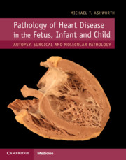
Pathology of Heart Disease in the Fetus, Infant and Child
Autopsy, Surgical and Molecular Pathology
, pp. 118 - 154
Publisher: Cambridge University Press
Print publication year: 2019

Access options

Get access to the full version of this content by using one of the access options below. (Log in options will check for institutional or personal access. Content may require purchase if you do not have access.)

References

Frescura, C, Thiene, G. The new concept of univentricular heart. Front Pediatr 2014; 2: 62.CrossRefGoogle ScholarPubMed
Anderson, RH, Becker, AE, Tynan, M et al. The univentricular atrioventricular connection: getting to the root of a thorny problem. Am J Cardiol 1984; 54: 822828.Google Scholar
Meyer, SL, Jongbloed, MR, Ho, SY et al. Intracardiac anatomical relationships and potential for streaming in double inlet left ventricles. PLoS ONE 2017; 12: e0188048.Google Scholar
Kurosawa, H, Arai, T, Imai, Y, Matsumura, G. Ventricular septation for double inlet left ventricle. World J Pediatr Congenit Heart Surg 2012; 3: 337343.Google Scholar
Anderson, RH, McCarthy, K, Cook, AC. Double outlet right ventricle. Cardiol Young 2001; 11: 329344.Google Scholar
Yu, FF, Lu, B, Gao, Y, et al. Congenital anomalies of coronary arteries in complex congenital heart disease: diagnosis and analysis with dual-source CT. J Cardiovasc Comput Tomogr. 2013; 7: 383390.Google Scholar
Brown, JW, Ruzmetov, M, Okada, Y, Vijay, P, Turrentine, MW. Surgical results in patients with double outlet right ventricle: a 20-year experience. Ann Thorac Surg 2001; 72: 16301635.Google Scholar
Schwarz, F, Blaschczok, HC, Sinzobahamvya, N et al. The Taussig-Bing anomaly: long-term results. Eur J Cardiothorac Surg 2013; 44: 821827.Google Scholar
DeLisle, G, Ando, M, Calder, AL et al. Total anomalous pulmonary venous connection: report of 93 autopsied cases with emphasis on diagnostic and surgical considerations. Am Heart J 1976; 91: 99122.Google Scholar
Neill, CA, Ferencz, C, Sabiston, DC, Sheldon, H. The familial occurrence of hypoplastic right lung with systemic arterial supply and venous drainage: “scimitar syndrome”. Johns Hopkins Med J 1960; 107: 115.Google Scholar
Yoshimura, N, Fukahara, K, Yamashita, A et al. Current topics in surgery for isolated total anomalous pulmonary venous connection. Surg Today 2014; 44: 22212226.Google Scholar
Sun, C-CJ, Doyle, T, Ringel, RE. Pulmonary vein stenosis. Hum Pathol 1995; 26: 880886.CrossRefGoogle ScholarPubMed
Fong, LV, Anderson, RH, Park, SC, Zuberbuhler, JR. Morphologic features of stenosis of the pulmonary veins. Am J Cardiol 1988; 62: 11361138.Google Scholar
Anderson, RK, Lie, JT. Pathologic anatomy of Ebstein’s anomaly of the heart revisited. Am J Cardiol 1978; 41: 739745.Google Scholar
Walsh, EP. Interventional electrophysiology in patients with congenital heart disease. Circulation 2007; 115: 32243234.Google Scholar
Sharland, G, Tingay, R, Jones, A, Simpson, J. Atrioventricular and ventriculoarterial discordance (congenitally corrected transposition of the great arteries): echocardiographic features, associations, and outcome in 34 fetuses. Heart 2005; 91: 14531458.CrossRefGoogle ScholarPubMed
Chauvaud, SM, Mihaileanu, SA, Gaer, JAR, Carpentier, AC. Surgical treatment of Ebstein’s malformation – the “Hôpital Broussais” experience. Cardiol Young 2006; 6: 411.Google Scholar
Rigby, ML, Carvalho, JS, Anderson, RH et al. The investigation and diagnosis of tricuspid atresia. Int J Cardiol 1990; 27: 117.CrossRefGoogle ScholarPubMed
McMahon, CJ, Nihill, MR, Kovalchin, JP, Lewin, MB. Echocardiographic features of Chiari’s network in association with tricuspid atresia. Tex Heart Inst J 2000; 27: 312313.Google ScholarPubMed
Wilder, TJ, Ziemer, G, Hickey, EJ, et al. Surgical management of competing pulmonary blood flow affects survival before Fontan/Kreutzer completion in patients with tricuspid atresia type I. J Thorac Cardiovasc Surg 2015; 150: 12221230.Google Scholar
Anderson, RH, Silverman, NH, Zuberbuhler, JR. Congenitally unguarded tricuspid orifice: its differentiation from Ebstein’s malformation in association with pulmonary atresia and intact ventricular septum. Pediatr Cardiol 1990; 11: 8690.Google Scholar
Rosenquist, GC, Sweeny, LJ, McAllister, HA. Relationship of the tricuspid valve to the membranous septum in Down’s syndrome without endocardial cushion defect: study of 28 specimens, 14 with a ventricular septal defect. Am Heart J 1975; 90: 458462.CrossRefGoogle Scholar
Uhl, HSM. A previously undescribed congenital malformation of the heart: almost total absence of the myocardium of the right ventricle. Bull Johns Hopkins Hosp 1952; 91: 197209.Google ScholarPubMed
Dixon, DB, Mackey-Bojack, SM, Sivanandam, S. Uhl’s anomaly: perspective of fetal echocardiography and histopathological correlation. Cardiol Young 2017; 27: 388390.Google Scholar
Corazza, G, Soliani, M, Bava, GL. Uhl’s anomaly in a newborn. Eur J Pediatr 1981; 137: 347352.Google Scholar
Gerlis, LM, Schmidt-Ott, SC, Ho, SY, Anderson, RH. Dysplastic conditions of the right ventricular myocardium: Uhl’s anomaly v arrhythmogenic right ventricular dysplasia. Br Heart J 1993; 69: 142150.Google Scholar
Kalita, JP, Dutta, N, Awasthy, N et al. Surgical options for Uhl’s anomaly. World J Pediatr Congenit Heart Surg 2017; 8: 470474.Google Scholar
Yim, D, Nagata, H, Lam, CZ et al. Disharmonious patterns of heterotaxy and isomerism: how often are the classic patterns breached? Circ Cardiovasc Imaging 2018; 11: e006917.Google Scholar
Macartney, FJ, Zuberbuhler, JR, Anderson, RH. Morphological considerations pertaining to recognition of atrial isomerism. Consequences for sequential chamber localisation. Br Heart J 1980; 44: 657667.Google Scholar
Melhuish, BPP, Van Pragh, R. Juxtaposition of the atrial appendages. A sign of severe cyanotic congenital heart disease. Br Heart J 1968; 30: 269284.Google Scholar
Frescura, C, Thiene, G. Juxtaposition of the atrial appendages. Cardiovascular Pathol 2012; 21: 169179.Google Scholar
Buirsky, G, Jordan, SC, Joffe, HS, Wilde, P. Superior vena caval abnormalities: their occurrence rate, associated cardiac abnormalities and angiographic classification in a paediatric population with congenital heart disease. Clin Radiol 1986; 37: 131138.Google Scholar
Molz, G, Burri, B. Aberrant subclavian artery (arteria lusoria): sex differences in the prevalence of various forms of the malformation. Evaluation of 1378 observations. Virchows Arch A Pathol Anat Histol 1978; 380: 303315.CrossRefGoogle ScholarPubMed
Salomonowitz, E, Edwards, JE, Hunter, DW et al. The three types of aortic diverticula. AJR Am J Roentgenol. 1984; 142: 673679.CrossRefGoogle ScholarPubMed
Tanaka, A, Milner, R, Ota, T. Kommerell’s diverticulum in the current era: a comprehensive review. Gen Thorac Cardiovasc Surg 2015; 63: 245259.Google Scholar
Kim, KM, Cambria, RP, Isselbacher, EM et al. Contemporary surgical approaches and outcomes in adults with Kommerell diverticulum. Ann Thorac Surg 2014; 98: 13471354.Google Scholar
Backer, CL, Russell, HM, Wurlitzer, KC, Rastatter, JC, Rigsby, CK. Primary resection of Kommerell diverticulum and left subclavian artery transfer. Ann Thorac Surg 2012; 94: 16121617.Google Scholar
Cantrell, JR, Haller, JA, Ravitch, MM. A syndrome of congenital defects involving the abdominal wall, sternum, diaphragm, pericardium and heart. Surg Gynecol Obstet 1958; 107: 602614.Google Scholar
Aryal, MR, Hakim, FA, Ghimire, S et al. Left atrial appendage aneurysm: a systematic review of 82 cases. Echocardiography 2014; 31: 13121318.CrossRefGoogle ScholarPubMed
Van der Hauwaert, LG, Dumoulin, M, Daenen, W, Stalpaert, G. Aneurysm of the left atrial appendage. Clin Cardiol 1979; 2: 4951.Google Scholar
Moaddab, A, Tonni, G, Grisolia, G et al. Predicting outcome in 259 fetuses with agenesis of ductus venosus: a multicenter experience and systematic review of the literature. J Matern Fetal Neonatal Med 2016; 29: 36063614.Google Scholar
Volpe, P, Marasini, M, Caruso, G et al. Prenatal diagnosis of ductus venosus agenesis and its association with cytogenetic/congenital anomalies. Prenat Diagn 2002; 22: 9951000.Google Scholar
Strizek, B, Zamprakou, A, Gottschalk, I et al. Prenatal diagnosis of agenesis of ductus venosus: a retrospective study of anatomic variants, associated anomalies and impact on postnatal outcome. Ultraschall Med Sep 21 2017. doi: 10.1055/s-0043-115109 [Epub ahead of print].Google Scholar
Meyer, WW, Lind, J. The ductus venosus and the mechanism of its closure. Arch Dis Child 1966; 41: 597605.Google Scholar
Loberant, N, Herskovits, M, Barak, M et al. Closure of the ductus venosus in premature infants: findings on real-time gray-scale, color-flow Doppler, and duplex Doppler sonography. AJR Am J Roentgenol 1999; 172: 227229.Google Scholar
Franchi-Abella, S, Branchereau, S, Lambert, V et al. Complications of congenital portosystemic shunts in children: therapeutic options and outcomes. J Pediatr Gastroenterol Nutr 2010; 51: 322330.CrossRefGoogle ScholarPubMed
Papamichail, M, Pizanias, M, Heaton, N. Congenital portosystemic venous shunt. Eur J Pediatr 2018; 177: 285294.Google Scholar
Feldman, DN, Roman, MJ. Aneurysms of the sinuses of Valsalva. Cardiology 2006; 106: 7381.Google Scholar
Kawaguchi, A, Waki, K, Arakaki, Y, Baba, K. Huge aneurysms of the aortic sinuses of Valsalva with leaflet perforation in an infant: a case report. J Thorac Cardiovasc Surg 2010; 139: e54–6.Google Scholar
Moustafa, S, Mookadam, F, Cooper, L et al. Sinus of Valsalva aneurysms: 47 years of a single center experience and systematic overview of published reports. Am J Cardiol 2007; 99: 11591164.CrossRefGoogle ScholarPubMed
Lubaua, I, Priedite, I, Anderson, D. Giant sinus of Valsalva aneurysm in a foetus. Cardiol Young 2013; 23: 267268.Google Scholar
Perry, LW, Martin, GR, Galioto, FM Jr, Midgley, FM. Rupture of congenital sinus of Valsalva aneurysm in a newborn. Am J Cardiol 1991; 68: 12551256.Google Scholar
Takahashi, T, Koide, T, Yamaguchi, H, et al. Ehlers–Danlos syndrome with aortic regurgitation, dilation of the sinuses of Valsalva, and abnormal dermal collagen fibrilsAm Heart J 1992; 123: 17091712.Google Scholar
McKay, R. Aorto-ventricular tunnel. Orphanet J Rare Dis 2007; 2: 41.Google Scholar
McKay, R, Anderson, RH, Cook, AC. The aorto-ventricular tunnels. Cardiol Young 2002; 12: 563580.Google Scholar
Sadeghpour, A, Peighambari, M, Dalirrooyfard, M et al. Aorta-to-left ventricle tunnel associated with noncompaction left ventricle. J Am Soc Echocardiogr 2006; 19: 1073.e1–5.Google Scholar
Sousa-Uva, M, Touchot, A, Fermont, L, et al. Aortico-left ventricular tunnel in fetuses and infants.  Ann Thorac Surg 1996; 61: 18051810.Google Scholar
Terry, J, Tapas, M, Nowaczyk, MJ. Prenatal and pathologic features of aorto-left ventricular tunnel causing fetal hydrops and intrauterine demise. Pediatr Dev Pathol 2013; 16: 97101.Google Scholar
Bharucha, T, Spicer, DE, Mohun, TJ et al. Cor triatriatum or divided atriums: which approach provides the better understanding? Cardiol Young 2015; 25: 193207.Google Scholar
Krasemann, Z, Scheld, HH, Tjan, TD, Krasemann, T. Cor triatriatum: short review of the literature upon ten new cases. Herz 2007; 32: 506510.Google Scholar
Saxena, P, Burkhart, HM, Schaff, HV et al. Surgical repair of cor triatriatum sinister: the Mayo Clinic 50-year experience. Ann Thorac Surg 2014; 97: 16591663.Google Scholar
Ohlow, MA. Congenital left ventricular aneurysms and diverticula: definition, pathophysiology, clinical relevance and treatment. Cardiology 2006; 106: 6372.Google Scholar
Ohlow, MA, von Korn, H, Lauer, B. Characteristics and outcomes of congenital left ventricular aneurysm and diverticulum: analysis of 809 cases published since 1816. Int J Cardiol 2015; 185: 3445.Google Scholar
Kim, GB, Kim, WH, Min, BJ et al. Aneurysm of the left ventricular apex in a patient with pulmonary atresia, intact ventricular septum. J Am Coll Cardiol 2011; 57: e201.Google Scholar
Patel, AR, Farrel, P, Harris, M, Gaynor, JW, Gillespie, MJ. Acquired ventricular septal aneurysm in a patient with pulmonary atresia with intact ventricular septum. Cardiol Young 2012; 22: 227229.Google Scholar
Wagenvoort, CA, Mooi, WJ. Biopsy Pathology of the Pulmonary Vasculature. London: Chapman and Hall Medical; 1989.Google Scholar
Simonneau, G, Gatzoulis, MA, Adatia, I et al. Updated clinical classification of pulmonary hypertension. J Am Coll Cardiol 2013; 62: D34–41.Google Scholar
Tuder, RM, Abman, SH, Braun, T et al. Development and pathology of pulmonary hypertension. J Am Coll Cardiol 2009; 54 (1 Suppl): S3–9.Google Scholar
Heath, D, Edwards, JE. The pathology of hypertensive pulmonary vascular disease. A description of six grades of structural changes in the pulmonary arteries with special reference to congenital cardiac septal defects. Circulation 1958; 18: 533547.CrossRefGoogle ScholarPubMed
Hansmann, G. Pulmonary hypertension in infants, children, and young adults. J Am Coll Cardiol 2017; 69: 25512569.Google Scholar
Kaza, AK, Gruber, PJ. Surgical approaches for CHD: and update on success and challenges. Curr Opin Pediatr 2013; 25: 591596.CrossRefGoogle ScholarPubMed
Diller, GP, Kempny, A, Alonso-Gonzalez, R et al. Survival prospects and circumstances of death in contemporary adult congenital heart disease patients under follow-up at a large tertiary centre. Circulation 2015; 132: 21182125.Google Scholar
Baruteau, AE, Hascoët, S, Baruteau, J et al. Transcatheter closure of patent ductus arteriosus: past, present and future. Arch Cardiovasc Dis 2014; 107: 122132.Google Scholar
Holzer, RJ, Hijazi, ZM. Transcatheter pulmonary valve replacement: State of the art. Catheter Cardiovasc Interv 2016; 87: 117128.Google Scholar
Crystal, MA, Ing, FF. Pediatric interventional cardiology: 2009. Curr Opin Pediatr 2010; 22: 567572.Google Scholar
Donofrio, MT, Moon-Grady, AJ, Hornberger, LK et al.; American Heart Association Adults With Congenital Heart Disease Joint Committee of the Council on Cardiovascular Disease in the Young and Council on Clinical Cardiology, Council on Cardiovascular Surgery and Anesthesia, and Council on Cardiovascular and Stroke Nursing. Diagnosis and treatment of fetal cardiac disease: a scientific statement from the American Heart Association. Circulation 2014; 129: 21832242.Google Scholar
Kapadia, MR, Popowich, DA, Kibbe, MR. Modified prosthetic vascular conduits. Circulation 2008; 17: 18731882.Google Scholar
Petrucci, O, O’Brien, SM, Jacobs, ML et al. Risk factors for mortality and morbidity after the neonatal Blalock-Taussig shunt procedure. Ann Thorac Surg 2011; 92: 642651.Google Scholar
DeLeon, SY, Ilbawi, MN, Tubeszewski, K, Wilson, WR Jr, Idriss, FS. The Damus-Stansel-Kaye procedure: anatomical determinants and modifications. Ann Thorac Surg 1991; 52: 680687.Google Scholar
Ohye, RG, Schranz, D, D’Udekem, Y. Current therapy for hypoplastic left heart syndrome and related single ventricle lesions. Circulation 2016; 134: 12651279.Google Scholar
van der Ven, JPG, van den Bosch, E, Bogers, AJCC, Helbing, WA. State of the art of the Fontan strategy for treatment of univentricular heart disease. F1000Res. 2018 Jun 27;7. pii: F1000 Faculty Rev-935.Google Scholar
Brown, JW, Ruzmetov, M, Vijay, P, Rodefeld, MD, Turrentine, MW. The Ross–Konno procedure in children: outcomes, autograft and allograft function, and reoperations. Ann Thorac Surg 2006; 82: 13011306.Google Scholar
Mookhoek, A, de Heer, E, Bogers, AJ, Takkenberg, JJ, Schoof, PH. Pulmonary autograft valve explants show typical degeneration. J Thorac Cardiovasc Surg 2010; 139: 14161419.Google Scholar
Konno, S, Imai, J, Iida, Y, Nakajima, M, Tatsuno, K. A new method for prosthetic valve replacement in congenital aortic stenosis associated with hypoplasia of the aortic valve ring. J Thorac Cardiovasc Surg 1975; 70: 909917.Google Scholar
Lo Rito, M, Fittipaldi, M, Haththotuwa, R, et al. Long-term fate of the aortic valve after an arterial switch operation. J Thorac Cardiovasc Surg 2015; 149: 10891094.Google Scholar
Cleveland, DC, Kirklin, JK, Pavnica, JW et al. Late left ventricular outflow tract obstruction following the Rastelli operation: expectations out to 20 years. World J Pediatr Congenit Heart Surg 2016; 7: 605610.Google Scholar
Baysa, SJ, Olen, M, Kanter, RJ. Arrhythmias following the Mustard and Senning operations for dextro-transposition of the great arteries: clinical aspects and catheter ablation. Card Electrophysiol Clin 2017; 9: 255271.CrossRefGoogle ScholarPubMed
Haeffele, C, Lui, GK. Dextro-transposition of the great arteries: long-term sequelae of atrial and arterial switch. Cardiol Clin 2015; 33: 543558.Google Scholar
Schranz, D, Rupp, S, Müller, M et al. Pulmonary artery banding in infants and young children with left ventricular dilated cardiomyopathy: a novel therapeutic strategy before heart transplantation. J Heart Lung Transplant 2013; 32: 475481.Google Scholar
Butany, J, Collins, MJ. Analysis of prosthetic cardiac devices: a guide for the practising pathologist. J Clin Pathol 2005; 58: 113124.Google Scholar
Lee, AH, Gallagher, PJ. Post-mortem examination after cardiac surgery. Histopathology 1998; 33 : 399405.CrossRefGoogle ScholarPubMed
Hickling, MF, Pontefract, DE, Gallagher, PJ, Livesey, SA. Post mortem examinations after cardiac surgery. Heart 2007; 93: 761765.Google Scholar
Allwork, SP. Pathological Correlation after Cardiac Surgery. London: Butterworth Heinemann; 1991.Google Scholar

Save book to Kindle

To save this book to your Kindle, first ensure coreplatform@cambridge.org is added to your Approved Personal Document E-mail List under your Personal Document Settings on the Manage Your Content and Devices page of your Amazon account. Then enter the ‘name’ part of your Kindle email address below. Find out more about saving to your Kindle.

Note you can select to save to either the @free.kindle.com or @kindle.com variations. ‘@free.kindle.com’ emails are free but can only be saved to your device when it is connected to wi-fi. ‘@kindle.com’ emails can be delivered even when you are not connected to wi-fi, but note that service fees apply.

Find out more about the Kindle Personal Document Service.

Available formats
×

Save book to Dropbox

To save content items to your account, please confirm that you agree to abide by our usage policies. If this is the first time you use this feature, you will be asked to authorise Cambridge Core to connect with your account. Find out more about saving content to Dropbox.

Available formats
×

Save book to Google Drive

To save content items to your account, please confirm that you agree to abide by our usage policies. If this is the first time you use this feature, you will be asked to authorise Cambridge Core to connect with your account. Find out more about saving content to Google Drive.

Available formats
×