Skip to main content Accessibility help
×
Hostname: page-component-8448b6f56d-wq2xx Total loading time: 0 Render date: 2024-04-23T18:50:04.634Z Has data issue: false hasContentIssue false

Chapter 7 - Optical coherence tomography and electrophysiology of the visual pathway

Published online by Cambridge University Press:  05 May 2015

Peter A. Calabresi
Affiliation:
Department of Neurology, Johns Hopkins University Hospital, Baltimore
Laura J. Balcer
Affiliation:
Department of Neurology, NYU Langone Medical Center, New York
Elliot M. Frohman
Affiliation:
Department of Neurology, UT Southwestern Medical Center, Dallas
Get access

Summary

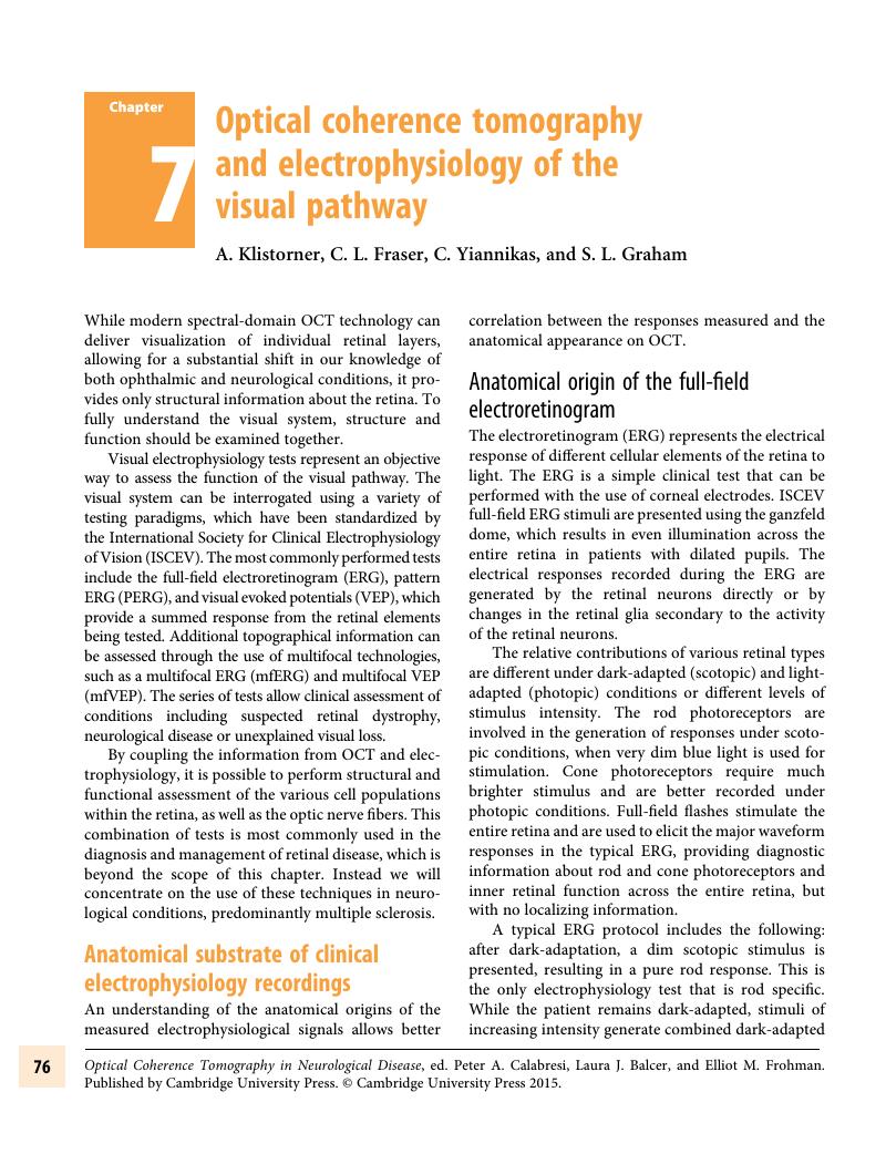
Image of the first page of this content. For PDF version, please use the ‘Save PDF’ preceeding this image.'
Type
Chapter
Information
Publisher: Cambridge University Press
Print publication year: 2015

Access options

Get access to the full version of this content by using one of the access options below. (Log in options will check for institutional or personal access. Content may require purchase if you do not have access.)

References

Holder, GE. Pattern electroretinography (PERG) and an integrated approach to visual pathway diagnosis. Prog Ret Eye Res 2001; 20: 531–61.CrossRefGoogle Scholar
Holder, GE, Gale, RP, Acheson, JF, Robson, AG. Electrodiagnostic assessment in optic nerve disease. Curr Opin Neurol 2009; 22: 310.CrossRefGoogle ScholarPubMed
Halliday, AM, McDonald, WI, Mushin, J. Delayed visual evoked response in optic neuritis. Lancet 1972; i: 982–5.Google Scholar
Fahle, M, Bach, M. Origin of the visual evoked potentials. In: Heckenlively, JR, Arden, GB, editors. Principles and practice of clinical electrophysiology of vision. Cambridge: The Mit Press 2006. p. 207234.Google Scholar
Jones, SJ, Brusa, A. Neurophysiological evidence for long-term repair of MS lesions: implications for axon protection. J Neurolog Sci 2003; 206: 193–8.CrossRefGoogle ScholarPubMed
Waxman, SG. Altered distributions and functions of multiple sodium channel subtypes in multiple sclerosis and its models. In: Waxman, SG, editor. Multiple sclerosis as a neuronal disease. Amsterdam: Elsevier, 2005.Google Scholar
Klistorner, A, Graham, SL, Fraser, C, Garrick, R, Nguyen, T, Paine, M, et al. Electrophysiological evidence for heterogeneity of lesions in optic neuritis. Invest Ophthalmol Vis Sci. 2007; 48: 4549–56.CrossRefGoogle ScholarPubMed
Brusa, A, Jones, SJ, Plant, GT. Long-term remyelination after optic neuritis. Brain. 2001; 124: 468–79.CrossRefGoogle ScholarPubMed
Yiannikas, C, Walsh, JC. The variation of the pattern shift visual evoked response with the size of the stimulus field. Electroencephalography & Clinical Neurophysiology 1983; 55: 427435.CrossRefGoogle ScholarPubMed
Halliday, AM, Darbett, G, Blumhardt, LD, Kriss, A. The macular and submacular subcomponents of the pattern evoked response. In: LDaG, B, editor. Human evoked potentials. New York: Plenum Publishing; 1979. p. 135451.CrossRefGoogle Scholar
Klistorner, AI, Graham, SL, Grigg, JR, Billson, FA. Multifocal topographic visual evoked potential: improving objective detection of local visual field defects. Invest Ophthalmol Vis Sci. 1998; 39(6): 937–50.Google ScholarPubMed
Klistorner, A, Fraser, C, Garrick, R, Graham, SL, Arvind, H. Correlation between full-field and multifocal VEPs in optic neuritis. Doc Ophthal. 2008; 116: 1927.CrossRefGoogle ScholarPubMed
Hood, DC, Odel, JG, Zhang, X. Tracking the recovery of local optic nerve function after optic neuritis: a multifocal VEP study. Invest Ophthalmol Vis Sci 2000; 41: 4032–8.Google ScholarPubMed
Graham, SL, Klistorner, A, Goldberg, I. Clinical Application of objective perimetry using multifocal VEP in Glaucoma Practice. Arch Ophthalmol 2005; 123: 729–39.CrossRefGoogle ScholarPubMed
Laron, M, Cheng, H, Xhang, B, Schiffman, JS, Tang, RA, Frishman, LJ. Assessing visual pathway function in multiple sclerosis patients with multifocal visual evoked potentials. Mult Scler 2009; 15: 1431–41.CrossRefGoogle ScholarPubMed
Fredericksen, JL, Petrera, J. Serial visual evoked potentials in 90 untreated patients with acute optic neuritis. Surv Ophthalmol 1999; 44: S5462.CrossRefGoogle Scholar
Ghezzi, A, Martinelli, V, Torri, V et al. Long-term follow-up of isolated optic neuritis: the risk of developing multiple sclerosis, its outcome, and the prognostic role of paraclinical tests. J Neurol 1999; 246: 770–5.CrossRefGoogle ScholarPubMed
Hickman, SJ, Toosy, AT, , SJJ, Altman, DR, Mizszkiel, KA, MacManus, DG, et al. A serial MRI study following optic nerve mean area in acute optic neuritis. Brain 2004; 127: 2498–505.CrossRefGoogle ScholarPubMed
Youl, BD, Turano, G, Miller, DH, Towell, AD, MacManus, DG, Moore, SG, et al. The pathophysiology of acute optic neuritis. An association of gadolinium leakage with clinical and electrophysiological deficits. Brain 1991; 114: 2437–50.Google ScholarPubMed
Miller, DH. Natural history of multiple sclerosis: when do axons degenerate? In: Waxman, SG, editor. Multiple sclerosis as a neuronal disease. Amsterdam: Elsevier 2005. p. 185200.CrossRefGoogle Scholar
Frohman, EM, Fujimoto, JG, Frohman, TC, Calabresi, PA, Cutter, GR, Balcer, LJ. Optical coherence tomography: a window into the mechanisms of multiple sclerosis. Nature Neurol 2008; 4: 664775.Google ScholarPubMed
Parisi, V, Manni, G, Centofanti, M, Gandolfi, SA, Olzi, D, Bucci, MG. Correlation between optical coherence tomography, pattern electroretinogram, and visual 7evoked potentials in open-angle glaucoma patients. Ophthalmology 2001; 108(5): 905–12.CrossRefGoogle Scholar
Trip, A, Schlottmann, PG, Jones, SJ, Altman, DR, Garway-Heath, DF, Thompson, AJ, et al. Retinal nerve fiber layer loss and visual disfunction in optic neuritis. Ann Neurol 2005; 58: 383–91.CrossRefGoogle Scholar
Pueyo, V, Martin, J, Fernandez, J, Almarcegui, C, Ara, J, Egea, C, et al. Axonal loss in the retinal fiber layer in patients with multiple sclerosis. Mult Scer 2008; 14: 609–14.Google ScholarPubMed
Almarcegui, C, Dolz, I, Pueyo, V, Garcia, E, Fernandez, FJ, Martin, J, et al. Correlation between functional and structural assessments of the optic nerve and retina in multiple sclerosis patients. Clin Neurophysiol 2010; 40: 129–35.CrossRefGoogle ScholarPubMed
Klistorner, A, Arvind, H, Nguyen, T, Garrick, R, Paine, M, Graham, S, et al. Inflammation, demyelination and axonal loss in post-acute optic neuritis. Ann Neurol 2008; 61: 325–31.Google Scholar
Klistorner, A, Arvind, H, Nguyen, T, Garrick, R, Paine, M, Graham, S, et al. Multifocal VEP and OCT in optic neuritis: a topographical study of the structure–function relationship. Doc Ophthal 2009; 118: 129–37.CrossRefGoogle ScholarPubMed
Smith, KJ, Waxman, SG. The conduction properties of demyelinated and remyelinated axons. In: Waxman, SG, editor. Multiple sclerosis as neuronal disease. Amsterdam: Elsevier Academic Press; 2005, p. 85100.CrossRefGoogle Scholar
Lassmann, H. New Concepts on progressive multiple sclerosis. Cur Neurol Neurosci Reports 2007; 7: 239–44.Google ScholarPubMed
Foster, RE, Whalen, CC, Waxman, SG. Reorganisation of the axonal membrane of demyelinated nerve fibres: morphological evidence. Science 1980; 210: 661–3.CrossRefGoogle Scholar
Bostock, H, Sears, TA. The internodal axon membrane: electrical excitability and continious conduction in segmental demyelination. J Physiol (London) 1978; 280: 273301.CrossRefGoogle Scholar
Shrager, P, Simon, W, Kazarinova-Noyes, K. Na+ channel reorganisation in demyelinated axons. In: SG, W, editor. Multiple sclerosis as a neuronal disease. Amsterdam: Elsevier; 2005.Google Scholar
Peterson, J, Kidd, D, Trapp, BD. Axonal degeneration in multiple sclerosis: the histopathological evidence. In: Waxman, SG, editor. Multiple sclerosis as a neuronal disease. Amsterdam: Elsevier; 2005. p. 165184.CrossRefGoogle Scholar
Pro, MJ, Pons, ME, Liebmann, JM, Ritch, R, Zafar, S, Lefton, D, et al. Imaging of the optic disk and retinal nerve fiber layer in acute optic neuritis. J Neurolog Sci. 2006; 250: 114–9.CrossRefGoogle ScholarPubMed
Petzold, A, de Boer, JF, Schippling, S, Vermersch, P, Kardon, R, Green, A, et al. Optical coherence tomography in multiple sclerosis: a systematic review and meta-analysis. The Lancet (Neurol) 2010; 9: 921–32.Google ScholarPubMed
Talman, LS, Bisker, ER, Sackel, BS, Long, DA, Galetta, KM, Ratchford, JN, et al. Longitudinal study of vision and retinal nerve fiber layer thickness in multiple sclerosis. Ann Neurol 2010; 67: 749–60.CrossRefGoogle ScholarPubMed
Klistorner, A, Arvind, H, Garrick, R, Graham, SL, Paine, M, Yiannikas, C. Interrelationship of optical coherence tomography and multifocal visual-evoked potentials after optic neuritis. Invest Ophthalmol Vis Sci 2010; 51(5): 2770–7.CrossRefGoogle ScholarPubMed
Klistorner, A, Garrick, R, Paine, M, Graham, SL, Arvind, H, Van Der Walt, A, et al. Relationship between chronic demyelination of the optic nerve and short term axonal loss. J Neurol Neurosurg Psychiatry. 2011; Epub ahead of print.CrossRefGoogle Scholar
Klistorner, A, Garrick, R, Barnett, MH, Graham, SL, Arvind, H, Sriram, P, et al. Axonal loss in non-optic neuritis eyes of patients with multiple sclerosis linked to delayed visual evoked potential. Neurol 2013; 15: 242–5.Google Scholar
Bruck, W. Inflammatory demyelination is not central to the pathogenesis of multiple sclerosis. J Neurol. 2005; 252(Suppl 5): V/10-V/5.Google ScholarPubMed
Patrikios, P, Stadelmann, C, Kutzelnigg, A, Rauschka, H, Schmidbauer, M, Laursen, H, et al. Remyelination is extensive in a subset of multiple sclerosis patients. Brain 2006; 129: 3165–72.CrossRefGoogle Scholar
Prineas, JW, Barnard, RO, Kwon, EE, Sharer, LR, Cho, ES. Multiple sclerosis: remyelination of nascent lesions. Ann Neurol 1993; 33: 137–51.CrossRefGoogle ScholarPubMed
Prineas, JW, Kwon, EE, Goldenberg, PZ, Ilyas, AA, Quarles, RH, Benjamins, JA, et al. Multiple sclerosis. Oligodendrocyte proliferation and differentiation in fresh lesions. Lab Invest 1989; 61: 489503.Google ScholarPubMed
Blakemore, WF, Chari, DM, Gilson, JM, Crang, AJ. Modelling large areas of demyelination in the rat reveals the potential and possible limitations of transplanted glial cells for remyelination in the CNS. Glia 2002; 38: 155–68.CrossRefGoogle ScholarPubMed
Klistorner, A, Arvind, H, Garrick, R, Yiannikas, C, Graham, SL. Remyelination of optic nerve lesions: spatial and temporal factors. Mult Scler 2010; 16: 786–95.CrossRefGoogle ScholarPubMed
Klistorner, A, Arvind, H, Nguyen, T, Garrick, R, Paine, M, Graham, S, et al. Axonal loss and myelin in early ON loss in postacute optic neuritis. Ann Neurol 2008; 64(3): 325–31.CrossRefGoogle ScholarPubMed
You, Y, Klistorner, A, Thie, J, Graham, SL. Latency delay of visual evoked potential is a real measurement of demyelination ina rat model of optic neuritis. Invest Ophthalmol Vis Sci 2011; 52: 6911–8.CrossRefGoogle Scholar
Raz, N, Chokron, S, Ben-Hur, T, Levin, N. Temporal reorganization to overcome monocular demyelination. Neurology. August 20, 2013; 81(8): 702–9. doi: 10.1212/WNL.0b013e3182a1aa3e. Epub July 19, 2013. PubMed PMID: 23873970.CrossRefGoogle ScholarPubMed
Costello, F, Hodge, W, Pan, YI, Eggenberger, E, Coupland, S, Kardon, R. Tracking retinal nerve fiber layer loss after optic neuritis: a prospective study using optical coherence tomography. Mult Scler 2008; 14: 893905.CrossRefGoogle ScholarPubMed
Hickman, SJ, Brierley, CMH, Brex, PA, MacManus, DG, Scolding, NJ, Compston, DAS, et al. Continuing optic nerve atrophy following optic neuritis: a serial MRI study. Multiple sclerosis 2002; 8: 339–42.CrossRefGoogle ScholarPubMed
Toosy, AT, Hickman, SJ, Mizszkiel, KA, Jones, SJ, Plant, GT, Altman, DR, et al. Adaptive cortical plasticity in higher visual areas after optic neuritis. Ann Neurol 2005; 57: 622–33.CrossRefGoogle ScholarPubMed
Raz, N, Dotan, S, Benoliel, T, Chokron, S, Ben-Hur, T, Levin, N. Sustained motion perception deficit following optic neuritis: Behavioral and cortical evidence. Neurology. June 14, 2011; 76(24): 2103–11. doi: 10.1212/WNL.0b013e31821f4602. PubMed PMID: 21670440.CrossRefGoogle ScholarPubMed
Korsholm, K, Madsen, KH, Fredericksen, JL, Skimmingr, A, Lund, TE. Recovery from optic neuritis: anROI-based analysis of LGN and visual cortical areas. Brain 2007; 130: 1244–53.CrossRefGoogle ScholarPubMed
Werring, DJ, Bullmore, ET, Toosy, AT, Miller, DH, Barker, GJ, MacManus, DG, et al. Recovery from optic neuritis is associated with a change in the distribution of cerebral response to visual stimulation: a functional magnetic resonance imaging study. Journal of Neurology, Neurosurgery & Psychiatry 2000; 68(4): 441–9.CrossRefGoogle ScholarPubMed
Archibald, NK, Clarke, MP, Mosimann, UP, Burn, DJ. The retina in Parkinson’s disease. Brain 2009; 132: 1128–45.CrossRefGoogle ScholarPubMed
Tagliati, M, Bodis-Wollner, I, Yahr, MD. The pattern electroretinogram in Parkinson’s disease reveals lack of retinal spatial tuning. Electroencephalogr Clin Neurophysiol 1996; 100: 111.CrossRefGoogle ScholarPubMed
Kirbas, S, Turkyilmaz, K, Tufekci, A, Durmus, M. Retinal nerve fiber layer thickness in Parkinson disease. J Neuroophthalmol 2013; 33: 62–t5.Google ScholarPubMed
Adam, CR, Shrier, E, Ding, Y, Glazman, S, Bodis-Wollner, I. Correlation of inner retinal thickness evaluated by spectral-domain optical coherence tomography and contrast sensitivity in Parkinson disease. J Neuroophthalmol 2013.CrossRefGoogle Scholar
Nowacka, B, Lubinski, W, Karczewicz, D. Ophthalmological and electrophysiological features of Parkinson’s disease. Klin Oczna 2010; 112: 247–52.Google ScholarPubMed
Moschos, MM, Tagaris, G, Markopoulos, I, Margetis, I, Tsapakis, S, Kanakis, M, et al. Morphologic changes and functional retinal impairment in patients with Parkinson disease without visual loss. 2011; 21: 2429.Google Scholar
Kirbas, S, Turkyilmaz, K, Anlar, O, Tufekci, A, Durmus, M. Retinal nerve fiber layer thickness in patients with Alzheimer disease. J Neuroophthalmol 2013; 33: 5861.CrossRefGoogle ScholarPubMed
Moschos, MM, Markopoulos, I, Chatziralli, I, Rouvas, A, Papageorgiou, SG, Ladas, I, et al. Structural and functional impairment of the retina and optic nerve in Alzheimer’s disease. Curr Alzheimer Res 2012; 9: 782–8.CrossRefGoogle ScholarPubMed
Meier-Ruge, W. Drug induced retinopathy. CRC Toxicol 1972; 1: 325–60.Google Scholar
Lyons, JS, Severns, ML. Using multifocal ERG ring ratios to detect and follow Plaquenil retinal toxicity: a review. Doc Ophthalmol 2009; 118: 2936.CrossRefGoogle ScholarPubMed
Marmor, MF, Kellner, U, Lai, TY, Lyons, JS, Mieler, WF. Revised recommendations on screening for chloroquine and hydroxychloroquine retinopathy. Ophthalmology 2011; 118: 415–22.CrossRefGoogle ScholarPubMed
Kjellstrom, U, Andreasson, S, Ponjavic, V. Attenuation of the retinal nerve fibre layer and reduced retinal function assessed by optical coherence tomography and full-field electroretinography in patients exposed to vigabatrin medication. Acta Ophthalmol 2013.CrossRefGoogle Scholar
Tjoa, CW, Benedict, RH, Dwyer, MG, Carone, DA, Zivadinov, R. Regional specificity of magnetization transfer imaging in multiple sclerosis. J Neuroimaging April 2008; 18(2): 130–6. doi: 10.1111/j.1552-6569.2007.00198.x.CrossRefGoogle ScholarPubMed
Gallo, A, Rovaris, M, Benedetti, B, Sormani, MP, Riva, R, Ghezzi, A, et al. A brain magnetization transfer MRI study with a clinical follow up of about four years in patients with clinically isolated syndromes suggestive of multiple sclerosis. J Neurol. January 2007; 254(1): 7883. Epub February 14, 2007. PubMed PMID: 17508141.CrossRefGoogle ScholarPubMed
Tortorella, P, Rocca, MA, Mezzapesa, DM, Ghezzi, A, Lamantia, L, Comi, G, et al. MRI quantification of gray and white matter damage in patients with early-onset multiple sclerosis. J Neurol. July 2006; 253(7): 903–7. Epub March 6, 2006. PubMed PMID: 16511645.CrossRefGoogle ScholarPubMed
Deloire-Grassin, MS, Brochet, B, Quesson, B, Delalande, C, Dousset, V, Canioni, P, et al. In vivo evaluation of remyelination in rat brain by magnetisation transfer imaging. J Neurol Sci 2000; 178: 1016.CrossRefGoogle Scholar
Zaaraoui, W, Deloire, M, Merie, M, Girard, C, Raffard, G, Biran, M, et al. Monitoring demyelination and remyelination by magnetization transfer imaging in the mouse brain at 9.4 T. Magn Res Mat Phys. 2008; 21: 357–62.Google Scholar
Klistorner, A, Chaganti, J, Garrick, R, Moffat, K, Yiannikas, C. Magnetisation transfer ratio in optic neuritis is associated with axonal loss, but not with demyelination. Neuroimage 2011; 56(1): 21–6.Google Scholar

Save book to Kindle

To save this book to your Kindle, first ensure coreplatform@cambridge.org is added to your Approved Personal Document E-mail List under your Personal Document Settings on the Manage Your Content and Devices page of your Amazon account. Then enter the ‘name’ part of your Kindle email address below. Find out more about saving to your Kindle.

Note you can select to save to either the @free.kindle.com or @kindle.com variations. ‘@free.kindle.com’ emails are free but can only be saved to your device when it is connected to wi-fi. ‘@kindle.com’ emails can be delivered even when you are not connected to wi-fi, but note that service fees apply.

Find out more about the Kindle Personal Document Service.

Available formats
×

Save book to Dropbox

To save content items to your account, please confirm that you agree to abide by our usage policies. If this is the first time you use this feature, you will be asked to authorise Cambridge Core to connect with your account. Find out more about saving content to Dropbox.

Available formats
×

Save book to Google Drive

To save content items to your account, please confirm that you agree to abide by our usage policies. If this is the first time you use this feature, you will be asked to authorise Cambridge Core to connect with your account. Find out more about saving content to Google Drive.

Available formats
×