Skip to main content Accessibility help
×
Hostname: page-component-76fb5796d-9pm4c Total loading time: 0 Render date: 2024-04-27T00:00:51.842Z Has data issue: false hasContentIssue false

9 - Mature T-Cell Neoplasms and Natural Killer-Cell Malignancies

Published online by Cambridge University Press:  01 February 2018

Anna Porwit
Affiliation:
Lunds Universitet, Sweden
Marie Christine Béné
Affiliation:
Université de Nantes, France
Get access

Summary

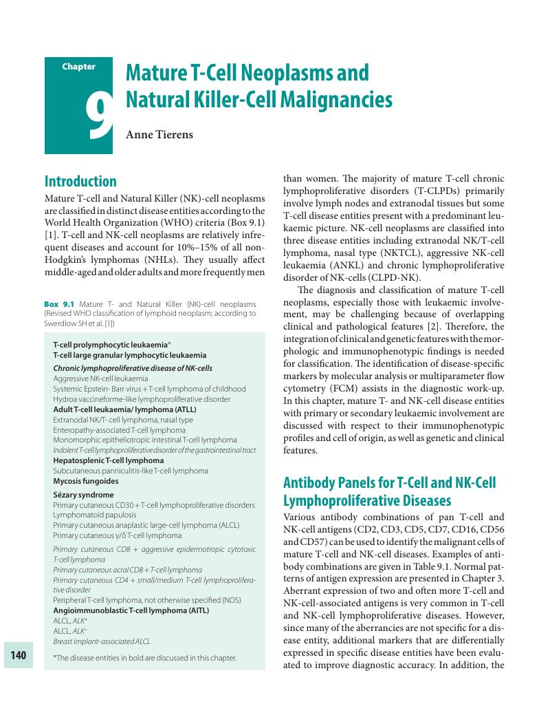
Image of the first page of this content. For PDF version, please use the ‘Save PDF’ preceeding this image.'
Type
Chapter
Information
Publisher: Cambridge University Press
Print publication year: 2018

Access options

Get access to the full version of this content by using one of the access options below. (Log in options will check for institutional or personal access. Content may require purchase if you do not have access.)

References

Swerdlow, S.H., Campo, E., Pileri, S.A., et al. The 2016 revision of the World Health Organization classification of lymphoid neoplasms. Blood, 127 (2016), 2375–90.CrossRefGoogle ScholarPubMed
Herling, M., Khoury, J.D., Washinghton, L.T., et al. A systematic approach to diagnosis of mature T-cell leukemias reveals heterogeneity among WHO categories. Blood, 104 (2004), 328–35.CrossRefGoogle Scholar
Aggarwal, N., Fischer, J., Swerdlow, S.H. and Craig, F.E.. Splenic lymphoid subsets with less-recognized phenotypes mimic aberrant antigen expression. Am J Clin Pathol, 140 (2013), 787–94.CrossRefGoogle Scholar
van Dongen, J.J., Lhermitte, L., Böttcher, S., et al. Euroflow antibody panels for standardized n-dimensional flow cytometric immunophenotyping of normal, reactive and malignant leukocytes. Leukemia, 26 (2012), 1908–75.CrossRefGoogle Scholar
Baseggio, L., Traverse-Giehen, A., Berger, F., et al. CD10 and ICOS expression by multiparameter flow cytometry in angioimmunoblastic T-cell lymphoma. Mod Pathol, 24 (2011), 9931003.CrossRefGoogle Scholar
Chen, X. and Cherian, S.. Immunophenotypic characterization of T-cell prolymphocytic leukemia. Am J Clin Pathol, 140 (2013), 727–35.CrossRefGoogle ScholarPubMed
Porwit, A.. Immunophenotyping of selected hematologic disorders-focus on lymphoproliferative disorders with more than one malignant cell population. Int J Lab Hematol, 35 (2013), 275–82.CrossRefGoogle Scholar
Loghavi, S., Wang, S.A., Medeiros, J., et al. Immunophenotypic and diagnostic characterization of angioimmunoblastic T-cell lymphoma by advanced flow cytometric technology. Leuk Lymphoma, 57 (2016), 2804–12.CrossRefGoogle ScholarPubMed
Lima, M., Spínola, A., Fonseca, S., et al. Aggressive mature natural killer cell neoplasms, report on a series of 12 European patients with emphasis on flow cytometry based immunophenotype and DNA content of neoplastic natural killer cells. Leuk Lymphoma, 56 (2015), 103–12.CrossRefGoogle Scholar
Kalina, T., Flores-Montero, J., van der Velden, V.H.J., et al. EuroFlow Consortium (EU-FP6, LSHB-CT-2006-018708). Euroflow standardization of flow cytometer instrument settings and immunophenotyping protocols. Leukemia, 26 (2012), 19862010.CrossRefGoogle ScholarPubMed
Costa, E.S., Pedreira, C.E., Barrena, S., et al. Automated pattern-guided principal component analysis vs expert-based immunophenotypic classification of B-cell chronic lymphoproliferative disorders, a step forward in the standardization of clinical immunophenotyping. Leukemia, 24 (2010), 1927–33.CrossRefGoogle ScholarPubMed
Pedreira, C.E., Costa, E.S., Almeida, J., et al. EuroFlow Consortium. A probabilistic approach for the evaluation of minimal residual disease by multiparameter flow cytometry in leukemic B-cell chronic lymphoproliferative disorders. Cytometry A, 73A (2008), 1141–50.CrossRefGoogle Scholar
Pedreira, C.E., Costa, E.S., Barrena, S., et al. EuroFlow Consortium. Generation of flow cytometry data files with a potentially infinite number of dimensions. Cytometry A, 73A (2008), 834–46.CrossRefGoogle Scholar
Lima, M., Almeida, J., Montero, A.G., et al. Clinicobiological, immunophenotypic, and molecular characterization of monoclonal CD56-/+dim chronic natural killer cell large granular lymphocytosis. Am J Pathol, 165 (2004), 1117–27.CrossRefGoogle Scholar
Lima, M., Almeida, J., dos Anjos Teixera, M., et al. The “ex vivo” patterns of CD2/CD7, CD57/CD11c, CD38/CD11b, CD45RA/CD45RO and CD11a/HLA-DR expression identify acute/early and chronic/late NK-cell activation states. Blood Cells Mol Dis, 28 (2002), 181–90.CrossRefGoogle Scholar
Kelemen, K., Guitart, J., Kuzel, T.M., et al. The usefulness of CD26 in flow cytometric analysis PB in Sézary syndrome. Am J Clin Pathol, 129 (2008), 146–56.CrossRefGoogle Scholar
Sokolowska-Wojdylo, M., Wenzel, J., Gaffal, E., et al. Absence of CD26 expression on skin-homing CLA+ CD4+ T lymphocytes in PB is a highly sensitive marker for early diagnosis and therapeutic monitoring of patients with Sézary syndrome. Clin Exp Dermatol, 30 (2005), 702–6.CrossRefGoogle Scholar
Novelli, M., Fava, P., Sarda, C., et al. Blood flow cytometry in Sézary syndrome. New insights on prognostic relevance and immunophenotypic changes during follow-up. Am J Clin Pathol, 143 (2015), 5769.CrossRefGoogle ScholarPubMed
Moins-Teisserenc, H., Daubord, M., Clave, E., et al. CD158k is a reliable marker for diagnosis of Sézary syndrome and reveals an unprecedented heterogeneity of circulating malignant cells. J Invest Dermatol, 135 (2015), 247–57.CrossRefGoogle ScholarPubMed
Shao, H., Yuan, C.M., Xi, L., et al. Minimal residual disease detection by flow cytometry in adult T-cell leukemia/lymphoma. Am J Clin Pathol, 133 (2010), 592601.CrossRefGoogle ScholarPubMed
Tian, Y., Kobayashi, S., Ohno, N., et al. Leukemic T cells are specifically enriched in a unique CD3 (dim) CD7 (low) subpopulation of CD4 (+) T cells in adult-type T-cell leukemia. Cancer Sci, 102 (2011), 569–77.CrossRefGoogle Scholar
Singh, A., Schabath, R., Ratei, R., et al. PB sCD3-CD4+ T cells, a useful diagnostic tool in angioimmunoblastic T cell lymphoma. Hematol Oncol, 32 (2014), 1621.CrossRefGoogle Scholar
Marafioti, T., Paterson, J.C., Ballabio, E., et al. The inducible T-cell co-stimulator molecule is expressed on subsets of T-cells and is a new marker of lymphomas of T follicular helper cell-derivation. Haematologica, 95 (2010), 432–39.CrossRefGoogle Scholar
Lundell, R., Hartung, L., Hill, S., et al. T-cell large granular lymphocyte leukemias have multiple phenotypic abnormalities involving pan T-cell antigens and receptors for MHC molecules. Am J Clin Pathol, 124 (2005), 937–46.CrossRefGoogle ScholarPubMed
Morice, W.G., Kurtin, P.J., Leibson, P.J., et al. Demonstration of aberrant T-cell and natural killer-cell antigen expression in all cases of granular lymphocytic leukemia. Br J Haematol, 120 (2003), 1026–36.CrossRefGoogle Scholar
Lima, M., Almeida, J., Dos Anjos Teixera, M., et al. TCRalphabeta+/CD4+ large granular lymphocytosis, a new clonal T-cell lymphoproliferative disorder. Am J Pathol, 163 (2003), 763–71.Google ScholarPubMed
Chen, Y.H., Chadburn, A., Evens, A.M., et al. Clinical, morphologic, immunophenotypic, and molecular cytogenetic assessment of CD4-/CD8-γδ T-cell large granular lymphocytic leukemia. Am J Clin Pathol, 36 (2011), 289–99.Google Scholar
Chan, J.K.C., Quintanilla-Martinez, L., Ferry, J.A. and Peg, S.C.. Extranodal NK/T- cell lymphoma, nasal type. In, Swerdlow, SH, Campo, E, Harris, NL, Jaffe, ES, Pileri, SA, Stein, H, Thiele, J, Vardiman, JW, editors. Pathology and Genetics of Tumours of Haematopoietic and Lymphoid Tissues. (4th ed. Lyon, France, IARC Press, 2009), 285–8.Google Scholar
Morice, W.G., Jevremovic, D., Olteanu, H., et al. Chronic lymphoproliferative disorders of natural killer cells, a distinct entity with subtypes correlating with normal natural killer subsets. Leukemia, 24 (2010), 881–4.CrossRefGoogle Scholar
Langerak, A.W., Beemd, R. van Den, Wolvers-Tettero, I.L., et al. Molecular and flow cytometric analysis of the Vbeta repertoire for clonality assessment in mature TCRalphabeta T-cell proliferations. Blood, 98 (2001), 165–73.CrossRefGoogle ScholarPubMed
Lima, M., Almeida, J., Santos, A.H., et al. Immunophenotypic analysis of the TCR-Vbeta repertoire in 98 persistent expansions of CD3 (+) TCR-alphabeta (+) large granular lymphocytes, utility in assessing clonality and insights into the pathogenesis of the disease. Am J Pathol, 159 (2001), 1861–8.CrossRefGoogle ScholarPubMed
Beck, R.C., Tubbs, R.R., Hussein, M., et al. Detection of mature T-cell leukemias by flow cytometry using anti-T-cell receptor V beta antibodies. Am J Clin Pathol, 120 (2003), 785–94.CrossRefGoogle Scholar
Morice, W.G., Kimlinger, T., Katzmann, J.A., et al. Flow cytometric assessment of TCR-Vb expression in the evaluation of PB involvement by T-cell lymphoproliferative disorders. Am J Clin Pathol, 121 (2004), 373–83.CrossRefGoogle Scholar
Tembhare, P., Yuan, C.M., Xi, L., et al. Flow cytometric immunophenotypic assessment of T-cell clonality by Vβ repertoire analysis, detection of T-cell clonality at diagnosis and monitoring of minimal residual disease following therapy. Am J Clin Pathol, 135 (2011), 890900.CrossRefGoogle ScholarPubMed
Scala, E., Abeni, D., Pomponi, D., et al. Single TCR-Vbeta evaluation discloses the circulating T-cell clone in Sezary syndrome, one family fits all. Arch Dermatol Res, 307 (2015), 487–93.CrossRefGoogle ScholarPubMed
Isobe, M., Russo, G., Haluska, F.G., et al. Cloning of the gene encoding the delta subunit of the human T-cell receptor reveals its physical organization within the alpha-subunit locus and its involvement in chromosome translocations in T-cell malignancy. Proc Natl Acad Sci U S A, 85 (1988), 3933–7.CrossRefGoogle ScholarPubMed
Russo, G., Isobe, M., Gatti, R., et al. Molecular analysis of a t(14,14) translocation in leukemic T-cells of an ataxia telangiectasia patient. Proc Natl Acad Sci U S A, 96 (1989), 602–6.Google Scholar
Madani, A., Choukroun, V., Soulier, J., et al. Expression of p13MTCP1 is restricted to mature T-cell proliferation with t(X,14) translocations. Blood, 87 (1996), 1923–7.CrossRefGoogle Scholar
Catovsky, D., Muller-Hermelink, H.K. and Ralfkiaer, E.. T prolymphocytic leukemia. In, Swerdlow, SH, Campo, E, Harris, NL, Jaffe, ES, Pileri, SA, Stein, H, Thiele, J, Vardiman, JW, editors. Pathology and Genetics of Tumours of Haematopoietic and Lymphoid Tissues. (4th ed. Lyon, France, IARC Press, 2009), 270–1.Google Scholar
Thick, J., Metcalfe, J.A., Mak, Y.F., et al. Expression of either the TCL1 oncogene, or transcripts from its homologue MTCP1/c6.1B, in leukaemic and non-leukaemic T cells from ataxia telangiectasia patients. Oncogene, 12 (1996), 379–86.Google ScholarPubMed
Brito-Babapulle, V. and Catovsky, D.. Inversions and tandem translocations involving chromosome 14q11 and 14q32 in T- prolymphocytic leukemia and T-cell leukemias in patients with ataxia telangiectasia. Cancer Genet Cytogenet, 55 (1991), 19.CrossRefGoogle Scholar
Delgado, P., Starshak, P., Rao, N., et al. A comprehensive update on molecular and cytogenetic abnormalities in T-cell prolymphocytic leukemia. J Assoc Genet Technol, 38 (2012), 193–8.Google Scholar
Stengel, A., Kern, W., Zenger, M., et al. Genetic characterization of T-PLL reveals two major biologic subgroups and JAK3 mutations as prognostic marker. Genes, Chromosomes & Cancer, 55 (2016), 8294.CrossRefGoogle ScholarPubMed
Kiel, M.J., Velusamy, T., Rolland, D., et al. Integrated genomic sequencing reveals mutational landscape of T-cell prolymphocytic leukemia. Blood, 124 (2014), 1460–72.CrossRefGoogle ScholarPubMed
Matutes, E., Brito-Babapulle, V., Swansbury, J., et al. Clinical and laboratory features of 78 cases of T-prolymphocytic leukemia. Blood, 78 (1991), 3269–74.CrossRefGoogle ScholarPubMed
De Schouwer, P.J., Dyer, M.J., Brito-Babapulle, V., et al. T-cell prolymphocytic leukemia, antigen receptor gene rearrangement and a novel mode of MTCP1 B1 activation. Br J Haematol, 110 (2000), 831–8.CrossRefGoogle Scholar
Foucar, K.. Mature T-cell leukemias including T-prolymphocytic, adult T-cell leukemia/lymphoma, and Sézary syndrome. Am J Clin Pathol, 127 (2007), 496510.CrossRefGoogle ScholarPubMed
Herling, M., Patel, K.A., Teitell, M.A., et al. High TCL1 expression and intact T-cell receptor signaling define a hyperproliferative subset of T-cell prolymphocytic leukemia. Blood, 111 (2008), 328–37.CrossRefGoogle Scholar
Uchiyama, T., Yodoi, J., Sagawa, K., et al. Adult T-cell leukemia, clinical and hematologic features of 16 cases. Blood, 50 (1977), 481–92.CrossRefGoogle Scholar
Ohshima, K., Jaffe, E.S. and Kikuchi, M.. Adult T-cell leukemia/ lymphoma. In, Swerdlow, SH, Campo, E, Harris, NL, Jaffe, ES, Pileri, SA, Stein, H, Thiele, J, Vardiman, JW, editors. Pathology and Genetics of Tumours of Haematopoietic and Lymphoid Tissues. (4th ed. Lyon, France, IARC Press, 2009), 281–4.Google Scholar
Takemoto, S., Mulloy, J.C., Cereseto, A., et al. Proliferation of adult T cell leukemia/lymphoma cells is associated with the constitutive activation of JAK/STAT proteins. Proc Natl Acad Sci U S A, 94 (1997), 13897–902.CrossRefGoogle ScholarPubMed
Grassmann, R., Aboud, M. and Jeang, K.T.. Molecular mechanism of cellular transformation by HTLV-1 Tax. Oncogene, 24 (2005), 5976–85.CrossRefGoogle ScholarPubMed
Baydoun, H.H., Pancewicz, J. and Nicot, C.. Human T-lymphotropic type 1 virus p30 inhibits homologous recombination and favors unfaithful DNA repair. Blood, 177 (2011), 5897–906.Google Scholar
Kataoka, K., Nagata, Y., Kitanaka, A., et al. Integrated molecular analysis of adult T-cell leukemia/lymphoma. Nat Genet, 47 (2015), 1304–15.CrossRefGoogle Scholar
Nagata, Y., Kontani, K., Enami, T., et al. Variegated RHOA mutations in adult T-cell leukemia/lymphoma. Blood, 93 (2016), 1318.Google Scholar
Oshiro, A., Tagawa, H., Ohshima, K., et al. Identification of subtype-specific genomic alterations in aggressive adult T-cell leukemia/lymphoma. Blood, 127 (2006), 596604.Google Scholar
Hatta, Y., Yamada, Y., Tomonaga, M., et al. Detailed deletion mapping of the long arm of chromosome 6 in adult T-cell leukemia. Blood, 93 (1999), 813–16.CrossRefGoogle Scholar
Matutes, E.. Adult T-cell leukemia. J Clin Pathol, 60 (2007), 1373–7.Google Scholar
Karube, T., Ohshima, K., Tsuchiya, T., et al. Expression of FoxP3, a key molecule in CD24CD25 regulatory T cells, in adult T-cell leukaemia/lymphoma cells. Br J Haematol, 126 (2004), 81–4.CrossRefGoogle ScholarPubMed
Roncador, G., Garcia, J.F., Garcia, J.F., et al. FOXP3, a selective marker for a subset of adult T-cell leukaemia/lymphoma. Leukemia, 19 (2005), 2247–53.CrossRefGoogle ScholarPubMed
Yokote, T., Akioka, T., Oka, S., et al. Flow cytometric immunophenotyping of adult T-cell leukemia/lymphoma using CD3 gating. Am J Clin Pathol, 124 (2005), 199204.CrossRefGoogle ScholarPubMed
Ralfkiaer, E., Cerroni, L., Sander, C.A., et al. Mycosis fungoides. In, Swerdlow, SH, Campo, E, Harris, NL, Jaffe, ES, Pileri, SA, Stein, H, Thiele, J, Vardiman, JW, editors. Pathology and Genetics of Tumours of Haematopoietic and Lymphoid Tissues. (4th ed. Lyon, France, IARC Press, 2009), 296–8.Google Scholar
Ralfkiaer, E., Willemze, R. and Whittaker, S.L.. Sézary syndrome. In, Swerdlow, SH, Campo, E, Harris, NL, Jaffe, ES, Pileri, SA, Stein, H, Thiele, J, Vardiman, JW, editors. Pathology and Genetics of Tumours of Haematopoietic and Lymphoid Tissues. (4th ed. Lyon, France, IARC Press, 2009), 299.Google Scholar
Olsen, E., Vonderheid, E., Pimpinelli, N., et al. Revisions to the staging and classification of mycosis fungoides and Sézary syndrome, a proposal of the International Society for Cutaneous Lymphomas (ISCL) and the cutaneous lymphoma task force of the European Organization of Research and Treatment of Cancer (EORTC). Blood, 110 (2007), 1713–22.CrossRefGoogle Scholar
Mao, X., Lillington, D., Scarisbrick, J.J., et al. Molecular cytogenetic analysis of cutaneous T-cell lymphomas, identification of common genetic alterations in Sézary syndrome and mycosis fungoides. Br J Dermatol, 147 (2002), 464–75.CrossRefGoogle ScholarPubMed
Batista, D.A., Vonderheid, E.C., Hawkins, A., et al. Multicolor fluorescence in situ hybridization (SKY) in mycosis fungoides and Sézary syndrome search for recurrent chromosome abnormalities. Genes Chromosomes Cancer, 45 (2006), 383–91.CrossRefGoogle ScholarPubMed
Vermeer, M.H., van Doorn, R., Dijkman, R., et al. Novel and highly recurrent chromosomal alterations in Sézary syndrome. Cancer Res, 68 (2008), 2689–98.CrossRefGoogle Scholar
da Silva Almeida, A.C., Abate, F., Khiabanian, H., et al. The mutational landscape of cutaneous T cell lymphoma and Sézary syndrome. Nat Genet, 47 (2015), 1465–70.CrossRefGoogle Scholar
Ungewickell, A., Bhaduri, A., Rios, E., et al. Genomic analysis of mycosis fungoides and Sézary syndrome identifies recurrent alterations in TNFR2. Nat Genet, 47 (2015), 1056–60.CrossRefGoogle ScholarPubMed
Kiel, M.J., Sahasrabuddhe, A., Rolland, D.C., et al. Genomic analyses reveal recurrent mutations in epigenetic modifiers and the JAK-STAT pathway in Sézary syndrome. Nat Commun, 6 (2015), 8470.CrossRefGoogle ScholarPubMed
Wang, L., Ni, X., Covington, K.R., et al. Genomic profiling of Sézary syndrome identifies alterations of key T cell signaling and differentiation genes. Nat Genet, 47 (2015), 1426–34.CrossRefGoogle Scholar
Sokolowska-Wojdylo, M., Wenzel, J., Gaffal, E., et al. Circulating clonal CLA(+) and CD4(+) T cells in Sézary syndrome express the skin-homing chemokine receptors CCR4 and CCR10 as well as the lymph node-homing receptor CCR7. Br J Dermatol, 152 (2005), 258–64.CrossRefGoogle Scholar
Campbell, J.J., Clark, R.A., Watanabe, R. and Kupper, T.S.. Sézary syndrome and mycosis fungoides arise from distinct T-cell subsets, a biologic rational for their distinct clinical behaviors. Blood, 116 (2010), 767–71.CrossRefGoogle Scholar
Wood, G.S., Hong, S.R., Sasaki, D.T., et al. Leu-8/CD7 antigen expression by CD3+ T cells, comparative analysis of skin and blood in mycosis fungoides/ Sézary syndrome relative to normal blood values. J Am Acad Dermatol, 22 (1990), 602–7.CrossRefGoogle ScholarPubMed
Harmon, C.B., Witzig, T.E., Katzmann, J.A. and Pittelkow, M.R.. Detection of circulating T cells with CD3+CD7- immunophenotype in patients with benign and malignant lymphoproliferative dermatoses. J Am Acad Dermatol, 35 (1996), 404–10.CrossRefGoogle Scholar
Vonderheid, E.C., Bigler, R.D., Kotecha, A., et al. Variable CD7 expression on T cells in the leukemic phase of cutaneous T cell lymphoma (Sézary syndrome). J Invest Dermatol, 117 (2001), 654–62.CrossRefGoogle Scholar
Jones, D., Dang, N.H., Duvic, M., et al. Absence of CD26 expression is a useful marker for diagnosis of T-cell lymphoma in PB. Am J Clin Pathol, 115 (2001), 885–92.CrossRefGoogle Scholar
Bernengo, M.G., Novelli, M., Quaglino, P., et al. The relevance of CD4+CD26- subset in the identification of circulating Sézary cells. Br J Dermatol, 144 (2001), 125–35.CrossRefGoogle ScholarPubMed
Lima, M., Almeida, J., dos Anjos Teixeira, M., et al. Utility of flow cytometry immunophenotyping and DNA ploidy studies for diagnosis and characterization of blood involvement in CD4+ Sézary's syndrome. Haematologica, 88 (2003), 874–87.Google Scholar
Morice, W.G., Katzman, J.A., Pittelkow, M.R., et al. A comparison of morphologic features, flow cytometry, TCR-Vbeta and TCR-PCR in qualitatative and quantitative assessment of PB involvement by Sézary syndrome. Am J Clin Pathol, 125 (2006), 364–74.CrossRefGoogle Scholar
Hristov, A.C., Vonderheid, E.C. and Borowitz, M.J.. Simplified flow cytometric assessment in mycosis fungoides and Sézary syndrome. Am J Clin Pathol, 136 (2011), 944–53.CrossRefGoogle ScholarPubMed
Bagot, M., Moretta, A., Sivori, S., et al. CD4 (+) cutaneous T-cell lymphoma cells express the p140-killer immunoglobulin-like receptor. Blood, 97 (2001), 1388–91.CrossRefGoogle Scholar
Poszepczynska-Guigné, E., Schiavon, V., D'Incan, M., Ortonne, N. et al. CD158k/KIR3DL2 is a new phenotypic marker of Sézary cells, relevance for diagnosis and follow-up of Sézary syndrome. J Invest Dermatol, 122 (2004), 820–3.CrossRefGoogle ScholarPubMed
Bahler, D.W., Hartung, L., Hill, S., et al. CD158k/KIR3DL is a useful marker for identifying neoplastic T-cells in Sézary syndrome by flow cytometry. Cytometry B Clin Cytom, 74 (2008), 156–62.Google Scholar
Bouaziz, J.D., Remtoula, N., Bensussan, A., et al. Absolute CD3+CD158k+ lymphocyte count is reliable and more sensitive than cytomorphology to evaluate blood tumour burden in Sézary syndrome. Br J Dermatol, 162 (2010), 123–8.CrossRefGoogle Scholar
Scala, E., Abeni, D., Pomponi, D., et al. The role of 9-O-acetylated ganglioside D3 (CD60) and (alpha)4(beta)1)CD49d expression in predicting the survival of patients with Sézary syndrome. Haematologica, 95 (2010), 1905–12.CrossRefGoogle Scholar
Chung, J.S., Shiue, L.H., Duvic, M., et al. Sézary syndrome cells overexpress syndecan-4, bearing distinct heparan sulfate moieites that suppress T-cell activation by binding DC-HIL and trapping TGF-beta on the cell surface. Blood, 117 (2011), 3382–90.CrossRefGoogle Scholar
Wysocka, M., Kossenkov, A.V., Benoit, B.M., et al. CD164 and FCRL3 are highly expressed on CD4+CD26- T-cells in Sézary syndrome patients. J Invest Dermatol, 134 (2014), 229–36.CrossRefGoogle ScholarPubMed
Frizzera, G., Moran, E.M. and Rappaport, H.. Angioimmunoblastic lymphadenopathy with dysproteinemia. Lancet, 1 (1974), 1070–3.Google Scholar
Frizzera, G., Moran, E.M. and Rappaport, H.. Angio-immunoblastic lymphadenopathy, diagnosis and clinical course. Am J Med, 59 (1975), 803–18.CrossRefGoogle ScholarPubMed
Dogan, A., Gaulard, P., Jaffe, E.S., et al. Angioimmunoblastic T-cell lymphoma. In, Swerdlow, SH, Campo, E, Harris, NL, Jaffe, ES, Pileri, SA, Stein, H, Thiele, J, Vardiman, JW, editors. Pathology and Genetics of Tumours of Haematopoietic and Lymphoid Tissues. (4th ed. Lyon, France, IARC Press, 2009), 309–11.Google Scholar
de Leval, L., Rickman, D.S., Thielen, C., et al. The gene expression profile of nodal peripheral T-cell lymphoma demonstrates a molecular link between angioimmunoblastic T cell lymphoma (AITL) and follicular helper T (TFH) cells. Blood, 109 (2007), 4952–63.CrossRefGoogle Scholar
Piccaluga, P.P., Agostinelli, C., Califano, A., et al. Gene expression analysis of angioimmunoblastic lymphoma indicates derivation from T follicular helper cells and vascular endothelial growth factor deregulation. Cancer Res 67 (2007), 10703–10.CrossRefGoogle Scholar
Laurent, C., Fazilleau, N. and Brousset, P.. A novel subset of T-helper cells, follicular T-helper cells and their markers. Haematologica, 95 (2010), 356–8.CrossRefGoogle ScholarPubMed
Cairns, R.A., Iqbai, J., Lemonnier, F., et al. IDH2 mutations are frequent in angioimmunoblastic T-cell lymphoma. Blood, 119 (2012), 1901–3.CrossRefGoogle ScholarPubMed
Wang, C., McKeithan, T.W., Gong, Q., et al. IDH2R172 mutations define a unique subgroup of patients with angioimmunoblastic T-cell lymphoma. Blood, 126 (2015), 1741–52.Google ScholarPubMed
Serke, S., van Lessen, A., Hummel, M., et al. Circulating CD4+ T lymphocytes with intracellular but no surface CD3 antigen in five of seven patients consecutively diagnosed with angioimmunoblastic T+cell lymphoma. Cytometry, 42 (2000), 180–7.3.0.CO;2-0>CrossRefGoogle ScholarPubMed
Attygalle, A., Al-Jehani, R., Diss, T.C., et al. Neoplastic T cells in angioimmunoblastic T-cell lymphoma express CD10. Blood, 99 (2002), 627–33.CrossRefGoogle ScholarPubMed
Yuan, C.M., Vergilio, J.A., Zhao, X.F., et al. CD10 and BCL6 expression in the diagnosis of angioimmunoblastic T-cell lymphoma, utility of detecting CD10 T cells by flow cytometry. Hum Pathol, 36 (2005), 784–91.CrossRefGoogle Scholar
Baseggio, L., Berger, F., Morel, D., et al. Identification of circulating CD10 positive T cells in angioimmunoblastic T-cell lymphoma. Leukemia, 20 (2006), 296303.CrossRefGoogle ScholarPubMed
Stacchini, A., Demurtas, A., Aliberti, S., et al. The usefulness of flow cytometric CD10 detection in the differential diagnosis of peripheral T-cell lymphomas. Am J Clin Pathol, 128 (2007), 854–64.CrossRefGoogle ScholarPubMed
Chen, W., Kesler, M.V., Karandikar, N.J., et al. Flow cytometric features of angioimmunoblastic T-cell lymphoma. Cytometry B Clin Cytom, 70 (2006), 142–8.Google ScholarPubMed
Mao, Z.J., Surowiecka, M., Linden, M.A. and Singleton, TP. Abnormal immunophenotype of the T-cell receptor beta chain in follicular helper T cells and angioimmunoblastic T cell lymphoma. Cytometry B Clin Cytom, 88 (2015), 190–3.CrossRefGoogle ScholarPubMed
Roncador, G., García Verdes-Montenegro, J.F., Tedoldi, S., et al. Expression of two markers of germinal center T cells (SAP and PD-1) in angio-immunoblastic T-cell lymphoma. Haematologica, 92 (2007), 1059–66.CrossRefGoogle Scholar
Kadin, M.E., Kamoun, M. and Lamberg, J.. Erythrophagocytic T gamma lymphoma, a clinicopathologic entity resembling malignant histiocytosis. N Engl J Med, 304 (1981), 648–53.CrossRefGoogle ScholarPubMed
Farcet, J.P., Gaulard, P., Marolleau, J.P., et al. Hepatosplenic T-cell lymphoma, sinusal/ sinusoidal localization of malignant cells expressing the T-cell receptor gamma delta. Blood, 75 (1990), 2213–19.CrossRefGoogle ScholarPubMed
Gaulard, P., Bourquelot, P., Kanavaros, P., et al. Expression of the alpha/beta and gamma/delta T-cell receptors in 57 cases of peripheral T-cell lymphomas, identification of a subset of gamma/delta T-cell lymphomas. Am J Pathol, 137 (1990), 617–28.Google ScholarPubMed
Gaulard, P., Jaffe, E.S., Krenacs, L. and Macon, W.R.. Hepatosplenic T cell lymphoma. In, Swerdlow, SH, Campo, E, Harris, NL, Jaffe, ES, Pileri, SA, Stein, H, Thiele, J, Vardiman, JW, editors. Pathology and Genetics of Tumours of Haematopoietic and Lymphoid Tissues. (4th ed. Lyon, France, IARC Press, 2009), 292–93.Google Scholar
Alonsozana, E.L., Stamberg, J., Kumar, D., et al. Isochromosome 7q, the primary cytogenetic abnormality in hepatosplenic gamma delta T cell lymphoma. Leukemia, 11 (1997), 1367–72.CrossRefGoogle Scholar
Wlodarska, I., Martin-Garcia, N., Achten, R., et al. Fluorescence in situ hybridization study of chromosome 7 aberrations in hepatosplenic T-cell lymphoma, isochromosome 7q as a common abnormality accumulating in forms with features of cytologic progression. Genes Chromosomes Cancer, 33 (2002), 243–51.CrossRefGoogle Scholar
Vega, F., Medeiros, L.J. and Gaulard, P.. Hepatosplenic and other gammadelta T-cell lymphomas. Am J Clin Pathol, 127 (2007), 869–80.CrossRefGoogle ScholarPubMed
Haedicke, W., Ho, F.C., Chott, A., et al. Expression of CD94/NKG2A and killer immunoglobulin-like receptors in NK cells and a subset of extranodal cytotoxic T-cell lymphomas. Blood, 95 (2000), 3628–30.CrossRefGoogle Scholar
Morice, W.G., Macon, W.R., Dogan, A., et al. NK-cell associated receptor expression in hepatosplenic T-cell lymphoma, insights into pathogenesis. Leukemia, 20 (2006), 883–6.CrossRefGoogle ScholarPubMed
Loughran, T.P., Kadin, M.E., Starkebaum, G., et al. Leukemia of large granular lymphocytes, association with clonal chromosomal abnormalities and autoimmune neutropenia, thrombocytopenia, and hemolytic anemia. Ann Intern Med, 102 (1985), 169–75.CrossRefGoogle ScholarPubMed
Chan, W.C., Foucar, K., Morice, W.G. and Catovksy, D. T-cell large granular lymphocytic leukemia. In, Swerdlow, SH, Campo, E, Harris, NL, Jaffe, ES, Pileri, SA, Stein, H, Thiele, J, Vardiman, JW, editors. Pathology and Genetics of Tumours of Haematopoietic and Lymphoid Tissues. (4th ed. Lyon, France, IARC Press, 2009), 272–3.Google Scholar
Steinway, S.N., LeBlanc, F. and Loughran, T.P. Jr. The pathogenesis and treatment of large granular lymphocyte leukemia. Blood Reviews, 28 (2014), 8794.CrossRefGoogle ScholarPubMed
Sokol, L. and Loughran, T.P. Jr. Large granular lymphocyte leukemia. Oncologist, 11 (2006), 263–73.CrossRefGoogle ScholarPubMed
Rossi, D., Franceschetti, S., Capello, D., et al. Transient monoclonal expansion of CD8+/CD57+ T cell large granular lymphocytes after primary cytomegalovirus infection. Am J Hematol, 82 (2007), 1103–5.CrossRefGoogle ScholarPubMed
Wolniak, K.L., Goolsby, C.L., Chen, Y.H., et al. Expansion of a clonal CD8+CD57+ large granular lymphocyte population after autologous stem cell transplant in multiple myeloma. Am J Clin Pathol, 139 (2013), 231–41.CrossRefGoogle ScholarPubMed
Jerez, A., Clemente, M.J., Makishima, H., et al. STAT3 mutations unify the pathogenesis of chronic lymphoproliferative disorders of NK cells and T-cell large granular lymphocytic leukemia. Blood, 120 (2012), 3048–57.CrossRefGoogle Scholar
Koskela, H.L., Eldfors, S., Ellonen, P., et al. Somatic STAT3 mutations in large granular lymphocytic leukemia. N Engl J Med, 366 (2012), 1905–13.CrossRefGoogle ScholarPubMed
Rajala, H.L., Olson, T., Clemente, M.J., et al. The analysis of clonal diversity and therapy responses using STAT3 mutations as a molecular marker in large granular lymphocytic leukemia. Haematologica, 100 (2015), 91–9.CrossRefGoogle ScholarPubMed
Rajala, H.L., Eldfors, S., Kuusanmäki, H., et al. Discovery of STAT5b mutations in large granular lymphocytic leukemia. Blood, 121 (2013), 4541–50.CrossRefGoogle ScholarPubMed
Bareau, B., Rey, J., Hamidou, M., et al. Analysis of a French cohort of patients with large granular lymphocyte leukemia, a report on 229 cases. Haematologica, 95 (2010), 1534–41.CrossRefGoogle Scholar
Cambiaggi, A., Orengo, A.M., Meazza, R., et al. The natural killer-related receptor for HLA-C expressed on T cells from CD3+ lymphoproliferative disease of granular lymphocytes displays either inhibitory or stimulatory function. Blood, 87 (1996), 2369–75.CrossRefGoogle ScholarPubMed
Bigouret, V., Hoffmann, T., Arlettaz, L., et al. Monoclonal T-cell expansions in asymptomatic individuals and in patients with large granular leukemia consist of cytotoxic effector T cells expressing the activating CD94, NKG2 C/E and NKD2D killer receptors. Blood, 101 (2003), 3198–204.CrossRefGoogle Scholar
Epling-Burnette, P.K., Painter, J.S., Chaurasia, P., et al. Dysregulated NK receptor expression in patients with lymphoproliferative disease of granular lymphocytes. Blood, 103 (2004), 3431–9.CrossRefGoogle ScholarPubMed
Fischer, L., Hummel, M., Burmeister, T., et al. Skewed expression of natural-killer (NK)-associated antigens on lymphoproliferations of large granular lymphocytes (LGL). Hematol Oncol, 24 (2006), 7885.CrossRefGoogle ScholarPubMed
Sandberg, Y., Almeida, J., Gonzalez, M., et al. TCRgammadelta+ large granular lymphocyte leukemias reflect the spectrum of normal antigen-selected TCRgammadelta+ T-cells. Leukemia, 20 (2006), 505–13.CrossRefGoogle ScholarPubMed
Bourgault-Rouxel, A.S., Loughran, T.P. Jr., Zambello, R., et al. Clinical spectrum of gammadelta + T cell LGL leukemia, analysis of 20 cases. Leuk Res, 32 (2008), 45–8.CrossRefGoogle ScholarPubMed
Garrido, P., Ruiz-Cabello, F., Bárcena, P., et al. Monoclonal TCR-Vbeta13.1+/CD4+/NKa+/CD8-/+dim T-LGL lymphocytosis, evidence for an antigen-driven chronic T-cell stimulation origin. Blood, 109 (2007), 4890–8.CrossRefGoogle ScholarPubMed
Rodriguez-Caballero, A., Garcia-Montero, A.C., Barcena, P., et al. Expanded cells in monoclonal TCR-alphabeta+/CD4+/NKa+/CD8-/+ dim T-LGL lymphocytosis recognize hCMV antigens. Blood, 112 (2008), 4609–16.CrossRefGoogle ScholarPubMed
Villamor, N., Morice, W.G., Chan, W.C. and Foucar, K.. Chronic lymphoproliferative disorders of NK cells. In, Swerdlow, SH, Campo, E, Harris, NL, Jaffe, ES, Pileri, SA, Stein, H, Thiele, J, Vardiman, JW, editors. Pathology and Genetics of Tumours of Haematopoietic and Lymphoid Tissues. (4th ed. Lyon, France, IARC Press, 2009), 274–5.Google Scholar
Poullot, E., Zambello, R., Leblanc, F., et al. Chronic natural killer lymphoproliferative disorders, characteristics of an international cohort of 70 patients. Ann Oncol, 25 (2014), 2030–5.CrossRefGoogle ScholarPubMed
Gattazzo, C., Teramo, A., Passeri, F., et al. Detection of monoclonal T populations in patients with KIR-restricted chronic lymphoproliferative disorder of NK cells. Haematologica, 99 (2014), 1826–33.CrossRefGoogle ScholarPubMed
Mori, K.L., Egashira, M. and Oshimi, K.. Differentiation stage of natural killer cell-lineage lymphoproliferative disorders based on phenotypic analysis. Br J Haematol 115 (2001), 225–8.CrossRefGoogle ScholarPubMed
Zambello, R., Falco, M., Della Chiesa, M., et al. Expression and function of KIR and natural cytotoxicity receptors in NK-type lymphoproliferative diseases of granular lymphocytes. Blood, 102 (2003), 1797–805.CrossRefGoogle ScholarPubMed
Freud, A.G., Zhao, S., Wei, S., et al. Expression of the activating receptor, NKp46 (CD335) in human natural killer and T-cell neoplasia. Am J Clin Pathol, 140 (2013), 853–66.CrossRefGoogle ScholarPubMed
Barcena, P., Jara-Acevedo, M., Tabernero, M.D., et al. Phenotypic profile of expanded NK cells in chronic lymphoproliferative disorders, a surrogate marker for NK-cell clonality. Oncotarget, 6 (2015), 42938–51.CrossRefGoogle ScholarPubMed
Chan, J.K.C., Jaffe, E.S., Ralfkiaer, E. and Ko, Y.H.. Agressive NK-cell leukemia. In, Swerdlow, SH, Campo, E, Harris, NL, Jaffe, ES, Pileri, SA, Stein, H, Thiele, J, Vardiman, JW, editors. Pathology and Genetics of Tumours of Haematopoietic and Lymphoid Tissues. (4th ed. Lyon, France, IARC Press, 2009), 276–7.Google Scholar
Lima, M., Almeida, J., Teixera, M.A, et al. Reactive phenotypes after acute and chronic NK-cell activation. J Biol Regul Homeost Agents, 18 (2004), 331–4.Google ScholarPubMed
Krause, D.S., Delelys, M.E. and Preffer, F.I.. Flow cytometry for hematopoietic cells. Methods Mol Biol, 1109 (2014), 2346.CrossRefGoogle ScholarPubMed
Bendall, S.C., Nolan, G.P., Roederer, M. and Chattopadhyay, P.K.. A deep profiler's guide to cytometry. Trends Immunol, 33 (2012), 323–32.CrossRefGoogle ScholarPubMed

Save book to Kindle

To save this book to your Kindle, first ensure coreplatform@cambridge.org is added to your Approved Personal Document E-mail List under your Personal Document Settings on the Manage Your Content and Devices page of your Amazon account. Then enter the ‘name’ part of your Kindle email address below. Find out more about saving to your Kindle.

Note you can select to save to either the @free.kindle.com or @kindle.com variations. ‘@free.kindle.com’ emails are free but can only be saved to your device when it is connected to wi-fi. ‘@kindle.com’ emails can be delivered even when you are not connected to wi-fi, but note that service fees apply.

Find out more about the Kindle Personal Document Service.

Available formats
×

Save book to Dropbox

To save content items to your account, please confirm that you agree to abide by our usage policies. If this is the first time you use this feature, you will be asked to authorise Cambridge Core to connect with your account. Find out more about saving content to Dropbox.

Available formats
×

Save book to Google Drive

To save content items to your account, please confirm that you agree to abide by our usage policies. If this is the first time you use this feature, you will be asked to authorise Cambridge Core to connect with your account. Find out more about saving content to Google Drive.

Available formats
×