Skip to main content Accessibility help
×
Hostname: page-component-7c8c6479df-nwzlb Total loading time: 0 Render date: 2024-03-29T11:46:07.569Z Has data issue: false hasContentIssue false

9 - Transcranial insonation

Published online by Cambridge University Press:  05 May 2016

László Csiba
Affiliation:
Department of Neurology, Debreceni Egyetem, Hungary
Claudio Baracchini
Affiliation:
Department of Neuroscience, Università degli Studi di Padova, Italy
Get access

Summary

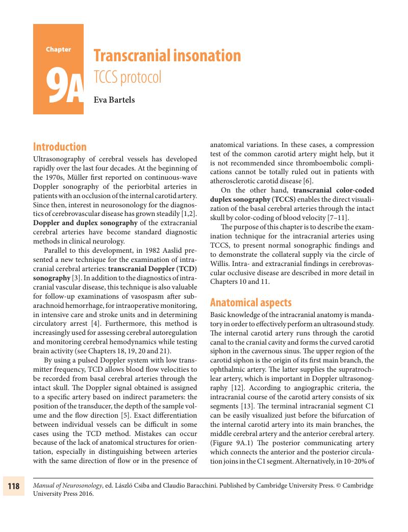
Image of the first page of this content. For PDF version, please use the ‘Save PDF’ preceeding this image.'
Type
Chapter
Information
Publisher: Cambridge University Press
Print publication year: 2016

Access options

Get access to the full version of this content by using one of the access options below. (Log in options will check for institutional or personal access. Content may require purchase if you do not have access.)

References

References

Müller, HR. Direktionell Doppler-Sonographie der A. frontalis medialis. EEG-EMG. 1971;2:2432.Google Scholar
Müller, HR. The diagnosis of internal carotid artery occlusion by directional Doppler sonography of the ophthalmic artery. Neurology. 1972;22:816823.Google Scholar
Aaslid, R, Markwalder, T-M, Nornes, H. Noninvasive transcranial Doppler ultrasound recording of velocity in basal cerebral arteries. J Neurosurg. 1982;57:769774.CrossRefGoogle ScholarPubMed
Reutern, GM. Zerebraler Zirkulationsstand. Diagnostik mit der Dopplersonographie. Dt. Ärzteblatt. 1991;49: 5565.Google Scholar
Arnolds, BJ, von Reutern, G-M. Transcranial Doppler sonography, examination technique and normal reference values. Ultrasound Med Biol. 1986;12: 115123.CrossRefGoogle ScholarPubMed
Khaffaf, N, Karnik, R, Winkler, W-B, Valentin, A. Embolic stroke by compression maneuver during transcranial Doppler sonography. Stroke. 1994;25:10561057.Google Scholar
Furuhata, H. New evolution of transcranial tomography (TCT) and transcranial color Doppler tomography (TCDT). Neurosonology. 1989;2:815.CrossRefGoogle Scholar
Schöning, M, Grunert, D, Stier, B. Transkranielle Duplexsonographie durch den intakten Knochen: Ein neues diagnostisches Verfahren. Ultraschall Med. 1989; 10:6671.CrossRefGoogle Scholar
Bogdahn, U, Becker, G, Winkler, J, et al. The transcranial colour coded real-time sonography in adults. Stroke. 1990;21:16801688.CrossRefGoogle ScholarPubMed
Bartels, E. Transcranial color-coded duplex ultrasonography in routine cerebrovascular diagnostics. In: Bartels, E, Bartels, S, Poppert, H, eds. New trends in neurosonology and cerebral hemodynamics – an update. Perspect Med. 2012;1:325330. http://dx.doi.org/10.1016/j.permed.2012.06.001CrossRefGoogle Scholar
Baumgartner, RW. Transcranial color duplex sonography in cerebrovascular disease: a systematic review. Cerebrovasc Dis. 2003;16:413.CrossRefGoogle ScholarPubMed
Bartels, E. Color-Coded Duplex Ultrasonography of the Cerebral Vessels: Atlas and Manual [Farbduplexsonographie der hirnversorgenden Gefäße: Atlas und Handbuch]. Stuttgart: Schattauer; 1999.Google Scholar
Valdueza, JM, Schreiber, SJ, Roehl, J, Klingebiel, R. Neurosonology and Neuroimaging of Stroke. New York: Thieme Stuttgart; 2008.CrossRefGoogle Scholar
Schlachetzki, F. Technical aspects of the clinical examination. In: Bogdahn, U, Becker, G, Schlachetzki, F, eds. Echoenhancers and Transcranial Color Duplex Sonography. Berlin: Blackwell; 1998, 100111.Google Scholar
Bartels, E, Fuchs, H-H, Flügel, KA. Color Doppler imaging of basal cerebral arteries. Examination technique and normal reference values. Angiology. 1995;10:877844.Google Scholar
Walter, U. Transcranial sonography of the cerebral parenchyma: update on clinically relevant applications. In: Bartels, E, Bartels, S, Poppert, H, eds. New trends in neurosonology and cerebral hemodynamics – an update. Perspect Med. 2012;1:334343. http://dx.doi.org/10.1016/j.permed.2012.02.014Google Scholar
Bartels, E. The axial imaging plane – the main domain of the transcranial color-coded duplex ultrasonography? Eur J Ultrasound. 2002;16:4757.CrossRefGoogle ScholarPubMed
Nedelmann, M, Stolz, E, Gerriets, T, et al. Consensus recommendations for transcranial color-coded duplex sonography for the assessment of intracranial arteries in clinical trials in acute stroke. Stroke. 2009; 40:32383244.CrossRefGoogle ScholarPubMed
Baumgartner, RW. Transcranial color-coded duplex sonography. J Neurol. 1999;246:637647.Google Scholar
Krejza, J, Baumgartner, RW. Clinical applications of transcranial color-coded duplex sonography. J Neuroimaging. 2004;14:215225.Google ScholarPubMed
Eggers, J, Pade, O, Rogge, A, Schreiber, SJ, Valdueza, JM. Transcranial color-coded sonography successfully visualizes all intracranial parts of the internal carotid artery using the combined transtemporal axial and coronal approach. AJNR Am J Neuroradiol. 2009; 30:15891593.CrossRefGoogle ScholarPubMed
Stolz, E. Ultrasound examination techniques of extra- and intracranial veins. In: Bartels, E, Bartels, S, Poppert, H, eds. New trends in neurosonology and cerebral hemodynamics – an update. Perspect Med. 2012;1:366370. http://dx.doi.org/10.1016/j.permed.2012.02.030Google Scholar
Kaps, M, Seidel, G, Bauer, T, Behrmann, B. Imaging if the intracranial vertebrobasilar system using colour-coded ultrasound. Stroke. 1992;23:15771582.Google Scholar
Schöning, M, Walter, J. Evaluation of the vertebrobasilar- posterior system by transcranial color duplex sonography in adults. Stroke. 1992;23: 12801286.Google Scholar
Becker, G, Lindner, A, Bogdahn, U. Imaging of the vertebrobasilar system by transcranial colour-coded real-time sonography. J Ultrasound Med. 1993;12:395401.CrossRefGoogle Scholar
Stolz, E, Nückel, M, Mendes, I, Gerriets, T, Kaps, M. Vertebrobasilar transcranial colour-coded duplex ultrasonography: improvement with echo enhancement. Am J Neuroradiol. 2002;23:10511054.Google Scholar
Bäuerle, J, Nedelmann, M. B-mode sonography of the optic nerve in neurological disorders with altered intracranial pressure. In: Bartels, E, Bartels, S, Poppert, H, eds. New trends in neurosonology and cerebral hemodynamics – an update. Perspect Med. 2012;1:404407. http://dx.doi.org/10.1016/j.permed.2012.01.001Google Scholar
Lovrencic-Huzjan, A, Sodec-Simicevic, D, Popovic, IM, et al. Ultrasonography of the optic nerve sheath in brain death. In: Bartels, E, Bartels, S, Poppert, H, eds. New trends in neurosonology and cerebral hemodynamics – an update. Perspect Med. 2012;1:414416. http://dx.doi.org/10.1016/j.permed.2012.02.060Google Scholar
Ertl, M, Altmann, M, Torka, E, et al. The retrobulbar spot sign in sudden blindness – Sufficient to rule out vasculitis? In: Bartels, E, Bartels, S, Poppert, H, eds. New trends in neurosonology and cerebral hemodynamics – an update. Perspect Med. 2012;1:408413. http://dx.doi.org/10.1016/j.permed.2012.02.048CrossRefGoogle Scholar
Bartels, E, Flügel, KA. Quantitative measurements of blood flow velocity in basal cerebral arteries with transcranial color Doppler imaging. J Neuroimag. 1994:4;7781.CrossRefGoogle Scholar
Martin, P, Evans, D, Naylor, A. Transcranial colour-coded sonography of the basal cerebral circulation. Reference data from 115 volunteers. Stroke. 1994;25:390396.CrossRefGoogle Scholar
Baumgartner, RW, Baumgartner, I, Mattle, HP, Schroth, G. Transcranial color-coded duplex sonography in the evaluation of collateral flow through the circle of Willis. Am J Neuroradiol. 1997;18:127133.Google Scholar
Stolz, E, Kaps, M, Kern, A, Babacan, S, Dorndorf, W. Transcranial colour-coded duplex sonography of intracranial veins and sinuses in adults. Reference data from 130 volunteers. Stroke. 1999;30:10701075.CrossRefGoogle Scholar
Zipper, SG, Stolz, E. Clinical application of transcranial colour-coded duplex sonography – review. Eur J Neurol. 2002;9:18.CrossRefGoogle ScholarPubMed
Grolimund, P, Seiler, RW. Age dependence of the flow velocity in the basal cerebral arteries: a transcranial Doppler ultrasound study. Ultrasound Med Biol. 1988;14:191198.CrossRefGoogle ScholarPubMed
Ringelstein, EB, Kahlscheurer, B, Niggermeyer, E, et al. Transcranial Doppler sonography: anatomical landmarks and normal velocity values. Ultrasound Med Biol. 1990;16:745761.Google Scholar
Droste, DW, Boehm, T, Ritter, MA, Dittrich, R, Ringelstein, EB. Benefit of echocontrast-enhanced transcranial arterial color-coded duplex ultrasound. Cerebrovasc Dis. 2005;20:332336.Google Scholar
Krejza, J, Swiat, M, Pawlak, MA, et al. Suitability of temporal bone acoustic window: conventional TCD versus transcranial color-coded duplex sonography. J Neuroimaging. 2007;17:311314.CrossRefGoogle ScholarPubMed
Kaps, M. Extra- und intrakranielle Farbduplexsonographie. Berlin: Springer; 1994.CrossRefGoogle Scholar
Delcker, A, Diener, H-C. 3D-Ultrasound of the carotid arteries. Eur J Ultrasound. 1994;1:337344.Google Scholar
Klötzsch, C, Bozzato, A, Lammers, G, Mull, M, Noth, J. Three-dimensonal transcranial color-coded sonography of cerebral aneurysms. Stroke. 1999;30: 22852290.CrossRefGoogle Scholar
Giller, CA. Is angle correction correct? J Neuroimaging. 1994;4:5152.CrossRefGoogle ScholarPubMed
Griewing, B, Becker, G. Vascular anatomy and flow velocities. In: Bogdahn, U, Becker, G, Schlachetzki, F, eds. Echoenhancers and Transcranial Color Duplex Sonography. Berlin: Blackwell; 1998, 251259.Google Scholar
Riggs, HE, Rupp, C. Variation in form of circle of Willis. Arch Neurol. 1963;8:2430.Google Scholar
Hoksbergen, AWJ, Legemate, DA, Ubbink, DT, Jacobs, MJHM. Collateral Variations in circle of Willis in atherosclerotic population assessed by means of Transcranial color-coded duplex sonography. Stroke. 2000;31:16561660.Google Scholar

References

Bartels, E. Color-coded Duplex Ultrasonography of the Cerebral Vessels, Stuttgart: Schattauer; 1999.Google Scholar
Schöning, M, Walter, J, Scheel, P. Estimation of cerebral blood flow through color duplex sonography of the carotid and vertebral arteries in healthy adults. Stroke. 1994;25:1722.CrossRefGoogle ScholarPubMed
Schöning, M, Walter, J. Evaluation of the vertebrobasilar-posterior system by transcranial color duplex sonography in adults. Stroke. 1992;23: 12801286.CrossRefGoogle ScholarPubMed
Martin, PJ, Evans, DH, Naylor, AR. Transcranial color- coded sonography of the basal cerebral circulation. Stroke. 1994;25:390396.Google Scholar
Valdueza, JM, Schreiber, SJ, Roehl, J, Klingebiel, R. Neurosonology and Neuroimaging of Stroke. Stuttgart-New York: Thieme, 2008.CrossRefGoogle Scholar
Stolz, E, Kaps, M, Kern, A, Dorndorf, W. Frontal bone windows for transcranial color-coded duplex sonography. Stroke. 1999; 30:814820.Google Scholar
Krejza, J, Swiat, M, Pawlak, MA, et al. Suitability of temporal bone acoustic window: conventional TCD versus transcranial color-coded duplex sonography. J Neuroimaging. 2007;17:311314.Google Scholar
Gerriets, T, Goertler, M, Stolz, E, et al. Feasibility and validity of transcranial duplex sonography in patients with acute stroke. J Neurol Neurosurg Psychiatry. 2002;73:1720.CrossRefGoogle ScholarPubMed
Giller, CA. Is angle correction correct? J Neuroimaging. 1994;4:5152.Google Scholar
Eggers, J, Pade, O, Rogge, A, Schreiber, SJ, Valdueza, JM. Transcranial color-coded sonography successfully visualizes all intracranial parts of the internal carotid artery using the combined transtemporal axial and coronal approach. AJNR Am J Neuroradiol. 2009;30:15891593.Google Scholar
Jurgita, V, Felix, S, Thilo, H, et al. Transcranial color-coded duplex sonography of the carotid siphon: the coronal plane approach. Clin Imaging. 2002;26: 8185.Google Scholar
Valaikiene, J, Schuierer, G, Ziemus, B, et al. Transcranial color-coded duplex sonography for detection of distal internal carotid artery stenosis. AJNR Am J Neuroradiol. 2008;29:347353.Google Scholar
Kurre, W, Berkefeld, J, Brassel, F, et al; INTRASTENT Study Group. In-hospital complication rates after stent treatment of 388 symptomatic intracranial stenoses: results from the INTRASTENT multicentric registry. Stroke. 2010;41:494498.Google Scholar
Hayreh, SS, Dass, R. The ophthalmic artery. Br J Ophthalmol. 1962;46: 6598.Google Scholar
Lang, J, Kageyama, I. The ophthalmic artery and its branches, measurements and clinical importance. Surg Radiol Anat. 1990;12:8390.Google Scholar
Lasjaunias, P, Bernstein, A, Ter Brugge, KG. The ophthalmic artery and its branches. In: Surgical Neuroangiography (vol. 1). New York: Springer; 2001: 435440.Google Scholar
Schreiber, SJ, Angstwurm, K, Doepp, F, Valdueza, JM. Transcranial duplex ultrasound of the ophthalmic artery. Ultrasound Med Biol. 2006;32:309313.Google Scholar
Huber, P. Cerebral Angiography (2nd ed.). Stuttgart: Thieme; 1982.Google Scholar
Rogge, A, Schreiber, SJ, Doepp, F, Valdueza, JM. Transcranial color-coded duplex sonography of the middle cerebral artery: more than just the M1 segment. J Ultrasound Med. 2015;34:267273.CrossRefGoogle ScholarPubMed
Gibo, H, Carver, C, Rhoton, AL, Lenkey, C, Mitchell, RJ. Microsurgical anatomy of the middle cerebral artery. J Neurosurg. 1981;54:151169.CrossRefGoogle ScholarPubMed
Tanriover, N, Kawashima, M, Rhoton, A, Ulm, AJ, Mericle, RA. Microsurgical anatomy of the early branches of the middle cerebral artery: morphometric analysis and classification with angiographic correlation. J Neurosurg. 2003;98:12771290.Google Scholar
Umansky, F, Dujovny, M, Ausman, JI, Diaz, FG, Mirchandani, HG. Anomalies and variations of the middle cerebral artery: a microanatomical study. Neurosurgery. 1988;22:10231027.Google Scholar
Valdueza, JM, Schreiber, SJ, Roehl, J, Klingebiel, R. Case 10. M1 middle cerebral artery occlusion with prominent early temporal branch. In: Neurosonology and Neuroimaging of Stroke. Stuttgart: Thieme; 2008;269278.Google Scholar
Yasargil, MG. Microneurosurgery, Vol. I: Microsurgical Anatomy of the Basal Cisterns and Vessels of the Brain, Diagnostic Studies, General Operative Techniques and Pathological Considerations of the Intracranial Aneurysms. Stuttgart-New York: Thieme, 1984.Google Scholar
Caruso, G, Vincentelli, F, Rabehanta, P, Giudicelli, G, Grisoli, F. Anomalies of the P1 segment of the posterior cerebral artery: early bifurcation or duplication, fenestration, common trunk with the superior cerebellar artery. Acta Neurochir. 1991;109:6671.CrossRefGoogle ScholarPubMed
Hoksbergen, AWJ, Legemate, DA, Ubbink, DT, Jacobs, MJHM. Collateral variations in circle of Willis in atherosclerotic population assessed by means of transcranial color-coded duplex sonography. Stroke. 2000;31:16561660.Google Scholar
Saeki, N, Rhoton, AL Jr. Microsurgical anatomy of the upper basilar artery and the posterior circle of Willis. J Neurosurg. 1977;46:563578.Google Scholar
Zeal, AA, Rhoton, AL Jr. Microsurgical anatomy of the posterior cerebral artery. J Neurosurg. 1978;48:534559.Google Scholar
Frid, P, Schreiber, SJ, Pade, O, et al. The posterior cerebral artery and its main cortical branches identified with non-invasive transcranial color-coded duplex sonography. Ultrasound Internat Open. 2015;01(02):E53–E57.Google Scholar
Schulte-Altedorneburg, G, Droste, DW, Popa, V. Visualization of the basilar artery by transcranial colour-coded duplex sonography: comparison with postmortem results. Stroke. 2000;31:11231127.Google Scholar
Becker, G, Lindner, A, Bogdahn, U. Imaging of the vertebrobasilar system by transcranial colour-coded real-time sonography. J Ultrasound Med. 1993;12:395401.Google Scholar
Bogdahn, U, Becker, G, Winkler, J, Greiner, K, Perez, J, Meurers, B. Transcranial colour-coded real-time sonography in adults. Stroke. 1990; 21:16801688.CrossRefGoogle Scholar
Iglseder, B, Huemer, M, Staffen, W, Ladurner, G. Imaging the basilar artery by contrast-enhanced colour-coded ultrasound. J Neuroimaging. 2000;10:195199.Google Scholar
Postert, T, Braun, B, Federlein, J, Przuntek, H, Büttner, T. Comparison of transcranial power Doppler and contrast-enhanced colour-coded sonography in the identification of intracranial arteries. J Ultrasound Med. 1998;17:9196.Google Scholar
Stolz, E, Nückel, M, Mendes, I, Gerriets, T, Kaps, M. Vertebrobasilar transcranial colour-coded duplex ultrasonography: improvement with echo enhancement. Am J Neuroradiol. 2002;23:10511054.Google Scholar
Pade, O, Eggers, J, Schreiber, SJ, Valdueza, J. Complete basilar artery assessment by transcranial color-coded duplex sonography using the combined transforaminal and transtemporal approach. Ultraschall Med. 2011;32(Suppl 2):E63–68.Google Scholar
Kermer, P, Wellmer, A, Crome, O, et al. Transcranial colour-coded duplex sonography in suspected acute basilar artery occlusion. Ultrasound Med Biol. 2006;32: 315320.Google Scholar
Mani, RL, Newton, TH, Glickman, MG. The superior cerebellar artery: an anatomic-roentgenographic correlation. Radiology. 1968;91:11021108.Google Scholar
Hardy, DG, Peace, DA, Rhoton, AL Jr. Microsurgical anatomy of the superior cerebellar artery. Neurosurgery. 1980;6:1028.CrossRefGoogle ScholarPubMed
Baumgartner, RW, Heinrich, P, Mattle, HP, Schroth, G. Assessment of ≥ 50% and < 50% intracranial stenosis by transcranial colour-coded duplex sonography. Stroke. 1999;30:8792.Google Scholar
Pade, O, Schreiber, SJ, Eggers, J, Valdueza, J. Superior cerebellar artery – a potential pitfall in transcranial posterior cerebral artery insonation. Ultraschall Med. 2010;31:401404.CrossRefGoogle ScholarPubMed

References

Aaslid, R, Markwalder, TM, Nornes, H. Noninvasive transcranial Doppler ultrasound recording of flow velocity in basal cerebral arteries. Neurosurgery. 1982;57:769774.Google Scholar
Hennerici, M, Neuerburg-Heusler, D. Vascular Diagnosis with Ultrasound. Stuttgart: Thieme; 1998, 89123.Google Scholar
Alexandrov, AV, Sloan, MA, Tegeler, CH, et al. Practical standards for transcranial Doppler (TCD) ultrasound. Part II. Clinical indications and expected outcomes. J Neuroimaging. 2012;22:214224.Google Scholar
Tsivgoulis, G, Sharma, VK, Lao, AZ, et al. Validation of transcranial Doppler compared with computed angiography in acute cerebral ischemia. Stroke. 2007;38:12451249.Google Scholar
Brunsen, AM, Lavados, PM, Hoppe, A, et al. Accuracy of transcranial Doppler compared with CT angiography in diagnosis arterial obstructions in acute ischemic stroke. Stroke. 2009;40:20372041.Google Scholar
Lachtaw, RE, Alberts, MJ, Lev, MH, et al.; American Heart Association Council on Cardiovascular Radiology and Intervention, Stroke Council. Recommendations for imaging of acute ischemic stroke: a scientific statement from the American Heart Association. Stroke. 2009;40:36463678.Google Scholar
Feldmann, E, Wilterdink, JL, Kosinski, A, et al.; Stroke Outcomes and Neuroimaging of Intracranial Atherosclerosis (SONIA) Trial Investigators. The Stroke Outcomes and Neuroimaging of Intracranial Atherosclerosis (SONIA) trial. Neurology. 2007;68:20992106.CrossRefGoogle ScholarPubMed
Alexandrov, AV, Sloan, MA, Wong, LKS, et al. Practical standards for transcranial Doppler (TCD) ultrasound. Part I – test performance. J Neuroimaging. 2007;17:1118.Google Scholar
Postert, T, Federlein, J, Przuntek, H, Buttner, T. Insufficient and absent acoustic temporal bone window: potential and limitations of transcranial contrast-enhanced color-coded sonography and contrast-enhanced power-based sonography. Ultrasound Med Biol. 1997;23:857862.Google Scholar
American Institute of Ultrasound in Medicine (AIUM). AIUM practice guideline for documentation of an ultrasound examination. J Ultrasound Med. 2014; 33(6):10981102.Google Scholar
Aaslid, R. Transcranial Doppler Sonography. Wien: Springer-Verlag; 1986, 3959.Google Scholar
Arnolds, BJ, von Reutern, GM. Transcranial Doppler sonography: examination technique and normal reference values. Ultrasound Med Biol. 1986;20:115123.Google Scholar
Rossitti, S, Volkmann, R, Stephensen, H. The transoccipital approach for transcranial Doppler ultrasonography of the vertebrobasilar circulation. Neurochirurgia. 1993;36:148150.Google Scholar
Lindegaard, K, Bakke, S, Grolimund, P, et al. Assessment of intracranial hemodynamics in carotid artery disease by noninvasive transcranial Doppler ultrasound. J Neurosurg. 1985;63:890898.Google Scholar
Hennerici, M, Rautenberg, W, Sitzer, G, Schwartz, A. Transcranial Doppler ultrasound for the assessment of intracranial arterial flow velocity – Part I. Surg Neurol. 1987;27:439448.Google Scholar
Zanette, EM, Fieschi, C, Bozzao, L, et el. Comparison of cerebral angiography and transcranial Doppler sonography in acute stroke. Stroke. 1989;20:899903.Google Scholar
Titianova, E, Velcheva, I, Mateev, Р. Effects of aging and hematocrit on cerebral blood flow velocity in patients with unilateral cerebral infarctions: a Doppler ultrasound evaluation. Angiology. 1993;44:100106.CrossRefGoogle ScholarPubMed
Titianova, E. Diagnostic Value of Neurosonography in Patients with Ischemic Disturbances of Cerebral Circulation. PhD thesis, Sofia, 1990.Google Scholar
Titianova, E, Niederkorn, K, Christova, E, eds. Atlas of Neurosonology. Sofia: Coty Ltd; 2008, 102144.Google Scholar
Newell, DW, Winn, HR. Transcranial Doppler in cerebral vasospasm. Neurosurg Clin N Am. 1990;2:319328.Google Scholar
Diehl, RR, Henkes, H, Nahser, H-C, Kuhne, D, Berlit, P. Blood flow velocity and vasomotor reactivity in patients with arteriovenous malformations. A transcranial doppler study. Stroke. 1994;5:15741580.Google Scholar
Baumgartner, RW. Transcranial insonation. In: Baumgartner, RW, ed. Handbook on Neurovascular Ultrasound. Front. Neurol. Neurosci. Vol. 21. Basel: Karger; 2006, 105–16.Google Scholar
Alexandrov, AV, Sloan, MA, Wong, LKS, et al. Practice standards for transcranial Doppler ultrasound: Part I – test performance. J Neuroimaging. 2007;17:1118.Google Scholar
Wang, Q, Hong, YE, Yingying, SU. Transcranial Doppler sonography monitors cerebral blood flow of mannitol-treated patients with acute large hemispheric infarction. Turk Neurosurg. 2014;24:333336.Google Scholar
Mackinnon, AD, Aaslid, R, Markus, HS. Ambulatory transcranial Doppler cerebral embolic signal detection in symptomatic and asymptomatic carotid stenosis. Stroke. 2005;36(8):17261730.Google Scholar
Hynynen, K, McDannold, N, Vykhodtseva, N, et al. Focal disruption of the blood-brain barrier due to 260-kHz ultrasound bursts: a method for molecular imaging and targeted drug delivery. J Neurosurg. 2006;105:445454.Google Scholar
Marquet, F, Teichert, T, Wu, SY, et al. Real-time, transcranial monitoring of safe blood-brain barrier opening in non-human primates. PLoS One. 2014;9(2):e84310.CrossRefGoogle ScholarPubMed

Save book to Kindle

To save this book to your Kindle, first ensure coreplatform@cambridge.org is added to your Approved Personal Document E-mail List under your Personal Document Settings on the Manage Your Content and Devices page of your Amazon account. Then enter the ‘name’ part of your Kindle email address below. Find out more about saving to your Kindle.

Note you can select to save to either the @free.kindle.com or @kindle.com variations. ‘@free.kindle.com’ emails are free but can only be saved to your device when it is connected to wi-fi. ‘@kindle.com’ emails can be delivered even when you are not connected to wi-fi, but note that service fees apply.

Find out more about the Kindle Personal Document Service.

Available formats
×

Save book to Dropbox

To save content items to your account, please confirm that you agree to abide by our usage policies. If this is the first time you use this feature, you will be asked to authorise Cambridge Core to connect with your account. Find out more about saving content to Dropbox.

Available formats
×

Save book to Google Drive

To save content items to your account, please confirm that you agree to abide by our usage policies. If this is the first time you use this feature, you will be asked to authorise Cambridge Core to connect with your account. Find out more about saving content to Google Drive.

Available formats
×