Skip to main content Accessibility help
×
Hostname: page-component-8448b6f56d-wq2xx Total loading time: 0 Render date: 2024-04-25T04:10:25.671Z Has data issue: false hasContentIssue false

23 - Cervical venous ultrasound

Published online by Cambridge University Press:  05 May 2016

László Csiba
Affiliation:
Department of Neurology, Debreceni Egyetem, Hungary
Claudio Baracchini
Affiliation:
Department of Neuroscience, Università degli Studi di Padova, Italy
Get access

Summary

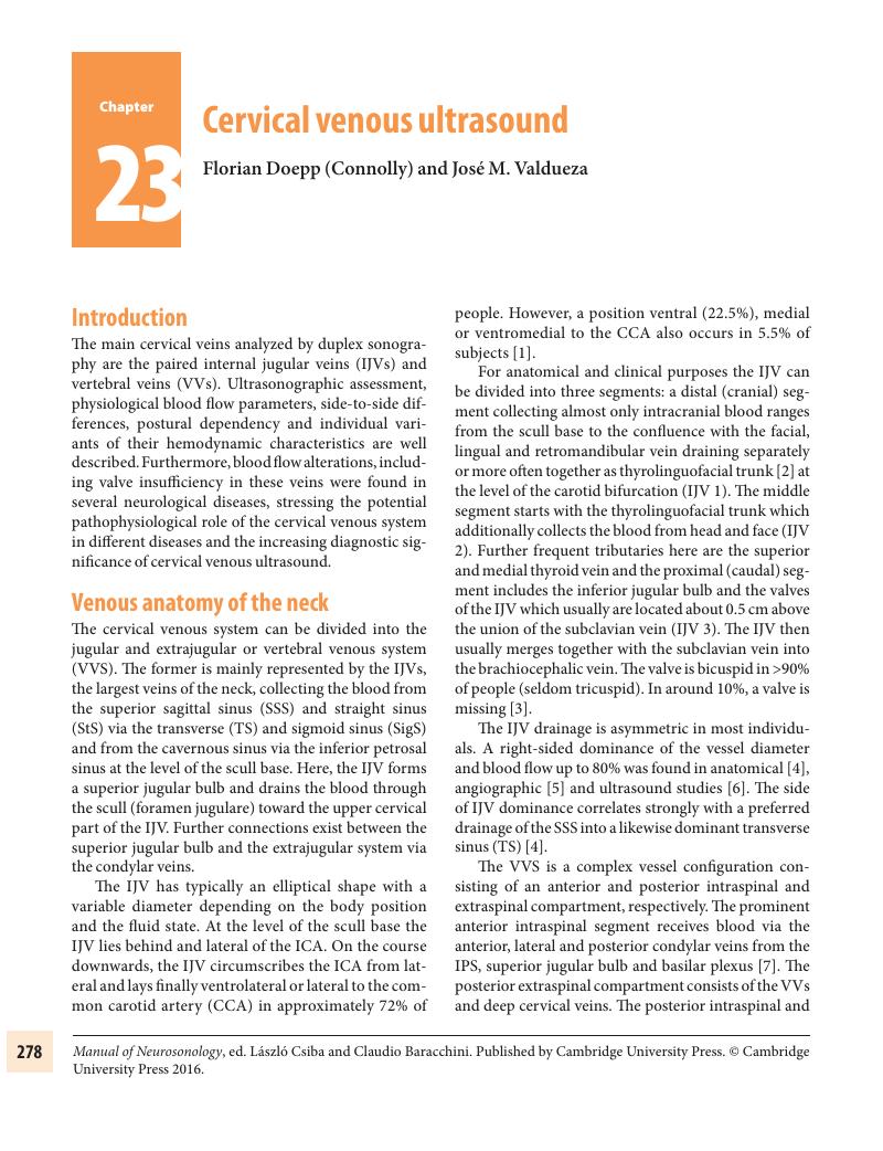
Image of the first page of this content. For PDF version, please use the ‘Save PDF’ preceeding this image.'
Type
Chapter
Information
Publisher: Cambridge University Press
Print publication year: 2016

Access options

Get access to the full version of this content by using one of the access options below. (Log in options will check for institutional or personal access. Content may require purchase if you do not have access.)

References

Troianos, CA, Hartmann, GS, Kathryn, EG, et al. Guidelines for performing ultrasound guided vascular cannulation: recommendations of the American Society of Echocardiography and the Society of Cardiovascular Anesthesiologists. J Am Soc Echocardiogr. 2011;24:12911318.Google Scholar
Shima, H, von Luedinghausen, M, Ohno, IK, et al. Anatomy of microvascular anastomosis in the neck. Plast Reconstr Surg. 1998;101:3341.Google Scholar
Lepori, D, Capasso, P, Fournier, D, et al. High-resolution ultrasound evaluation of internal jugular venous valve. Eur Radiol. 1999;9:12221226.Google Scholar
Saiki, K, Tsurumoto, T, Okamoto, K, et al. Relation between bilateral differences in internal jugular vein caliber and flow patterns of dural venous sinuses. Anat Sci Int. 2013;88:141150.Google Scholar
Durgung, B, Ilglt, ET, Cizmeli, MO. Evaluation by angiography of the lateral dominance of the drainage of the dural venous sinuses. Surg Radiol Anat. 1993;15:125130.CrossRefGoogle Scholar
Doepp, F, Hoffmann, O, Einhäupl, KM, et al. Normal blood flow velocities in the distal jugular vein and response during compression of the contralateral internal jugular vein. Cerebrovasc Dis. 1998;8(Suppl 3):121.Google Scholar
Ruiz, DSM, Gailloud, P, Rüfenacht, DA, et al. The craniocervical venous system in relation to cerebral venous drainage. AJNR. 2002;23:15001508.Google Scholar
Chou, CH, Chao, AC, Hu, HH. Ultrasonographic evaluation of vertebral venous valves. Am J Neuradiol. 2002;23:14181420.Google Scholar
Müller, HR, Hinn, G, Buser, MW. Internal jugular venous flow measurement by means of Duplex scanner. J Ultrasound Med. 1990;9:261265.Google Scholar
Pucheu, A, Evans, J, Thomas, D, et al. Doppler Ultrasonography of normal neck veins. J Clin Ultrasound. 1994; 22:367373.Google Scholar
Hoffmann, O, Weih, M, von Münster, T, et al. Blood flow velocities in the vertebral veins of healthy subjects: a duplex sonographic study. J Neuroimaging. 1999;9:198200.Google Scholar
Chung, CP, Hsu, HY, Chao, AC, et al. Flow volume in the jugular vein and related hemodynamics in the branches of the jugular vein. Ultrasound Med Biol. 2007;33:500505.Google Scholar
Doepp, F, Schreiber, SJ, von Münster, T, et al. How does the blood leave the brain? A systematic ultrasound analysis of cerebral venous drainage patterns. Neuroradiology. 2004;46:565570.Google Scholar
Hoffmann, O, Klingebiel, R, Braun, JS, et al. Diagnostic pitfall: atypical cerebral venous drainage via the vertebral venous system. Am J Neuroradiol. 2002;23:408411.Google Scholar
Schreiber, SJ, Lürtzing, F, Götze, R, et al. Extrajugular pathways of human cerebral venous blood drainage assessed by duplex ultrasound. J Appl Physiol. 2003;94: 18021805.Google Scholar
Peccola, J, Kangasniemi, M. Posterior fossa dural arteriovenous fistula: diagnostic and follow-up with time-resolved imaging of contrast kinetics (TRICKS) at 1.5T. Acta Radiologica. 2011;52:442447.CrossRefGoogle Scholar
Schreiber, SJ, Franke, U, Doepp, F, et al. Dopplersonographic measurement of global cerebral circulation time using echo contrast-enhanced ultrasound in normal individuals and patients with arteriovenous malformations. Ultrasound Med Biol. 2002;28:453458.Google Scholar
Schreiber, SJ, Kauert, A, Doepp, F, et al. Measurement of global cerebral circulation time using power duplex echo-contrast bolus tracking. Cerebrovasc Dis. 2003;15: 129132.Google Scholar
Müller, HR, Casty, M, Buser, M, et al. Ultrasonic jugular venous flow measurement. J Cardiovasc Ultrasonogr. 1988;7:2529.Google Scholar
Valdueza, JM, von Münster, T, Hoffmann, O, et al. Postural dependency of the cerebral venous outflow. Lancet. 2000;355:200201.CrossRefGoogle ScholarPubMed
Alperin, N, Lee, SH, Sivaramakrishnan, A. Quantifying the effect of posture on intracranial in humans by MRI flow studies. J Magn Reson Imaging. 2005;22:591596.CrossRefGoogle ScholarPubMed
Doepp, F, Paul, F, Valdueza, JM, et al. No cerebrocervical venous congestion in patients with multiple sclerosis. Ann Neurol. 2010;68:173183.Google Scholar
Lobato, EB, Sulek, CA, Moody, RL, et al. Cross-sectional area of the right and left internal jugular veins. J Cardiothorac Vasc Anesth. 1999;13:136138.Google Scholar
Chung, CP, Hu, HH. Jugular venous reflux. J Med Ultrasound. 2008;16:210222.Google Scholar
Hind, D, Calvert, N, McWilliams, R, et al. Ultrasonic location devices for central venous cannulation: meta-analysis. BMJ. 2003;327:361367.Google Scholar
Lordick, F, Hentrich, M, Decker, T, et al. Ultrasound screening for internal jugular vein thrombosis aids the detection of central venous catheter-related infections in patients with haemato-oncological diseases: a prospective observational study. Br J Haematol. 2003; 120:10731078.CrossRefGoogle ScholarPubMed
Lichtenstein, D, Saifi, R, Augarde, R, et al. The internal jugular veins are asymmetric. Usefulness of ultrasound before catheterization. Intensive Care Med. 2001;27:301305.Google Scholar
Lamperti, M, Subert, M, Cortelazzi, P, et al. Is a neutral head position safer than 45-degree neck rotation during ultrasound-guided internal jugular vein cannulation? Results of a randomized controlled clinical trial. Anesth Analg. 2012;114:777784.Google Scholar
Schreiber, SJ, Lambert, UK, Doepp, F, et al. Effects of prolonged head-down tilt on internal jugular vein cross-sectional area. Br J Anaesth. 2002;89:769771.Google Scholar
Brownlow, RL, McKinney, WM. Ultrasonographic evaluation of jugular venous valve competence. J Ultrasound Med. 1985;4:169172.Google Scholar
Akkawi, NM, Agosti, C, Borroni, B, et al. Jugular valve incompetence: a study using air contrast ultrasonography on a general population. J Ultrasound Med. 2002;21:747751.Google Scholar
Nedelmann, M, Eicke, BM, Dieterich, M. Functional and morphological criteria of internal jugular valve insufficiency as assessed by ultrasound. J Neuroimaging. 2005;15:7075.Google Scholar
Doepp, F, Bähr, D, John, M, et al. Internal jugular vein valve incompetence in COPD and primary pulmonary hypertension. J Clin Ultrasound. 2008;36:480484.Google Scholar
Schreiber, SJ, Doepp, F, Klingebiel, R, et al. Internal jugular vein valve incompetence and intracranial venous anatomy in transient global amnesia. J Neurol Neurosurg Psychiatry. 2005;76:509513.Google Scholar
Sander, D, Winbeck, K, Etgen, T, et al. Disturbance of venous flow patterns in patients with transient global amnesia. Lancet. 2000;356:19821984.Google Scholar
Doepp, F, Valdueza, JM, Schreiber, SJ. Incompetence of internal jugular valve in patients with primary exercional headache: a risk factor? Cephalalgia. 2008;28:182185.Google Scholar
Nedelmann, M, Kaps, M, Mueller-Forell, W. Venous obstruction and jugular valve insufficiency in idiopathic intracranial hypertension. J Neurol. 2009;256:964969.Google Scholar
Walter, C. Retrograde flow in the left internal jugular vein caused by compression of the left brachiocephalic vein while supine during moderate tidal volume ventilation: case report, evaluation by colour coded duplex sonography and review of literature. Ultraschall Med. 2002;23:260263.CrossRefGoogle ScholarPubMed
Donahue, SP, Wood, JP, Patel, BM, et al. Correlation of sonographic measurements of the internal jugular vein with central venous pressure. Am J Emerg Med. 2009;27:851855.Google Scholar
Simon, MA, Kliner, DE, Girod, JP, et al. Detection of elevated right atrial pressure using a simple bedside ultrasound measure. Am Heart J. 2010;159:421427.CrossRefGoogle ScholarPubMed
Zamboni, P, Galeotti, R, Menegatti, E, et al. Chronic cerebrospinal venous insufficiency in patients with multiple sclerosis. J Neurol Neurosurg Psychiatr. 2009;80:392399.CrossRefGoogle ScholarPubMed
Mayer, CA, Pfeilschifter, W, Lorenz, MW, et al. The perfect crime? CCSVI not leaving a trace in MS. J Neurol Neurosurg Psychiatr. 2011;82:436440.CrossRefGoogle Scholar
Comi, G, Battaglia, MA, Bertolotto, A, et al. Observational case-control study of the prevalence of chronic cerebrospinal venous insufficiency in multiple sclerosis: results from the CoSMo study. MSJ. 2013;19:15081517.Google Scholar
Valdueza, JM, Doepp, F, Schreiber, SJ, et al. What went wrong? The flawed concept of cerebrospinal venous insufficiency. J Cerebral Blood Flow Metab. 2013;33:657668.Google Scholar
Schurr, PH, Wickbom, I. Rapid serial angiography: further experience. J Neurol Psychiatr. 1952;15: 11231127.Google Scholar
Schreiber, SJ, Diehl, RR, Weber, W et al. Doppler sonographic evaluation of shunts in patients with dural arteriovenous fistulas. Am J Neuroradiol. 2004;25:775780.Google Scholar
Schreiber, SJ, Doepp, F, Spruth, E, et al. Ultrasonographic measurement of cerebral blood flow, cerebral circulation time and cerebral blood volume in vascular and Alzheimer’s dementia. J Neurol. 2005;252: 11711177.Google Scholar
Doepp, F, Valdueza, JM, Schreiber, SJ. Transcranial and extracranial ultrasound assessment of cerebral hemodynamics in vascular and Alzheimer’s dementia. Neurol Res. 2006;28:645649.Google Scholar
Mancini, M, Morra, VB, Di Donato, O, et al. Multiple sclerosis: cerebral circulation time. Radiology. 2012;262:947955.CrossRefGoogle ScholarPubMed
Sander, K, Poppert, H, Etgen, T, et al. Dynamics of intracranial venous flow patterns in patients with idiopathic intracranial hypertension. Eur Neurol 2011; 66: 334338.CrossRefGoogle ScholarPubMed

Save book to Kindle

To save this book to your Kindle, first ensure coreplatform@cambridge.org is added to your Approved Personal Document E-mail List under your Personal Document Settings on the Manage Your Content and Devices page of your Amazon account. Then enter the ‘name’ part of your Kindle email address below. Find out more about saving to your Kindle.

Note you can select to save to either the @free.kindle.com or @kindle.com variations. ‘@free.kindle.com’ emails are free but can only be saved to your device when it is connected to wi-fi. ‘@kindle.com’ emails can be delivered even when you are not connected to wi-fi, but note that service fees apply.

Find out more about the Kindle Personal Document Service.

Available formats
×

Save book to Dropbox

To save content items to your account, please confirm that you agree to abide by our usage policies. If this is the first time you use this feature, you will be asked to authorise Cambridge Core to connect with your account. Find out more about saving content to Dropbox.

Available formats
×

Save book to Google Drive

To save content items to your account, please confirm that you agree to abide by our usage policies. If this is the first time you use this feature, you will be asked to authorise Cambridge Core to connect with your account. Find out more about saving content to Google Drive.

Available formats
×